POU2AF1 / POU domain class 2-associating factor 1 · IHC design guide

Design Immunohistochemistry for POU2AF1

Plan POU2AF1 chromogenic IHC in paraffin sections using germinal center cells in tonsil or lymph node as positive controls (HPA tissue IHC). This guide covers fixation consistency, expected lymphoid staining, and interpretation of presumed off-target binding (HPA tissue IHC).

Evidence assembled Oct 2026 · For research use; verify linked source records and product datasheet before use
Immunohistochemistry protocol sheet for POU2AF1 (IHC for POU2AF1): expected localisation Nuclear expected (UniProt); nuclear and cytoplasmic staining observed (HPA tissue IHC), antibody M04431, validated IHC image, and IHC protocol steps
Printable POU2AF1 IHC protocol sheet — expected localisation Nuclear expected (UniProt); nuclear and cytoplasmic staining observed (HPA tissue IHC), antibody M04431, controls and protocol steps. Open the full POU2AF1 IHC guide →

POU2AF1 Immunohistochemistry Experimental Design Guide

Expected localisation, validated protocols, controls and antibodies — the at-a-glance facts below, then the full design guide.

Must know before staining
Expected localisation Nuclear expected (UniProt); nuclear and cytoplasmic staining observed (HPA tissue IHC)
Staining pattern Nuclear and cytoplasmic staining in lymphoid cell subsets (HPA tissue IHC)
Antigen retrieval EDTA pH 8.0 HIER, heat-mediated (datasheet M04431)
Positive control ⓘ Appendix+4 more · see all
Negative control ⓘ Adipose tissue+4 more · see all
Important caveats
Reasons your staining may differ from the expected pattern.
Fixation Keep fixation consistent across sections. (standard IHC practice; not target-specific)
Caveat Presumed off-target binding may mimic positive cells (HPA tissue IHC)
Regulation B-cell-specific expression (UniProt)
Isoform / epitope No annotated isoforms, signal peptide or propeptide (UniProt)
Section 1

Recommended POU2AF1 IHC & IF Protocols

Compare the catalog antibody’s IHC-P protocol (datasheet M04431) with three published POU2AF1 IHC protocols (PMC9035466; PMC8806937; PMC12833344).

Recommended immunohistochemistry (IHC-P) protocol parameters
SampleParaffin-embedded human spleen tissue; fixative not specified (datasheet M04431)
FixationImage fixative and duration unreported (datasheet M04431); verify before use.
Sectioning4–5 µm sections on charged slides (standard)
DeparaffinisationXylene, graded ethanol series to water (standard)
Antigen retrievalHeat retrieval: EDTA pH 8.0 (datasheet M04431); 20 min, 95–100 °C (standard)
Peroxidase block3% H2O2, 10 min, room temperature (standard)
Blocking10% goat serum (datasheet M04431)
Primary antibodyRabbit monoclonal (clone ADEH-16) anti-POU2AF1, 1:50 (datasheet M04431)
Primary incubationOvernight at 4 °C (datasheet M04431)
DetectionHRP-conjugated secondary, DAB chromogen (datasheet M04431)
CounterstainHematoxylin, blue, dehydrate and mount (standard)
Expected resultPOU2AF1-positive staining in lymphoid tissue of appendix (HPA tissue IHC: High). HPA tissue profile: Nuclear and cytoplasmic expression in subsets of lymphoid cells in several tissues. No signal in the no-primary control.
💡Decision noteStart with heat-mediated EDTA pH 8.0 for the catalog antibody (datasheet M04431). Published protocols using other antibodies report citrate pH 6.0 (PMC8806937; PMC12833344).
Section 2

What Is the Expected POU2AF1 Staining Pattern?

POU2AF1 is a nuclear transcriptional coactivator with no transmembrane segment (UniProt Q16633: location, function and topology). In tissue IHC, expect staining in subsets of lymphoid cells, especially germinal center cells; HPA also reports cytoplasmic staining in some lymphoid cells (HPA: tissue IHC). HPA rates its IHC evidence Enhanced but notes low consistency with RNA data and presumed off-target staining that was disregarded (HPA: reliability description).

What am I looking at on my slide?
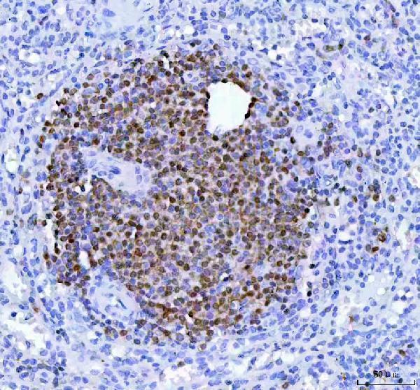
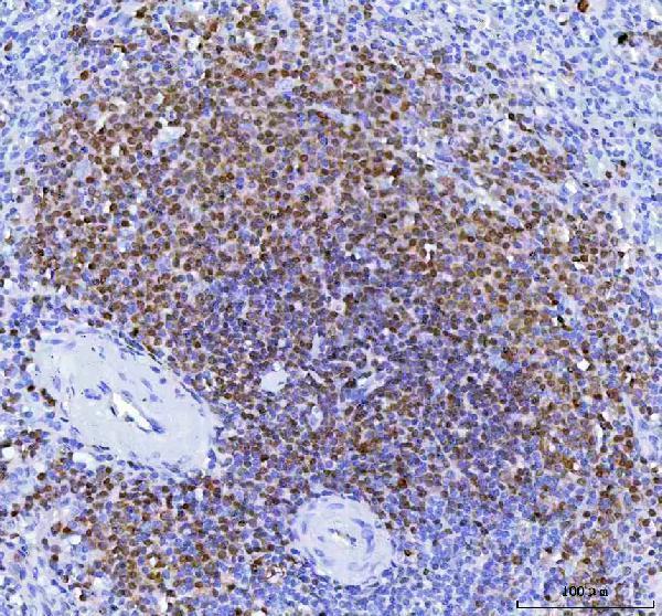
Germinal center cells stain in lymph node or tonsil; surrounding cells are less uniformly stained.This supports the expected cell distribution: HPA reports High staining in germinal center cells in both tissues (HPA: tissue IHC). Assess nuclear staining first, while allowing the cytoplasmic component reported in lymphoid subsets (UniProt Q16633: nucleus; HPA: tissue IHC).
Staining is confined to membranes, or appears cytoplasmic without discernible nuclear staining.A membrane-only pattern conflicts with the nuclear location and absence of a transmembrane segment (UniProt Q16633: location and topology). Cytoplasmic staining alone is less decisive because HPA reports a cytoplasmic component; compare it with nuclei and tissue controls before calling it specific (HPA: tissue IHC).
Broad staining appears in cells outside the expected lymphoid distribution.Question specificity, particularly if adipocytes or respiratory epithelial cells stain: HPA reports these cells as not detected (HPA: adipose tissue and bronchus IHC). Check a no-primary control for detection-related signal; HPA also notes presumed off-target staining in its tissue assessment (HPA: reliability description).
A uniform haze covers lymphoid and surrounding tissue, obscuring cell boundaries.The slide cannot establish the reported subset pattern (HPA: tissue IHC). In chromogenic IHC, background may arise from detection activity or inadequate blocking or washing (general IHC practice); evaluate controls before interpreting weak staining as POU2AF1.
No staining is visible in germinal center cells of lymph node or tonsil.This conflicts with the High staining reported at both sites (HPA: tissue IHC). Check tissue preservation and run controls for primary antibody and detection performance (general IHC practice); a blank slide alone cannot distinguish assay failure from a true negative.
💡Expected POU2AF1 appearanceCall a convincing positive when germinal center cells show clear nuclear staining, with possible cytoplasmic staining, against less uniformly stained neighbors; HPA reports High staining there, whereas membrane-only or widespread non-lymphoid signal is suspect (HPA: tissue IHC; UniProt Q16633: nucleus and topology).
How each factor affects the staining
Tissue and cell selectionLymph node and tonsil germinal centers are High; splenic white-pulp cells and bone-marrow hematopoietic cells are Medium (HPA: tissue IHC). Match the scored cell population to the atlas annotation rather than treating an entire section as uniformly positive.
Localization and topologyUniProt places POU2AF1 in the nucleus and records no transmembrane segment (UniProt Q16633: location and topology). HPA observes nuclear and cytoplasmic staining in lymphoid subsets (HPA: tissue IHC); require cell-context evidence before accepting cytoplasmic signal.
Antibody evidence and limitsThe listed IHC antibody, CAB011193, has an Enhanced IHC validation label (HPA: antibody record). HPA nevertheless reports low staining–RNA consistency and disregarded presumed off-target staining (HPA: reliability description); the label does not validate every stained cell.
Processing and target formThe recorded chain spans 1–256 aa, with no signal peptide or propeptide; UniProt lists no glycosylation sites or isoforms (UniProt Q16633: processing and features). These records offer no basis to interpret membrane or extracellular staining as a known processed POU2AF1 form.
IF/ICC: what can be inferred?Use the nuclear location as the localization reference (UniProt Q16633: location). HPA supplies no main ICC/IF location or cell-line images, so this record cannot establish an observed IF/ICC pattern or an IF/ICC antibody protocol (HPA: subcellular and antibody records).
Why is my staining missing, weak or wrong?
SituationLikely causeNext action
Positive tissue is blank.Assay failure is possible when expected germinal center staining is absent (HPA: lymph node and tonsil IHC).Verify tissue morphology, primary-antibody application and detection with appropriate controls; review retrieval against the antibody's IHC-P instructions (general IHC practice). Do not infer a POU2AF1-specific retrieval requirement from HPA.
Nuclei are hard to distinguish from cytoplasmic color.The reported pattern can include both compartments in lymphoid subsets (HPA: tissue IHC); dense chromogen or counterstain can obscure boundaries (general IHC practice).Inspect a lighter-developed section and the counterstain, then score nuclear and cytoplasmic signal separately (general IHC practice).
Membrane outlines dominate the slide.A membrane-only pattern conflicts with nuclear localization and the lack of a transmembrane segment (UniProt Q16633: location and topology).Check no-primary and known-positive controls, inspect the cell boundaries, and withhold a specific positive call unless the expected lymphoid nuclear pattern is also present (general IHC practice; HPA: tissue IHC).
Chromogen appears in many non-lymphoid cells.This may reflect nonspecific binding or endogenous detection activity (general IHC practice); HPA reports disregarded presumed off-target staining (HPA: reliability description).Compare with a no-primary control and an HPA-listed not-detected cell population; review blocking and detection controls before assigning cell-specific positivity (general IHC practice; HPA: tissue IHC).
Staining is weak in spleen or bone marrow.HPA reports Medium staining in splenic white-pulp cells and bone-marrow hematopoietic cells, compared with High in lymph node and tonsil germinal centers (HPA: tissue IHC).Judge the annotated cells, then compare the run with a germinal center positive control before changing assay conditions (HPA: tissue IHC; general IHC practice).
Leydig cells stain in testis.HPA lists Medium Leydig-cell staining but also describes low staining–RNA consistency and presumed off-target staining (HPA: testis IHC; reliability description).Record the observation separately from the lymphoid pattern and seek independent specificity evidence before treating it as established POU2AF1 expression (HPA: tissue IHC; general IHC practice).

Sample controls for POU2AF1 IHC & IF

🧪Run tonsil first: germinal center cells should show the strongest signal (HPA: High in tonsil germinal center cells). Use adipose tissue as a negative, where adipocytes are not detected (HPA: Not detected in adipocytes); non-B-cell stromal nuclei within the tonsil should lack specific nuclear staining (UniProt Q16633: B-cell specific; nucleus).
Positive control tissue: Appendix (Lymphoid tissue, HPA High)
Negative control tissue: Adipose tissue (HPA Not detected)
ICC-IF cell lines (HPA subcellular resource): HPA carries no ICC-IF cell line for POU2AF1; derive a cell-line control from the positive tissue's cell type (Lymphoid tissue) and confirm it by RNA or western blot first.
Technical controls: Include a no-primary, secondary-only control and an isotype control matched to the rabbit primary antibody’s clonality (caption: rabbit anti-POU2AF1; standard IHC practice), plus a validated POU2AF1 knockout sample as a biological negative (standard IHC practice). Quench endogenous peroxidase in tonsil sections before HRP/DAB detection (standard IHC practice).
⚠️Feasibility: Paraffin-section staining is documented in spleen with heat retrieval in EDTA at pH 8.0 and primary antibody at 1:50 overnight at 4°C (M04431 tissue-IHC caption); the caption does not establish whether retrieval is required. A target-specific fixation window and fixation effect are unreported, and the selected SKU caption does not state the fixative (M04431 tissue-IHC caption). Relative ease of frozen-section IHC or IF cannot be established from the supplied evidence; endogenous peroxidase can add background in lymphoid tissue (standard IHC practice).

HPA tissue IHC evidence for POU2AF1

Comprehensive Human Protein Atlas IHC scoring per tissue (reliability: Enhanced — Low consistency between antibody staining and RNA expression data. Presumed off target binding observed and disregarded. Antibody staining in cells/structures not annotated, view images.). Rows are taken directly from the HPA tissue chart — click any row's HPA link to view the source.

Positive expression · recommended positive controls

TissueCell typeLevelEvidenceSource
Appendix Lymphoid tissue High Protein (IHC) HPA →
Lymph node Germinal center cells High Protein (IHC) HPA →
Tonsil Germinal center cells High Protein (IHC) HPA →
Bone marrow Hematopoietic cells Medium Protein (IHC) HPA →
Spleen Cells in white pulp Medium Protein (IHC) HPA →

Undetected expression · recommended negative controls

TissueCell typeLevelEvidenceSource
Adipose tissue Adipocytes Not detected Protein (IHC) HPA →
Adrenal gland Glandular cells Not detected Protein (IHC) HPA →
Breast Adipocytes Not detected Protein (IHC) HPA →
Bronchus Respiratory epithelial cells Not detected Protein (IHC) HPA →
Caudate Glial cells Not detected Protein (IHC) HPA →
Section 3

Advanced POU2AF1 IHC Tips

Troubleshoot POU2AF1 chromogenic IHC by checking retrieval, cell identity and nuclear staining against the documented spleen protocol and tissue profile.

What retrieval should I start with for weak POU2AF1 staining in paraffin sections?
Start with heat mediated antigen retrieval in EDTA at pH 8.0 (datasheet M04431). The documented paraffin section workflow used this retrieval before staining human spleen with the catalog antibody at 1:50 overnight at 4°C (datasheet M04431). If staining is weak, check heating, cooling and buffer preparation across a small control series before changing antibody concentration; keep a known positive section in every run (standard IHC practice). Score cells in splenic white pulp separately from surrounding tissue, because that compartment showed staining while a whole section average could obscure it (HPA: spleen cells in white pulp, Medium).
Could fixation explain variable POU2AF1 staining between paraffin blocks?
Target specific fixation sensitivity is unknown: the catalog image identifies a paraffin section but does not state its fixative (datasheet M04431). Record each block’s actual fixative and processing history, then compare sections using the same retrieval, antibody incubation and detection conditions (standard IHC practice; datasheet M04431: EDTA pH 8.0, 1:50 overnight at 4°C). Include a positive control section in each run and examine whether the change affects the control as well as the test block (standard IHC practice). Do not assign weak staining to a POU2AF1 specific fixation effect without a controlled comparison (datasheet M04431: fixative unreported).
How should I assess nuclear versus cytoplasmic POU2AF1 staining?
Prioritize a nuclear signal when assessing POU2AF1, which is annotated as nuclear and lacks a transmembrane segment (UniProt Q16633: subcellular location and topology). Cytoplasmic staining needs closer scrutiny because tissue IHC reports nuclear and cytoplasmic expression in subsets of lymphoid cells, while also flagging presumed off target staining (HPA: tissue profile and Enhanced reliability description). Use a light nuclear counterstain and compare the signal with individual cell boundaries at high magnification (standard IHC practice). Record nuclear and cytoplasmic scores separately, and check whether each pattern follows expected lymphoid cells rather than every cell in the section (UniProt Q16633: B cell specificity; HPA: tissue profile).
Could an isoform or epitope difference explain discordant POU2AF1 staining?
No alternative isoform is listed in this record, so an isoform switch is not an evidence based explanation for discordant staining here (UniProt Q16633: 0 isoforms). The recorded protein is a 256 residue chain with an OCA domain at residues 16–38, no signal peptide, no propeptide and no recorded glycosylation sites (UniProt Q16633: processing, domains and modifications). The supplied catalog caption does not identify the antibody epitope, so do not attribute retrieval behavior to a particular residue or domain (datasheet M04431: epitope unspecified). Compare the staining pattern with another independently characterized antibody if available, alongside the same positive and negative tissue controls (standard IHC practice).
How can I investigate POU2AF1 by multiplex IF after seeing an IHC signal?
Treat multiplex IF as a separate assay to optimize: this payload supplies a paraffin section IHC caption but no catalog IF protocol or subcellular IF images (datasheet M04431; HPA: subcellular images unavailable). Pair POU2AF1 with a validated B cell marker and use a nuclear counterstain to test whether the signal occupies expected cells and nuclei (UniProt Q16633: B cell specificity and nuclear location; standard IF practice). Choose fluorophores after checking unstained tissue autofluorescence, and reserve a spectrally distinct channel for the weaker signal (standard IF practice). Because POU2AF1 is intracellular with no transmembrane segment, include an optimized permeabilisation step after fixation and verify nuclear access with controls (UniProt Q16633: location and topology; standard IF practice).
What should I check when brown staining appears outside expected lymphoid cells?
First inspect a no primary control for endogenous peroxidase or nonspecific detection, and review whether the peroxidase block, serum block and washes were effective (standard IHC practice; datasheet M04431: 10% goat serum block and HRP/DAB detection). The catalog spleen protocol used rabbit primary antibody at 1:50 overnight at 4°C; titrate from that documented starting point if diffuse staining persists (datasheet M04431). Compare suspected background with an expected positive compartment and a separately processed negative tissue (HPA: spleen white pulp, Medium; adipose adipocytes, Not detected). Flag widespread staining outside the expected cells for review, since presumed off target binding was reported in tissue images (HPA: Enhanced reliability description).
How should I quantify POU2AF1 staining across germinal centers? ⚠ ANSWER MARKED FOR VERIFICATION
Define the region and cell population before scoring; germinal center cells in lymph node and tonsil have high reported staining (HPA: lymph node and tonsil germinal center cells, High). For chromogenic IHC, report the percentage of positive cells and, when intensity is reproducible, an H-score calculated from percentages at each intensity level (standard IHC practice). Normalize cell counts to all evaluable cells in the annotated compartment, or report positive cell density per mm² of that compartment rather than per whole section (standard IHC practice). Keep nuclear and cytoplasmic scores distinct, and use the same threshold and exclusion rules for every slide (UniProt Q16633: nuclear location; HPA: nuclear and cytoplasmic tissue profile).
How can I distinguish true POU2AF1 positivity from staining artefact?
Give greatest weight to staining in the expected cell population and compartment: POU2AF1 is B cell specific and annotated as nuclear, with high germinal center staining in lymph node and tonsil (UniProt Q16633: tissue specificity and location; HPA: lymph node and tonsil, High). Review cytoplasmic only signals or unexpected cell types against the tissue context, because presumed off target binding was observed and disregarded in the tissue dataset (HPA: profile and Enhanced reliability description). Exclude section edges, necrotic areas and pigment or precipitate when they distort cell level scoring (standard IHC practice). Use a no primary control to investigate residual brown signal from endogenous enzyme or detection reagents (standard IHC practice).
Boster reagents

Best POU2AF1 / POU domain class 2-associating factor 1 IHC Antibodies

The catalog includes a POU2AF1 IHC figure from paraffin-embedded human spleen (M04431 image caption) and lists a human-reactive IF/ICC antibody without an IF figure (M04431-3 catalog).

Real IHC data IHC analysis of POU2AF1 using anti-POU2AF1 antibody (M04431). POU2AF1 was detected in a paraffin-embedded section of human spleen tissue. Heat mediated antigen retrieval was performed in EDTA buffer (pH 8.0, epitope retrieval solution). The tissue section was blocked with 10% goat serum. The tissue section was then incubated with 1:50 rabbit anti-POU2AF1 Antibody (M04431) overnight at 4°C. Peroxidase Conjugated Goat Anti-rabbit IgG was used as secondary antibody and incubated for 30 minutes at 37°C. The tissue section was developed using HRP Conjugated Rabbit IgG Super Vision Assay Kit (Catalog # SV0002) with DAB as the chromogen.
Anti-BOB1 Monoclonal Antibody
Cat # M04431

M04431 has IHC captions for paraffin-embedded human spleen and tonsil and rat lymph tissue (M04431 image captions). M04431-3 lists human reactivity and IHC and ICC/IF applications, but provides no IHC or IF image (M04431-3 catalog).

Which to pick: For tissue IHC, choose M04431: its rabbit monoclonal antibody has a paraffin-section IHC caption using 1:50 dilution and EDTA retrieval (M04431 catalog; M04431 image caption). For human IF/ICC, M04431-3 lists those applications and a 1:50 IF dilution, but has no IF figure (M04431-3 catalog). For human and rat IHC work, M04431 lists both species and has captions for human and rat tissue; its caption does not report the fixative (M04431 catalog; M04431 image captions).

Each figure is that product's own IHC / IF validation image from its datasheet.

References

  1. UniProt Consortium. UniProt entry Q16633 (OBF1_HUMAN, POU domain class 2-associating factor 1).
  2. Human Protein Atlas. POU2AF1 tissue IHC expression (reliability: Enhanced).
  3. Human Protein Atlas. POU2AF1 subcellular location (ICC-IF): Highest expression in REH: 66.1 nTPM.
  4. Human Protein Atlas. POU2AF1 antibody validation summary (1 antibodies).
  5. CD19 and POU2AF1 are Potential Immune-Related Biomarkers Involved in the Emphysema of COPD: On Multiple Microarray Analysis. Journal of inflammation research 2022 — PMC9035466.
  6. POU class 2 homeobox associating factor 1 (POU2AF1) participates in abdominal aortic aneurysm enlargement based on integrated bioinformatics analysis. Bioengineered 2021 — PMC8806937.
  7. HLA-DRA-mediated inhibition of T-Cell proliferation by differentiated airway epithelium and its disruption in smoking-associated airway inflammation. Respiratory research 2026 — PMC13289115.
  8. Identification of a prognostic signature based on immunogenic adverse event-related genes to guide therapy for non-small cell lung cancer. Frontiers in immunology 2025 — PMC12833344.
  9. PubMed PMID:7859290 — UniProt-cited evidence.
  10. PubMed PMID:7779176 — UniProt-cited evidence.
  11. PubMed PMID:8574789 — UniProt-cited evidence.