This website uses cookies to ensure you get the best experience on our website.


- Table of Contents
Plan POU2F1 chromogenic IHC in paraffin sections around the nuclear staining reported in most cells (HPA tissue IHC). This guide highlights high staining in colon endocrine cells (HPA tissue IHC), fixation consistency (standard IHC practice), and isoform considerations (UniProt).

Expected localisation, validated protocols, controls and antibodies — the at-a-glance facts below, then the full design guide.
| Expected localisation | Nuclear staining in most cells (HPA tissue IHC) | |
| Staining pattern | Nuclear in most cells; high in colon endocrine cells (HPA tissue IHC) | |
| Antigen retrieval | EDTA pH 8.0 HIER, heat-mediated (datasheet M01766-1) | |
| Positive control | Colon+4 more · see all | |
| Negative control | Adipose tissue+4 more · see all |
| Fixation | Keep fixation consistent across sections. (standard IHC practice; not target-specific) | |
| Caveat | Staining has medium consistency with RNA expression (HPA tissue IHC) | |
| Regulation | Ubiquitous expression (UniProt) | |
| Isoform / epitope | 6 isoforms; isoform 2 is lymphocyte-specific (UniProt) |
The catalog antibody uses EDTA pH 8.0 retrieval (datasheet M01766-1). The published IHC protocols below cover lung tumor, cardiac, and tumor tissue sections (PMC10183409; PMC13543148; PMC13097874).
| Sample | Paraffin-embedded human breast cancer tissue; fixative not specified (datasheet M01766-1) |
| Fixation | Image fixative and duration unreported (datasheet M01766-1); verify before use. |
| Sectioning | 4–5 µm sections on charged slides (standard) |
| Deparaffinisation | Xylene, graded ethanol series to water (standard) |
| Antigen retrieval | Heat retrieval: EDTA pH 8.0 (datasheet M01766-1); 20 min, 95–100 °C (standard) |
| Peroxidase block | 3% H2O2, 10 min, room temperature (standard) |
| Blocking | 10% goat serum (datasheet M01766-1) |
| Primary antibody | Rabbit monoclonal (clone 19P20) anti-POU2F1, 1:50 recommended; image 2 μg/ml (datasheet M01766-1) |
| Primary incubation | Overnight at 4 °C (datasheet M01766-1) |
| Detection | HRP-conjugated secondary, DAB chromogen (datasheet M01766-1) |
| Counterstain | Hematoxylin, blue, dehydrate and mount (standard) |
| Expected result | POU2F1-positive staining in endocrine cells of colon (HPA tissue IHC: High). HPA tissue profile: Nuclear expression in most cells. No signal in the no-primary control. |
POU2F1 should stain nuclei in most cells, with prominent staining in selected endocrine, ductal, placental and germ cells (HPA: nuclear expression in most cells; tissue IHC). This agrees with its nuclear localisation and lack of a transmembrane segment (UniProt P14859: subcellular location and topology). Treat tissue IHC patterns as supported, with medium consistency between antibody staining and RNA expression (HPA: reliability Supported).
| Nuclear staining in colon or rectal endocrine cells, kidney collecting ducts, placental syncytiotrophoblast cell bodies, or testicular preleptotene spermatocytes. | These are reported high-staining cell populations and useful positive comparisons (HPA: tissue IHC, High). Score the named cells and their nuclei; staining elsewhere in the section does not replace that comparison (general IHC practice). |
| Predominantly cytoplasmic staining with little or no nuclear signal in a high-staining cell population. | This disagrees with the reported tissue IHC pattern (HPA: nuclear expression in most cells). Check morphology and controls before calling it POU2F1. Approved ER and vesicle localisation in ICC-IF does not establish the same pattern in paraffin IHC (HPA: subcellular ICC-IF). |
| Strong staining confined to a cell population reported as not detected, such as adipocytes or cardiomyocytes. | The result conflicts with those specific tissue observations (HPA: adipocytes and cardiomyocytes, Not detected). Consider antibody cross-reactivity or endogenous chromogen-generating activity, and compare with controls; neither cause follows from staining alone (general IHC practice). |
| Diffuse colour across tissue spaces or many cells without discernible nuclear boundaries. | This is difficult to score against the expected nuclear pattern (HPA: tissue IHC profile). Incomplete blocking, excess reagent, or detection background can produce diffuse staining (general IHC practice); assess a no-primary control and tissue morphology. |
| No nuclear staining in the named high-staining cells of a positive comparison section. | A negative test section is hard to interpret until the positive comparison works (HPA: tissue IHC, High). Review section quality and the IHC workflow, including retrieval and detection, without assuming a POU2F1-specific fixation effect (general IHC practice). |
| Cell population and tissue | POU2F1 is described as ubiquitous, while HPA reports low tissue RNA specificity and different IHC staining levels among cell populations (UniProt P14859: tissue specificity; HPA: tissue IHC). Compare like cells when judging signal. |
| Isoform context | Six isoforms are listed, and isoform 2 is lymphocyte-specific (UniProt P14859: isoforms and tissue specificity). Those facts alone do not predict which isoform an antibody detects or require visible staining in every lymphocyte. |
| Assay-specific localisation | Nucleoplasm is the approved main ICC-IF location; ER and vesicles are additional approved locations (HPA: subcellular ICC-IF). For paraffin IHC, use the reported predominantly nuclear tissue pattern to interpret the slide (HPA: tissue IHC). |
| Strength of antibody evidence | HPA064323 and CAB002608 have Supported IHC status; HPA028256 and HPA058559 have no supplied IHC status (HPA: antibody validation). The supplied statuses do not establish performance for a different catalog antibody. |
| Chromogenic detection background | Endogenous activity can contribute colour independent of primary-antibody binding, depending on the detection chemistry (general IHC practice). A no-primary control helps identify that contribution; HPA tissue staining levels do not diagnose its cause. |
| Situation | Likely cause | Next action |
|---|---|---|
| The positive comparison lacks nuclear signal. | The run may have failed at retrieval, primary incubation, or detection; the slide alone cannot isolate the step (general IHC practice). | Check section integrity and reagent controls, then review the validated IHC-P workflow used for the run (general IHC practice). Use the named high-staining cells as the readout (HPA: tissue IHC, High). |
| Colour is predominantly cytoplasmic in paraffin IHC. | The pattern conflicts with nuclear expression reported in tissue IHC (HPA: tissue IHC profile); its cause is unresolved. | Inspect nuclear counterstain and morphology, then compare positive and no-primary sections (general IHC practice). Do not transfer ICC-IF ER or vesicle observations directly to paraffin IHC (HPA: subcellular ICC-IF). |
| A supposedly negative cell population stains strongly. | Cross-reactivity or chromogenic background is possible; HPA's Not detected call is an observation, not proof that every cell or preparation must be blank (HPA: tissue IHC; general IHC practice). | Identify the stained cell type, inspect the no-primary control, and compare with a named high-staining population on a comparable section (HPA: tissue IHC, High; general IHC practice). |
| Staining is diffuse and nuclear borders cannot be scored. | Nonspecific reagent binding or detection background may obscure compartment assignment (general IHC practice). | Review blocking, reagent concentrations, washing, and detection controls within the chosen IHC workflow (general IHC practice). Judge the result only when nuclear signal can be distinguished from background (HPA: tissue IHC profile). |
| Nuclear staining varies across the section. | Cell populations have different reported staining levels, including High, Medium, Low, and Not detected (HPA: tissue IHC). Uneven staining alone does not identify a technical failure. | Map staining to the named cell populations before comparing intensity. If the same population varies by section area, inspect section quality and processing consistency (HPA: tissue IHC; general IHC practice). |
| Q: IF/ICC shows signal outside the nucleus; is it inconsistent? | A: Nucleoplasm is the approved main location, with ER and vesicles approved as additional locations in ICC-IF (HPA: subcellular ICC-IF). | Interpret the IF/ICC image against those compartment assignments and its controls (HPA: subcellular ICC-IF; general IF practice). Use the separate IF/ICC guide for assay procedure. |
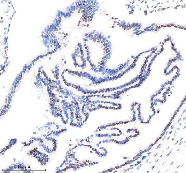
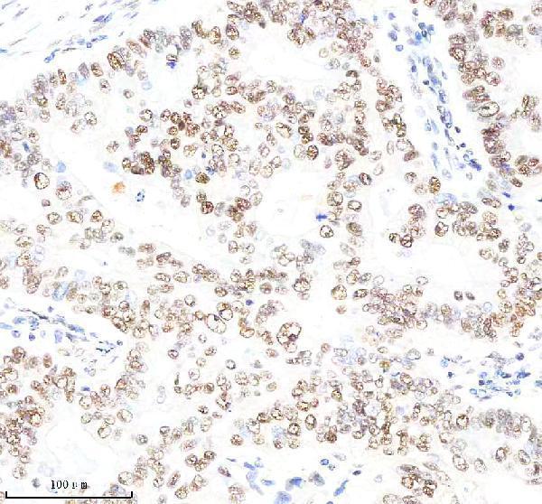


Comprehensive Human Protein Atlas IHC scoring per tissue (reliability: Supported — Medium consistency between antibody staining and RNA expression data.). Rows are taken directly from the HPA tissue chart — click any row's HPA link to view the source.
| Tissue | Cell type | Level | Evidence | Source |
|---|---|---|---|---|
| Colon | Endocrine cells | High | Protein (IHC) | HPA → |
| Kidney | Collecting ducts | High | Protein (IHC) | HPA → |
| Placenta | Syncytiotrophoblasts - cell body | High | Protein (IHC) | HPA → |
| Rectum | Endocrine cells | High | Protein (IHC) | HPA → |
| Testis | Preleptotene spermatocytes | High | Protein (IHC) | HPA → |
| Tissue | Cell type | Level | Evidence | Source |
|---|---|---|---|---|
| Adipose tissue | Adipocytes | Not detected | Protein (IHC) | HPA → |
| Bone marrow | Hematopoietic cells | Not detected | Protein (IHC) | HPA → |
| Bronchus | Respiratory epithelial cells | Not detected | Protein (IHC) | HPA → |
| Cerebellum | Cells in granular layer | Not detected | Protein (IHC) | HPA → |
| Heart muscle | Cardiomyocytes | Not detected | Protein (IHC) | HPA → |
Troubleshoot POU2F1 staining by checking retrieval, nuclear localisation, cell identity and matched controls before comparing signal across sections.
Two anti-POU2F1 antibodies have human tissue IHC images: breast cancer paraffin sections (M01766-1 image caption) and tonsil tissue (PB9317 image caption). Neither lists IF/ICC (catalog applications).
M01766-1 shows IHC in human breast cancer paraffin sections (M01766-1 image caption) and lists Human, Mouse and Rat reactivity (catalog). PB9317 shows IHC(P) in human tonsil tissue (PB9317 image caption) and lists Human reactivity (catalog).
Which to pick: For human paraffin-section IHC, M01766-1 has a breast cancer image with EDTA retrieval and DAB detection (M01766-1 image caption); PB9317 has a human tonsil IHC(P) image (PB9317 image caption). Choose M01766-1 when Mouse or Rat reactivity is needed, while recognizing that its shown IHC tissue is human (catalog reactivity; M01766-1 image caption). Neither SKU lists IF/ICC (catalog applications), and neither IHC caption reports the fixative (M01766-1 and PB9317 image captions).