PPA2 / Inorganic pyrophosphatase 2, mitochondrial · IHC design guide

Design Immunohistochemistry for PPA2

Plan PPA2 chromogenic IHC in paraffin sections using the catalog antibody's IHC conditions (datasheet A06587). Assess cytoplasmic staining with cell-type controls, since HPA reports high staining in some glandular cells and no detected staining in some muscle cells (HPA tissue IHC).

Evidence assembled Oct 2026 · For research use; verify linked source records and product datasheet before use
Immunohistochemistry protocol sheet for PPA2 (IHC for PPA2): expected localisation Cytoplasmic staining (HPA tissue IHC); mitochondrial protein (UniProt), antibody A06587, validated IHC image, and IHC protocol steps
Printable PPA2 IHC protocol sheet — expected localisation Cytoplasmic staining (HPA tissue IHC); mitochondrial protein (UniProt), antibody A06587, controls and protocol steps. Open the full PPA2 IHC guide →

PPA2 Immunohistochemistry Experimental Design Guide

Expected localisation, validated protocols, controls and antibodies — the at-a-glance facts below, then the full design guide.

Must know before staining
Expected localisation Cytoplasmic staining (HPA tissue IHC); mitochondrial protein (UniProt)
Staining pattern Cytoplasmic staining in most tissues (HPA tissue IHC)
Antigen retrieval EDTA pH 8.0 HIER, heat-mediated (datasheet A06587)
Positive control ⓘ Breast+4 more · see all
Negative control ⓘ Adipose tissue+3 more · see all
Important caveats
Reasons your staining may differ from the expected pattern.
Fixation Matched tissue-IHC evidence does not establish the fixation claim. Validate the specimen-specific method before use. (selected-SKU IHC image A06587)
Caveat Cell-type variation can confound tissue scores (HPA tissue IHC)
Regulation Stimulus-dependent changes unreported (UniProt)
Isoform / epitope Five isoforms; residues 1–32 are absent from the mature chain (UniProt)
Section 1

Recommended PPA2 IHC & IF Protocols

The catalog antibody has an IHC-P protocol (datasheet: A06587). One published KIRC study provides an additional staining workflow (PMC8428262: methods).

Recommended immunohistochemistry (IHC-P) protocol parameters
SampleParaffin-embedded human breast cancer tissue; fixative not specified (datasheet A06587)
FixationImage fixative and duration unreported (datasheet A06587); verify before use.
Sectioning4–5 µm sections on charged slides (standard)
DeparaffinisationXylene, graded ethanol series to water (standard)
Antigen retrievalHeat retrieval: EDTA pH 8.0 (datasheet A06587); 20 min, 95–100 °C (standard)
Peroxidase block3% H2O2, 10 min, room temperature (standard)
Blocking10% goat serum (datasheet A06587)
Primary antibodyRabbit anti-PPA2, 2-5 μg/ml (datasheet A06587)
Primary incubationOvernight at 4 °C (datasheet A06587)
DetectionHRP-conjugated secondary, DAB chromogen (datasheet A06587)
CounterstainHematoxylin, blue, dehydrate and mount (standard)
Expected resultPPA2-positive staining in glandular cells of breast (HPA tissue IHC: High). HPA tissue profile: Cytoplasmic expression in most tissues. No signal in the no-primary control.
💡Decision noteStart with heat-mediated EDTA pH 8.0 retrieval for the catalog antibody (datasheet: A06587). The published workflow specifies microwave retrieval without naming a buffer (PMC8428262: methods).
Section 2

What Is the Expected PPA2 Staining Pattern?

PPA2 localizes to mitochondria (UniProt Q9H2U2; HPA subcellular: supported). In paraffin-section IHC, expect cytoplasmic staining in many tissues (HPA tissue IHC: Supported), especially glandular cells in breast, endometrium, fallopian tube, gallbladder, parathyroid gland, prostate and rectum, and type II alveolar cells in lung (HPA tissue IHC: High). PPA2 has no annotated transmembrane segment (UniProt Q9H2U2 topology).

What am I looking at on my slide?
Cytoplasmic staining in breast glandular cells or lung type II alveolar cells (HPA tissue IHC: High).This fits the reported cell distribution. A granular cytoplasmic appearance is compatible with mitochondrial localization, although chromogenic IHC alone cannot prove organelle identity (HPA subcellular: mitochondria supported; general IHC interpretation).
Predominantly nuclear, membranous or extracellular staining.This conflicts with the reported mitochondrial location and absence of an annotated transmembrane segment (HPA subcellular: supported; UniProt Q9H2U2 topology). Check morphology and controls before assigning it to PPA2 (general IHC practice).
Strong staining in adipocytes or ovarian stromal cells.HPA reports PPA2 as not detected in those cell types (HPA tissue IHC). Consider cross-reactivity or endogenous detection activity, and compare a no-primary control; neither cause is established by the tissue profile (general IHC practice).
Uniform color across cells, stroma and empty spaces.Diffuse deposition is difficult to reconcile with the reported cellular, cytoplasmic pattern (HPA tissue IHC). Review the no-primary control and detection reagents before scoring specific staining (general IHC practice).
No staining in glandular cells of a reference breast section.HPA reports high staining there, so an absent result warrants a run check (HPA tissue IHC: High in breast glandular cells). It does not by itself establish absent PPA2 in the specimen (general IHC interpretation).
💡Expected PPA2 appearanceCall a positive result when appropriate cells show discernible cytoplasmic staining, potentially granular, with high staining in the listed HPA-positive cell types; dominant nuclear, membranous or tissue-wide diffuse color is suspect (HPA tissue IHC: High; HPA subcellular: mitochondria supported; general IHC interpretation).
How each factor affects the staining
Cell type within the sectionScore the named cells, not the tissue as a whole: HPA reports high staining in lung type II alveolar cells but no detection in ovarian stromal cells (HPA tissue IHC).
Reference tissue choiceBreast glandular cells offer a reported high-staining reference; adipocytes offer a reported not-detected comparison (HPA tissue IHC). HPA rates the overall tissue pattern Supported, pending external verification (HPA tissue IHC reliability).
Antibody evidenceHPA030888, HPA031671 and HPA031672 each have Supported IHC status (HPA antibodies). This supports comparison with HPA patterns, but does not validate an unlisted catalog antibody (HPA antibodies; general IHC interpretation).
Processing and isoformsUniProt lists a mature chain at residues 33–334 and five isoforms (UniProt Q9H2U2). The supplied record gives no antibody epitope, so it cannot predict which forms the IHC antibody detects.
IF/ICC: what should colocalization show?A mitochondrial pattern is expected (HPA subcellular: supported); HPA031671 has Supported ICC status (HPA antibodies). Those IF/ICC observations do not establish chromogenic IHC organelle resolution (general IHC interpretation).
Why is my staining missing, weak or wrong?
SituationLikely causeNext action
The reference breast section has no signal.A staining-run problem is possible because HPA reports high PPA2 in breast glandular cells (HPA tissue IHC). The supplied sources do not establish PPA2-specific fixation sensitivity.Confirm the intended glandular cells are present; review the catalog antibody's IHC-P instructions, reagent performance and run controls (general IHC practice).
The signal is chiefly nuclear or at cell borders.The compartment conflicts with mitochondrial localization and the lack of an annotated transmembrane segment (HPA subcellular: supported; UniProt Q9H2U2 topology).Check the no-primary control, section morphology and whether cytoplasmic signal appears in a HPA-positive reference cell type (general IHC practice; HPA tissue IHC).
Adipocytes stain as strongly as the reference cells.HPA reports PPA2 as not detected in adipocytes (HPA tissue IHC). Cross-reactivity or detection background is possible, but this comparison cannot identify the cause alone (general IHC interpretation).Compare no-primary and tissue controls, then assess whether staining follows cell boundaries or appears broadly across the section (general IHC practice).
Color covers stroma and blank areas.Broad deposition conflicts with the reported cytoplasmic cellular pattern (HPA tissue IHC); endogenous detection activity or excess background is possible (general IHC practice).Inspect the no-primary control and the detection and blocking steps, then score only interpretable cellular staining (general IHC practice).
Skeletal-muscle myocytes show little or no stain.HPA reports PPA2 as not detected in myocytes, while UniProt reports detection in skeletal muscle without assigning that signal to myocytes (HPA tissue IHC; UniProt Q9H2U2 tissue specificity).Do not treat myocytes alone as a failed-run control; assess a HPA high-staining cell type in the same run (HPA tissue IHC; general IHC practice).
IF/ICC appears mitochondrial, but IHC looks diffuse.HPA supports mitochondrial ICC localization, while its tissue IHC description is cytoplasmic; chromogenic IHC may not resolve individual mitochondria (HPA subcellular; HPA tissue IHC; general IHC interpretation).Judge IHC by cell type and cytoplasmic restriction, and check its own controls rather than requiring visible mitochondrial puncta (HPA tissue IHC; general IHC practice).

Sample controls for PPA2 IHC & IF

🧪Run breast first and score glandular cells for PPA2 staining (HPA: High in breast glandular cells). Use adipose tissue as the negative comparator, focusing on adipocytes (HPA: Not detected in adipocytes); compare adjacent nonglandular cells on the breast slide with background, without assuming they are PPA2-negative (HPA: breast annotation covers glandular cells).
Positive control tissue: Breast (Glandular cells, HPA High)
Negative control tissue: Adipose tissue (HPA Not detected)
ICC-IF cell lines (HPA subcellular resource): HPA ICC-IF images show PPA2 in A-431, U-251MG, U2OS, KOLF2.1J, with annotated localisation: Mitochondria (supported) (HPA subcellular).
Technical controls: Include a no-primary, secondary-only control; a concentration-matched rabbit IgG isotype control (caption: rabbit anti-PPA2 primary; clonality unreported); and a PPA2 knockout specimen as a biological negative. Block endogenous peroxidase and inspect breast sections for DAB-like background before scoring (caption: HRP/DAB detection).
⚠️Feasibility: A target-specific fixation window and fixation effect are unreported, and the selected A06587 tissue-IHC caption does not state a fixative (caption: fixative unreported). The reported paraffin-section procedure uses heat retrieval in EDTA at pH 8.0, so that is a starting condition rather than proof that retrieval is required (caption: EDTA retrieval). There is no supplied comparison showing frozen sections or IF to be easier; IF can assess mitochondrial localisation (HPA: supported mitochondrial ICC-IF localisation), while breast-section background should be checked under the reported HRP/DAB detection (caption: breast cancer section and HRP/DAB detection).

HPA tissue IHC evidence for PPA2

Comprehensive Human Protein Atlas IHC scoring per tissue (reliability: Supported — Medium consistency between antibody staining and RNA expression data. Pending external verification.). Rows are taken directly from the HPA tissue chart — click any row's HPA link to view the source.

Positive expression · recommended positive controls

TissueCell typeLevelEvidenceSource
Breast Glandular cells High Protein (IHC) HPA →
Endometrium Glandular cells High Protein (IHC) HPA →
Fallopian tube Glandular cells High Protein (IHC) HPA →
Gallbladder Glandular cells High Protein (IHC) HPA →
Lung Alveolar cells type II High Protein (IHC) HPA →

Undetected expression · recommended negative controls

TissueCell typeLevelEvidenceSource
Adipose tissue Adipocytes Not detected Protein (IHC) HPA →
Ovary Ovarian stroma cells Not detected Protein (IHC) HPA →
Skeletal muscle Myocytes Not detected Protein (IHC) HPA →
Smooth muscle Smooth muscle cells Not detected Protein (IHC) HPA →
Section 3

Advanced PPA2 IHC Tips

Troubleshoot PPA2 staining in paraffin sections by checking retrieval, compartment, cell type and controls before assigning biological meaning (UniProt Q9H2U2; HPA tissue IHC).

What retrieval should I try first when PPA2 staining is weak?
Start with heat-mediated antigen retrieval in EDTA at pH 8.0 (datasheet A06587). The selected paraffin-section example used that retrieval before overnight incubation with 2 μg/ml antibody at 4°C, so check both steps before changing the buffer (caption A06587). If staining remains weak, compare a carefully controlled alternative retrieval condition on serial sections while holding detection and exposure constant (standard IHC practice). Excessive heating can damage section morphology or increase nonspecific staining, so judge any gain against tissue preservation and a no-primary control (standard IHC practice). Score the same cell population across conditions; high staining in breast glandular cells provides a reference pattern (HPA tissue IHC).
How should I troubleshoot suspected fixation-related loss of PPA2 staining?
The selected image identifies a paraffin-embedded section but does not report its fixative, so target-specific fixation sensitivity is unknown (caption A06587). Record the fixative, fixation duration and processing history for each specimen, and compare sections with similar handling before attributing a weak signal to PPA2 biology (standard IHC practice). Keep the documented EDTA pH 8.0 retrieval and 2 μg/ml primary concentration constant during that comparison (caption A06587). Check whether tissue morphology and a known positive compartment remain interpretable after processing (standard IHC practice; UniProt Q9H2U2 localisation). Neither mitochondrial localisation nor the annotated modified residues establishes a preferred fixative for this assay (UniProt Q9H2U2).
Where should convincing chromogenic PPA2 staining appear?
Expect cellular cytoplasmic staining in tissue sections, consistent with the reported IHC profile, while recognising that PPA2 is a mitochondrial protein (HPA tissue IHC; UniProt Q9H2U2). Supported subcellular imaging places PPA2 in mitochondria, but chromogenic section resolution may make mitochondrial signal appear broadly granular or cytoplasmic (HPA subcellular; standard IHC practice). Compare signal within cells rather than interpreting extracellular deposits or isolated nuclear colour as expected PPA2 localisation (UniProt Q9H2U2 localisation; standard IHC practice). Breast glandular cells provide a reported high-staining tissue population for a positive reference (HPA tissue IHC). Review the counterstain and section morphology alongside DAB signal to identify misplaced precipitate or damaged regions (standard IHC practice).
Could isoforms or epitope accessibility explain discordant PPA2 staining?
PPA2 has 5 annotated isoforms and a processed chain spanning residues 33–334, so epitope position matters when comparing antibodies (UniProt Q9H2U2). The supplied IHC caption does not identify the A06587 epitope, leaving isoform coverage and recognition of the processed protein unresolved (caption A06587; UniProt Q9H2U2). Consult an available epitope map before interpreting differences between assays, and compare serial sections using the same retrieval and scoring rules (standard IHC practice). PPA2 has no annotated transmembrane segment, but that fact alone does not establish epitope accessibility after tissue processing (UniProt Q9H2U2). Annotated acetylation, succinylation and phosphorylation sites likewise should not be treated as proven causes of staining variation without epitope-specific evidence (UniProt Q9H2U2).
How can IF help assess ambiguous PPA2 IHC localisation?
Use IF as a separate localisation check: supported subcellular imaging places PPA2 in mitochondria, whereas tissue IHC reports a cytoplasmic pattern (HPA subcellular; HPA tissue IHC). Multiplex with a mitochondrial marker and, in breast sections, an epithelial marker such as pan-cytokeratin to identify the reported glandular cell population (HPA tissue IHC; standard IF practice). Select a far-red fluorophore if specimen autofluorescence compromises shorter-wavelength channels, and inspect single-stain controls for bleed-through (standard IF practice). PPA2 has no transmembrane segment, but its antibody epitope is unspecified; optimise permeabilisation for antibody access through cellular and mitochondrial membranes without assuming an epitope-facing side (UniProt Q9H2U2; caption A06587; standard IF practice). Do not carry the paraffin IHC retrieval conditions over as an IF fixation or permeabilisation prescription (caption A06587).
What should I change when DAB obscures cellular PPA2 signal?
First compare a no-primary control with the stained section to identify detection-system colour independent of the PPA2 antibody (standard IHC practice). For HRP/DAB detection, apply an appropriate endogenous peroxidase block and check whether colour persists in blood-rich or damaged areas (standard IHC practice). The selected image used 10% goat serum blocking and 2 μg/ml primary antibody overnight at 4°C; use these as documented starting conditions, then titrate if background remains high (caption A06587; standard IHC practice). Keep retrieval at EDTA pH 8.0 while assessing one background variable at a time (datasheet A06587; standard IHC practice). Evaluate whether remaining colour is cellular and cytoplasmic rather than diffuse across the section (HPA tissue IHC; standard IHC practice).
How should I score PPA2 staining across tissue sections? ⚠ ANSWER MARKED FOR VERIFICATION
Define the cell population and compartment before scoring; HPA reports cytoplasmic expression across most tissues and high staining in breast glandular cells (HPA tissue IHC). For chromogenic sections, record the percentage of positive target cells and staining intensity, then calculate an H-score using a consistent intensity scale (standard IHC practice). When cell counts are unreliable, quantify positive stained area or positive cells per mm² of viable, annotated tissue (standard IHC practice). Normalise to the number or area of the relevant cell population, and apply the same threshold, counterstain assessment and exclusion rules across specimens (standard IHC practice). Exclude necrosis, folds and section edges before comparing cases (standard IHC practice).
How can I distinguish true PPA2 staining from artefact?
A plausible positive is cellular and cytoplasmic in an appropriate population, such as breast glandular cells, with localisation consistent with mitochondrial PPA2 (HPA tissue IHC; UniProt Q9H2U2). Treat isolated nuclear, extracellular or edge-concentrated DAB deposits cautiously because they do not match the expected cellular pattern (HPA tissue IHC; standard IHC practice). Exclude necrotic regions and compare a no-primary control to assess nonspecific detection or endogenous peroxidase colour (standard IHC practice). HPA lists adipocytes and ovarian stromal cells as not detected, but those cell-specific observations do not establish that every cell in those tissues must be negative (HPA tissue IHC). Interpret a new pattern alongside morphology, controls and independent evidence rather than assigning it to PPA2 from colour alone (standard IHC practice).
Boster reagents

Best PPA2 / Inorganic pyrophosphatase 2, mitochondrial IHC Antibodies

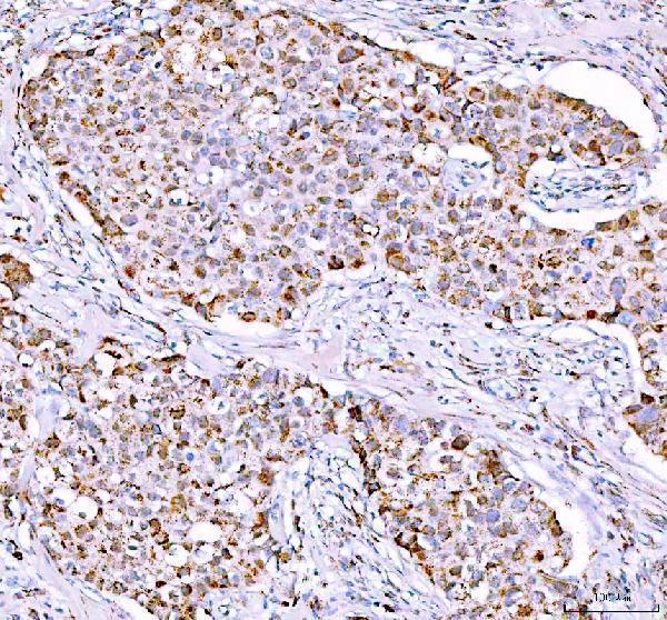
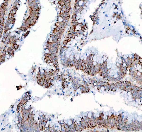
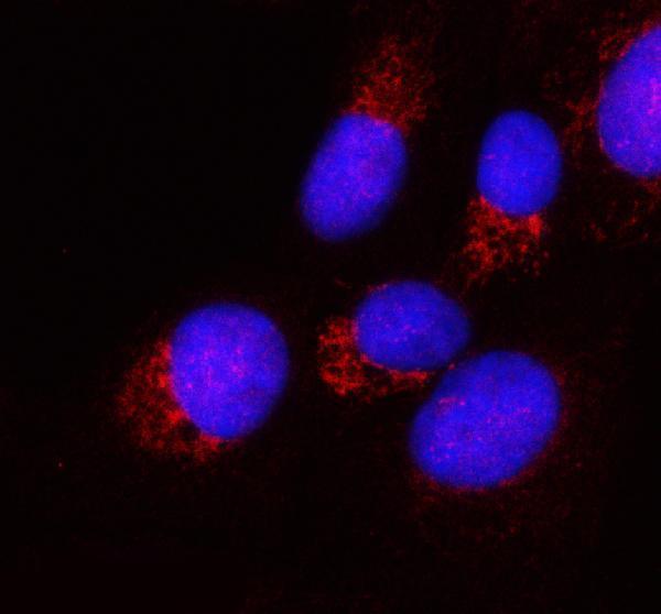
A06587 has real IHC images from paraffin-embedded human tumor tissues (catalog IHC captions) and IF data from U2OS cells (catalog IF caption); listed reactivity covers human and mouse (catalog).

Real IHC data IHC analysis of PPA2 using anti-PPA2 antibody (A06587). PPA2 was detected in a paraffin-embedded section of human breast cancer tissue. Heat mediated antigen retrieval was performed in EDTA buffer (pH 8.0, epitope retrieval solution). The tissue section was blocked with 10% goat serum. The tissue section was then incubated with 2 μg/ml rabbit anti-PPA2 Antibody (A06587) overnight at 4°C. Peroxidase Conjugated Goat Anti-rabbit IgG was used as secondary antibody and incubated for 30 minutes at 37°C. The tissue section was developed using HRP Conjugated Rabbit IgG Super Vision Assay Kit (Catalog # SV0002) with DAB as the chromogen.
Anti-PPA2 Antibody ®
Cat # A06587

A06587 was demonstrated by IHC in paraffin-embedded human breast cancer, colorectal adenocarcinoma, glioma, and intestinal diffuse large B-cell lymphoma sections (catalog IHC captions). A06587 was also demonstrated by IF in U2OS cells (catalog IF caption) and lists human and mouse reactivity (catalog).

Which to pick: For tissue IHC, choose A06587: its images show paraffin-embedded human sections using EDTA retrieval at pH 8.0 and 2 μg/ml primary antibody (catalog IHC captions); the fixative is unreported (catalog IHC captions). For IF/ICC, A06587 lists both applications (catalog) and shows IF in U2OS cells at 5 μg/ml (catalog IF caption). For cross-species work, A06587 lists human and mouse reactivity (catalog); it is rabbit-hosted, and its clone is unreported (catalog).

Each figure is that product's own IHC / IF validation image from its datasheet.

References

  1. UniProt Consortium. UniProt entry Q9H2U2 (IPYR2_HUMAN, Inorganic pyrophosphatase 2, mitochondrial).
  2. Human Protein Atlas. PPA2 tissue IHC expression (reliability: Supported).
  3. Human Protein Atlas. PPA2 subcellular location (ICC-IF): Localized to the mitochondria..
  4. Human Protein Atlas. PPA2 antibody validation summary (3 antibodies).
  5. Downregulation of PPA2 expression correlates with poor prognosis of kidney renal clear cell carcinoma. PeerJ 2021 — PMC8428262.
  6. Multi-omics pan-cancer analysis reveals the prognostic values and immunological functions of PPA2, with a spotlight on breast cancer. Frontiers in immunology 2024 — PMC11338811.
  7. PubMed PMID:14702039 — UniProt-cited evidence.
  8. PubMed PMID:15815621 — UniProt-cited evidence.
  9. PubMed PMID:15489334 — UniProt-cited evidence.