PPARA / Peroxisome proliferator-activated receptor alpha · IHC design guide

Design Immunohistochemistry for PPARA

Plan PPARA IHC on paraffin sections around the general nuclear staining reported in tissue (HPA tissue IHC). This guide covers fixation consistency, interpretation of tissue staining and the catalog antibody’s 1:25 IHC-P starting dilution (datasheet).

Evidence assembled Oct 2026 · For research use; verify linked source records and product datasheet before use
Immunohistochemistry protocol sheet for PPARA (IHC for PPARA): expected localisation General nuclear tissue staining (HPA tissue IHC); nuclear location (UniProt), antibody M00600, validated IHC image, and IHC protocol steps
Printable PPARA IHC protocol sheet — expected localisation General nuclear tissue staining (HPA tissue IHC); nuclear location (UniProt), antibody M00600, controls and protocol steps. Open the full PPARA IHC guide →

PPARA Immunohistochemistry Experimental Design Guide

Expected localisation, validated protocols, controls and antibodies — the at-a-glance facts below, then the full design guide.

Must know before staining
Expected localisation General nuclear tissue staining (HPA tissue IHC); nuclear location (UniProt)
Staining pattern Generally nuclear staining across cell types (HPA tissue IHC)
Antigen retrieval Tris-EDTA pH 9.0 HIER, 95–98 °C, 20 min (rule: nuclear antigen)
Positive control ⓘ Adipose tissue+4 more · see all
Negative control ⓘ None in HPA (detected in all 45 tissues); use no-primary + isotype controls
Important caveats
Reasons your staining may differ from the expected pattern.
Fixation Keep tissue fixation consistent across samples. (standard IHC practice; not target-specific)
Caveat Staining and RNA have medium consistency (HPA tissue IHC)
Regulation Tissue-enhanced RNA in tongue (HPA tissue RNA)
Isoform / epitope Two isoforms; epitope coverage is unspecified (UniProt; datasheet)
Section 1

Recommended PPARA IHC & IF Protocols

The catalog antibody’s IHC-P protocol is accompanied by 4 published PPARα IHC methods (datasheet; PMC8733009; PMC10763066; PMC13542957; PMC8990472).

Recommended immunohistochemistry (IHC-P) protocol parameters
SampleParaffin-embedded H. skeletal muscle tissue; fixative not specified (datasheet M00600)
FixationImage fixative and duration unreported (datasheet M00600); verify before use.
Sectioning4–5 µm sections on charged slides (standard)
DeparaffinisationXylene, graded ethanol series to water (standard)
Antigen retrievalHeat-induced epitope retrieval in Tris-EDTA buffer, pH 9.0, 20 min at 95–98 °C (standard rule: nuclear antigen)
Peroxidase block3% H2O2, 10 min, room temperature (standard)
Blocking10% normal serum of the secondary host, 30 min, room temperature (standard)
Primary antibodyMouse monoclonal (clone 1331CT894.186.143) anti-PPARA, 1:25 (datasheet M00600)
Primary incubationOvernight at 4 °C (standard)
DetectionHRP-polymer secondary, DAB chromogen 5–10 min (standard)
CounterstainHematoxylin, blue, dehydrate and mount (standard)
Expected resultPPARA-positive staining in adipocytes of adipose tissue (HPA tissue IHC: High). HPA tissue profile: General nuclear expression. No signal in the no-primary control.
💡Decision noteStart with Tris-EDTA pH 9.0 retrieval at 95–98 °C for 20 min for nuclear PPARα; citrate pH 6.0 is a published alternative (page retrieval rule; UniProt Q07869; PMC8733009).
Section 2

What Is the Expected PPARA Staining Pattern?

PPARA is nuclear (UniProt Q07869: no transmembrane segment); adipocytes stain High (HPA). IHC reliability: Supported (HPA).

What am I looking at on my slide?
Strong nuclear stain in adipocytes (HPA: High).Consistent with general nuclear IHC expression (HPA) and nuclear location (UniProt).
Cytoplasmic stain with blank nuclei.Suspect artefact; PPARA is nuclear (UniProt: Nucleus).
Stain only in cells outside the selected HPA-positive population.Check cross-reactivity or endogenous detection activity; adipocytes stain High (HPA).
Diffuse stain across the section.Check antibody concentration and blocking (general IHC practice).
No stain in adipocytes (HPA: High).Unexpected; check retrieval and detection with a positive control (general IHC practice).
💡Expected PPARA appearanceStrong adipocyte nuclear stain (HPA: High; UniProt: Nucleus); cytoplasmic-only stain is suspect.
How each factor affects the staining
CompartmentGeneral nuclear tissue IHC (HPA); primary cilium in ICC-IF is uncertain (HPA).
Antibody validationCAB075701 IHC: Supported; HPA066941 ICC: Uncertain (HPA).
IsoformsIsoforms 1 and 2 are reported (UniProt); antibody epitope coverage is unspecified.
Why is my staining missing, weak or wrong?
SituationLikely causeNext action
Adipocytes lack nuclear stain (HPA: High).Retrieval or detection may have failed (general IHC practice).Check retrieval conditions and a positive control.
Cytoplasmic-only stain.Possible nonspecific signal; PPARA is nuclear (UniProt).Compare with a no-primary control and an IHC-validated antibody.
Stain in unexpected cells.Possible cross-reactivity or endogenous detection activity (general IHC practice).Run a no-primary control; assess endogenous peroxidase blocking.
Diffuse background.Excess antibody or inadequate blocking (general IHC practice).Titrate antibody and optimize blocking.
Weak nuclear stain in an HPA-positive cell type.Dilution or detection sensitivity may be limiting (general IHC practice).Titrate antibody and verify the detection system.
IF/ICC shows ciliary signal?Primary cilium localization is uncertain (HPA: ICC-IF).Consult the separate IF/ICC guide; HPA066941 ICC validation is Uncertain (HPA).

Sample controls for PPARA IHC & IF

🧪Run adipose tissue first and score adipocytes, which HPA rates High for PPARA (HPA: High in adipocytes). HPA detects PPARA in all 45 scored tissues, so there is no HPA negative tissue; use no-primary and isotype controls as negatives, and treat unstained cells within the adipose section as background references rather than proven PPARA-negative cells (HPA: no negative tissues; standard IHC practice).
Positive control tissue: Adipose tissue (Adipocytes, HPA High)
Negative control tissue: None in HPA: PPARA is detected in all 45 scored tissues. Use a no-primary (secondary-only) and an isotype control instead.
ICC-IF cell lines (HPA subcellular resource): HPA ICC-IF images show PPARA in HEK293, Rh30, ASC52telo, RPTEC/TERT1, hTERT-RPE1 (serum starved), with annotated localisation: Primary cilium (uncertain) (HPA subcellular).
Technical controls: Include a no-primary, secondary-only slide and a control antibody matched to the primary antibody’s host species and isotype; confirm target-dependent staining with PPARA knockout material where available (standard IHC practice). For chromogenic adipose IHC, block endogenous peroxidase and check the control slides for background signal (standard IHC practice).
⚠️Feasibility: No target-specific fixation window or fixation effect is reported in the supplied evidence, and the selected M00600 paraffin-section caption does not state its fixative (M00600 caption). Antigen retrieval dependency is unreported, so optimize retrieval against the IHC controls; the supplied evidence does not establish whether frozen sections or IF would be easier (M00600 caption; standard IHC practice). In adipose sections, lipid extraction can leave empty spaces that complicate cell-level scoring (standard paraffin IHC practice).

HPA tissue IHC evidence for PPARA

Comprehensive Human Protein Atlas IHC scoring per tissue (reliability: Supported — Medium consistency between antibody staining and RNA expression data.). Rows are taken directly from the HPA tissue chart — click any row's HPA link to view the source.

Positive expression · recommended positive controls

TissueCell typeLevelEvidenceSource
Adipose tissue Adipocytes High Protein (IHC) HPA →
Bone marrow Hematopoietic cells High Protein (IHC) HPA →
Bronchus Respiratory epithelial cells High Protein (IHC) HPA →
Cerebellum Cells in granular layer High Protein (IHC) HPA →
Cerebral cortex Endothelial cells High Protein (IHC) HPA →

Undetected expression · recommended negative controls

TissueCell typeLevelEvidenceSource
None in HPA: PPARA is detected in all 45 scored tissues. Use a no-primary (secondary-only) and an isotype control instead.
Section 3

Advanced PPARA IHC Tips

Troubleshoot PPARA staining in paraffin sections by checking retrieval, nuclear localisation, controls and cell-specific scoring (UniProt Q07869; HPA tissue IHC).

What retrieval should I try when PPARA nuclear staining is weak?
Start with Tris-EDTA at pH 9.0, heated to 95–98 °C for 20 min (page retrieval rule: nuclear antigen). Allow sections to cool consistently, then compare staining with a matched control section; inconsistent heating or cooling can change apparent antigen exposure (standard IHC practice). PPARA is a nuclear protein, so assess nuclear signal before increasing antibody concentration (UniProt Q07869 subcellular location). If signal remains weak, test a milder retrieval condition on adjacent sections while holding detection settings constant, and retain the condition that gives interpretable nuclear staining with low background (standard IHC practice).
Could fixation explain weak PPARA staining in paraffin sections?
Target-specific sensitivity of PPARA staining to fixation is unknown from the supplied evidence; the selected skeletal-muscle caption identifies paraffin embedding but does not state a fixative (M00600 tissue-IHC caption). Record the actual fixative, fixation duration and processing history for each specimen before comparing staining intensity (standard IHC practice). If fixation varies, stain adjacent sections in one run with the same pH 9.0 retrieval and identical detection settings to isolate a possible processing effect (page retrieval rule; standard IHC practice). Interpret any difference as an observed batch effect until a controlled fixation comparison establishes its cause (standard IHC practice).
How should I assess PPARA staining outside nuclei?
Expect a nuclear pattern in paraffin-section IHC because PPARA is annotated in the nucleus and HPA reports general nuclear tissue expression (UniProt Q07869 subcellular location; HPA tissue IHC). Compare DAB deposits with a nuclear counterstain and score nuclei separately from cytoplasm or section debris (standard IHC practice). HPA also reports primary-cilium localisation as uncertain and nucleolar fibrillar-center and nuclear-body localisation as supported in subcellular imaging; those observations do not by themselves establish the expected chromogenic tissue pattern (HPA subcellular). If staining is predominantly extranuclear, check retrieval, secondary-only background and morphology before assigning it to PPARA (standard IHC practice).
Could PPARA isoforms or epitope accessibility change this IHC result?
PPARA has 2 listed isoforms and a nuclear-receptor ligand-binding domain spanning residues 239–466 (UniProt Q07869 isoforms and domains). The supplied evidence does not identify the M00600 epitope, so it cannot establish whether this antibody detects both isoforms or whether processing masks its binding site (M00600 tissue-IHC caption). When comparing specimens, use the same retrieval and staining run, and avoid attributing intensity differences to isoform abundance without an independent isoform-specific assay (standard IHC practice). Document the antibody identifier, lot and retrieval conditions alongside images so a change in staining can be investigated reproducibly (standard IHC practice).
How can IF help check a PPARA pattern seen by chromogenic IHC?
Treat IF/ICC as a separate validation experiment: the supplied M00600 tissue caption describes DAB IHC in paraffin-embedded skeletal muscle, with fixative unreported (M00600 tissue-IHC caption). Multiplex PPARA with a marker for the cell population being assessed and a nuclear stain, then inspect whether signals occupy the expected cells and compartments (UniProt Q07869 subcellular location; standard IF practice). Choose fluorophores after measuring tissue autofluorescence in unstained controls, favoring channels with clearer separation (standard IF practice). PPARA has no transmembrane segment and is nuclear, so permeabilise cellular and nuclear barriers sufficiently for antibody access while checking that morphology and control staining remain intact (UniProt Q07869 topology and subcellular location; standard IF practice).
What should I check when PPARA DAB staining is diffuse?
First inspect a secondary-only section for nonspecific detection and a section without primary antibody for residual endogenous peroxidase activity (standard IHC practice). The M00600 caption reports a 1:25 primary dilution, a 1:400 peroxidase-conjugated secondary dilution and DAB detection in a paraffin-embedded skeletal-muscle section (M00600 tissue-IHC caption). Use those values only as a documented starting point for that antibody and tissue, then titrate while keeping exposure and counterstain consistent (standard IHC practice). If diffuse staining persists, review blocking, wash stringency, tissue folds and drying artifacts before interpreting weak extranuclear color as PPARA (standard IHC practice; UniProt Q07869 subcellular location).
How should I quantify PPARA staining across paraffin sections? ⚠ ANSWER MARKED FOR VERIFICATION
Define the cell population and analysis area before scoring, then record the percentage of PPARA-positive nuclei and an intensity-based H-score if intensity is reproducible (standard IHC practice; UniProt Q07869 subcellular location). Normalise counts to the number of evaluable nuclei in that population; for spatial comparisons, report positive-cell density per mm² of evaluable tissue (standard IHC practice). Apply one positivity threshold to sections stained in the same run and exclude folds, damaged edges and necrotic regions by a predefined rule (standard IHC practice). Report cell identity because HPA tissue IHC describes high staining in specific cell populations across several tissues (HPA tissue IHC).
How do I distinguish genuine PPARA staining from artifact?
Give greatest weight to staining that tracks intact nuclei in the expected cell population and persists across comparable sections (UniProt Q07869 subcellular location; standard IHC practice). PPARA is reported in skeletal muscle, liver, heart, kidney and monocytes, while HPA describes high staining in several specified tissue cell populations; evaluate each specimen against its actual cell composition (UniProt Q07869 tissue specificity; HPA tissue IHC). Discount color concentrated at cut edges, folds or necrotic areas, and check a no-primary control for endogenous enzyme or detection background (standard IHC practice). Because HPA rates tissue-IHC reliability as Supported with medium staining–RNA consistency, treat an isolated unusual pattern as provisional pending independent corroboration (HPA tissue IHC).
Boster reagents

Best PPARA / Peroxisome proliferator-activated receptor alpha IHC Antibodies

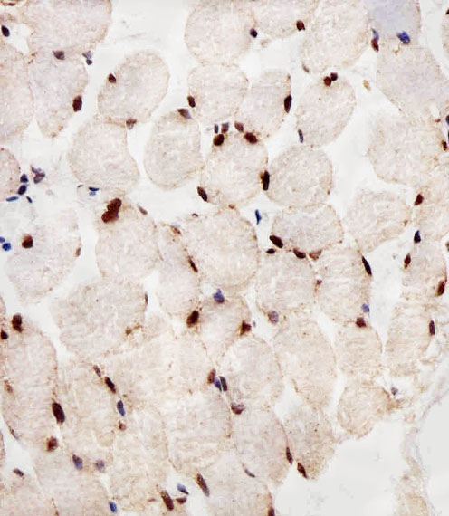
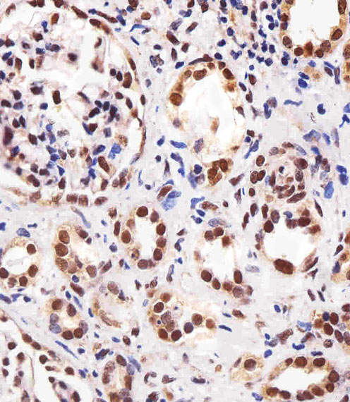
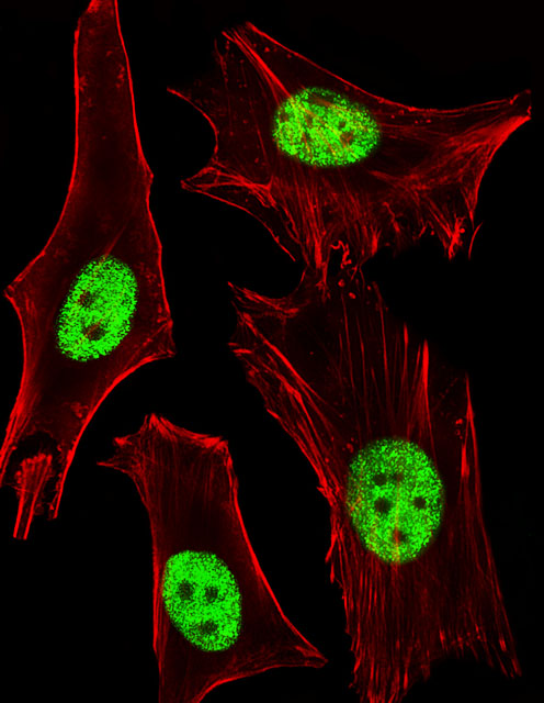
The IHC-validated anti-PPARA antibody M00600 has IHC images from paraffin-embedded human skeletal muscle and kidney and an IF image from HeLa cells (M00600 image captions).

Real IHC data Immunohistochemical analysis of paraffin-embedded H. skeletal muscle section using PPARA Antibody. M00600 was diluted at 1:25 dilution. A peroxidase-conjugated goat anti-mouse IgG at 1:400 dilution was used as the secondary antibody, followed by DAB staining.
Anti-PPARA Antibody
Cat # M00600

M00600 is listed for IHC-P and IF, with human and mouse reactivity (catalog: M00600 applications and reactivity). Its IHC images show paraffin-embedded human skeletal muscle and kidney; its IF image shows HeLa cells (M00600 image captions).

Which to pick: Choose M00600 for tissue IHC-P: its own captions show paraffin-embedded human skeletal muscle and kidney stained at 1:25 with peroxidase and DAB; the fixative is unreported (M00600 IHC image captions). For IF/ICC, M00600 has a HeLa fluorescence image at 1:25 (M00600 IF image caption). For human and mouse work, M00600 is a mouse monoclonal listed as reactive with both species, though the supplied IHC and IF images show human samples (catalog: M00600 host, clone and reactivity; M00600 image captions).

Each figure is that product's own IHC / IF validation image from its datasheet.

References

  1. UniProt Consortium. UniProt entry Q07869 (PPARA_HUMAN, Peroxisome proliferator-activated receptor alpha).
  2. Human Protein Atlas. PPARA tissue IHC expression (reliability: Supported).
  3. Human Protein Atlas. PPARA subcellular location (ICC-IF): Mainly localized to the primary cilium. In addition localized to the nucleoli fibrillar center, nuclear bodies, centriolar satellites and basal body..
  4. Human Protein Atlas. PPARA antibody validation summary (2 antibodies).
  5. PPARα-ACOT12 axis is responsible for maintaining cartilage homeostasis through modulating de novo lipogenesis. Nature communications 2022 — PMC8733009.
  6. Loss of Dec1 inhibits alcohol-induced hepatic lipid accumulation and circadian rhythm disorder. BMC molecular and cell biology 2024 — PMC10763066.
  7. Conditional overexpression of PPARα in intestinal epithelium diminishes GIP enteroendocrine cells and circulating hormone levels. Molecular metabolism 2026 — PMC13542957.
  8. Disruption of peroxisome proliferator-activated receptor α in hepatocytes protects against acetaminophen-induced liver injury by activating the IL-6/STAT3 pathway. International journal of biological sciences 2022 — PMC8990472.
  9. PubMed PMID:7684926 — UniProt-cited evidence.
  10. PubMed PMID:7981125 — UniProt-cited evidence.
  11. PubMed PMID:8993548 — UniProt-cited evidence.