This website uses cookies to ensure you get the best experience on our website.


- Table of Contents
Use catalog antibody A01856 at 1:50–1:200 for paraffin IHC (datasheet). Kidney tubules provide a high-staining reference, but interpret the observed cytoplasmic and nuclear pattern cautiously because HPA rates its tissue IHC reliability uncertain (HPA tissue IHC).

Expected localisation, validated protocols, controls and antibodies — the at-a-glance facts below, then the full design guide.
| Expected localisation | IHC: cytoplasm and nucleus (HPA tissue IHC) | |
| Staining pattern | Most tissues show cytoplasmic and nuclear staining (HPA tissue IHC) | |
| Antigen retrieval | Citrate pH 6.0 HIER, 95–98 °C, 20 min (rule: cytoplasmic / membrane antigen) | |
| Positive control | Adrenal gland+4 more · see all | |
| Negative control | Appendix+4 more · see all |
| Fixation | Keep fixation consistent across paraffin sections. (standard IHC practice; not target-specific) | |
| Caveat | Staining and RNA expression show low consistency (HPA tissue IHC) | |
| Regulation | No expression regulation reported (UniProt) | |
| Isoform / epitope | No annotated isoforms or processing (UniProt) |
The catalog antibody’s IHC-P protocol is accompanied by a published PPOX staining protocol for glioma tissue (PMC9157484).
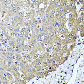
| Sample | Paraffin-embedded human colon carcinoma tissue; fixative not specified (datasheet A01856) |
| Fixation | Image fixative and duration unreported (datasheet A01856); verify before use. |
| Sectioning | 4–5 µm sections on charged slides (standard) |
| Deparaffinisation | Xylene, graded ethanol series to water (standard) |
| Antigen retrieval | Heat-induced epitope retrieval in citrate buffer, pH 6.0, 20 min at 95–98 °C (standard rule: cytoplasmic / membrane antigen) |
| Peroxidase block | 3% H2O2, 10 min, room temperature (standard) |
| Blocking | 10% normal serum of the secondary host, 30 min, room temperature (standard) |
| Primary antibody | Rabbit anti-PPOX, 1:50-1:200 (datasheet A01856) |
| Primary incubation | Overnight at 4 °C (standard) |
| Detection | HRP-polymer secondary, DAB chromogen 5–10 min (standard) |
| Counterstain | Hematoxylin, blue, dehydrate and mount (standard) |
| Expected result | PPOX-positive staining in glandular cells of adrenal gland (HPA tissue IHC: High). HPA tissue profile: Cytoplasmic and nuclear expression in most tissues. No signal in the no-primary control. |
PPOX is an inner mitochondrial membrane protein with no annotated transmembrane segment (UniProt P50336 topology). In paraffin IHC, expect cytoplasmic staining in glandular cells and kidney tubules reported as high by HPA (HPA tissue IHC). HPA also reports nuclear staining in most tissues, but rates its tissue IHC reliability Uncertain because staining and RNA expression have low consistency (HPA tissue IHC).
| Granular cytoplasmic signal in kidney tubule cells or adrenal glandular cells, with limited background. | This is a plausible positive IHC result: HPA reports High staining in both cell types (HPA tissue IHC). Mitochondrial localization supports cytoplasmic signal, but chromogenic IHC alone does not establish that each stained granule is a mitochondrion (UniProt P50336 subcellular location; HPA subcellular ICC-IF). |
| Predominantly nuclear signal with little cytoplasmic signal. | Review this as a compartment mismatch against PPOX's mitochondrial location (UniProt P50336 subcellular location). HPA describes cytoplasmic and nuclear tissue staining, but rates that IHC profile Uncertain; nuclear staining alone cannot confirm PPOX or establish an artefact (HPA tissue IHC). |
| Strong signal in alveolar cells or cardiomyocytes. | These cells are reported Not detected by HPA tissue IHC, so investigate cross-reactivity or endogenous detection activity (HPA tissue IHC; general IHC practice). Treat the discrepancy as a flag, not proof of nonspecific staining: UniProt reports PPOX expression in lung and heart (UniProt P50336 tissue specificity). |
| Diffuse color across tissue, stroma, or a reagent-only control. | A widespread signal that does not resolve to cells is difficult to interpret as PPOX staining (general IHC practice). Background can arise from detection chemistry or insufficient blocking and washing; compare a primary-antibody omission control before assigning cellular staining (general IHC practice). |
| No signal in kidney tubule cells despite a readable counterstain. | Kidney tubules are a useful expected-positive comparator because HPA reports High staining there (HPA tissue IHC). An absent signal calls for a run and control review; it cannot by itself establish absent PPOX because the HPA tissue IHC profile has Uncertain reliability (HPA tissue IHC). |
| Cell type and tissue | Compare like cells: HPA reports High staining in kidney tubules and several glandular epithelia, but Not detected in appendix glandular cells, cardiomyocytes, and alveolar cells (HPA tissue IHC). These are observed IHC patterns, not universal positive or negative controls (HPA tissue IHC reliability: Uncertain). |
| Subcellular interpretation | UniProt places PPOX at the inner mitochondrial membrane without an annotated transmembrane segment; HPA ICC-IF approves mitochondria as the main location and also reports vesicles and cytosol (UniProt P50336 topology; HPA subcellular ICC-IF). The ICC-IF localization informs interpretation but does not validate chromogenic IHC staining (HPA tissue IHC: Uncertain). |
| Antibody evidence | The listed antibody HPA030123 is Uncertain for IHC and Approved for ICC (HPA antibodies). Its ICC status does not upgrade its IHC validation; assess staining alongside cell-type patterns and controls (HPA antibodies; HPA tissue IHC; general IHC practice). |
| Processing and variants | UniProt annotates a single chain spanning residues 1–477, with no signal peptide, propeptide, glycosylation sites, or isoforms in this record (UniProt P50336 processing). These annotations provide no basis to predict a tissue-specific cleavage pattern or a fixation-dependent staining change (UniProt P50336 processing). |
| Situation | Likely cause | Next action |
|---|---|---|
| Kidney tubules fail to stain in the test run. | A missed positive may reflect a run or reagent problem; HPA reports kidney tubules as High, with Uncertain overall IHC reliability (HPA tissue IHC; general IHC practice). | Check a known working tissue control, primary-antibody dilution, retrieval conditions, detection reagents, and counterstain in the same run; avoid calling the sample PPOX-negative from this result alone (general IHC practice; HPA tissue IHC reliability: Uncertain). |
| Nuclei dominate the signal. | A nucleus-dominant pattern conflicts with the mitochondrial assignment, although HPA's uncertain tissue IHC profile includes nuclear staining (UniProt P50336 subcellular location; HPA tissue IHC). | Compare cytoplasmic staining in HPA high cell types and check omission and detection controls; report the nuclear pattern separately instead of using it alone to call PPOX positive (HPA tissue IHC; general IHC practice). |
| Color appears in alveolar cells or cardiomyocytes. | HPA reports these cells as Not detected in IHC, while UniProt reports PPOX expression in lung and heart; the evidence does not resolve that difference (HPA tissue IHC; UniProt P50336 tissue specificity). | Check cellular boundaries and an appropriate primary-antibody omission control for endogenous detection activity; describe the observed staining without treating the HPA negative as definitive (general IHC practice; HPA tissue IHC reliability: Uncertain). |
| Background obscures cytoplasmic staining. | Nonspecific binding or detection activity can create diffuse chromogenic color (general IHC practice). | Inspect omission controls, blocking, washes, and detection conditions; judge cell-specific signal only after background is low enough to distinguish stained cells (general IHC practice). |
| A whole section looks uniformly positive. | HPA describes staining in most tissues, but its IHC profile is Uncertain; uniform color alone cannot establish PPOX expression in every cell (HPA tissue IHC). | Score named cell populations separately, compare HPA reported high and not-detected cells where present, and inspect controls for background before assigning positivity (HPA tissue IHC; general IHC practice). |
| IHC appears nuclear while ICC-IF appears mitochondrial. | HPA approves mitochondrial ICC-IF localization but rates the tissue IHC profile Uncertain; the assays have different validation statuses (HPA subcellular ICC-IF; HPA tissue IHC). | Keep the IHC and ICC-IF observations distinct, verify IHC controls, and avoid using the ICC result as proof that the nuclear IHC signal identifies PPOX (HPA antibodies; general IHC practice). |


Comprehensive Human Protein Atlas IHC scoring per tissue (reliability: Uncertain — Low consistency between antibody staining and RNA expression data. Pending external verification.). Rows are taken directly from the HPA tissue chart — click any row's HPA link to view the source.
| Tissue | Cell type | Level | Evidence | Source |
|---|---|---|---|---|
| Adrenal gland | Glandular cells | High | Protein (IHC) | HPA → |
| Breast | Glandular cells | High | Protein (IHC) | HPA → |
| Bronchus | Respiratory epithelial cells | High | Protein (IHC) | HPA → |
| Endometrium | Cells in endometrial stroma | Low | Protein (IHC) | HPA → |
| Fallopian tube | Glandular cells | High | Protein (IHC) | HPA → |
| Tissue | Cell type | Level | Evidence | Source |
|---|---|---|---|---|
| Appendix | Glandular cells | Not detected | Protein (IHC) | HPA → |
| Cerebellum | Cells in granular layer | Not detected | Protein (IHC) | HPA → |
| Epididymis | Glandular cells | Not detected | Protein (IHC) | HPA → |
| Heart muscle | Cardiomyocytes | Not detected | Protein (IHC) | HPA → |
| Lung | Alveolar cells | Not detected | Protein (IHC) | HPA → |
Troubleshoot PPOX staining in paraffin sections using mitochondrial localisation, the reported tissue pattern, and controls that distinguish signal from background.
One anti-PPOX antibody has real IHC data from paraffin-embedded human colon carcinoma (A01856 image caption); the catalog lists human and mouse reactivity (A01856 catalog). No IF data are supplied (A01856 catalog).
A01856 is listed for IHC and has an image of paraffin-embedded human colon carcinoma stained at 1:100 after microwave retrieval in 10 mM PBS, pH 7.2 (A01856 image caption). The catalog lists human and mouse reactivity for A01856, but provides no mouse IHC image or IF image (A01856 catalog).
Which to pick: For paraffin-section IHC, choose A01856: its own image caption documents staining of human colon carcinoma, and the catalog lists an IHC dilution of 1:50–1:200 (A01856 image caption; A01856 catalog). The caption does not report the fixative (A01856 image caption). For IF/ICC, no SKU has listed validation; for mouse samples, A01856 lists mouse reactivity, although its supplied IHC image is human (A01856 catalog; A01856 image caption).