PPP1CA / Serine/threonine-protein phosphatase PP1-alpha catalytic subunit · IHC design guide

Design Immunohistochemistry for PPP1CA

Plan PPP1CA chromogenic IHC in paraffin sections using renal tubules and intestinal glands as high-staining reference tissues (HPA tissue IHC). Assess nuclear and cytoplasmic staining, and account for the catalog antibody’s PPP1CB reactivity when interpreting signal (HPA tissue IHC; datasheet M02801).

Evidence assembled Oct 2026 · For research use; verify linked source records and product datasheet before use
Immunohistochemistry protocol sheet for PPP1CA (IHC for PPP1CA): expected localisation Nuclear and cytoplasmic tissue staining (HPA tissue IHC), antibody M02801, validated IHC image, and IHC protocol steps
Printable PPP1CA IHC protocol sheet — expected localisation Nuclear and cytoplasmic tissue staining (HPA tissue IHC), antibody M02801, controls and protocol steps. Open the full PPP1CA IHC guide →

PPP1CA Immunohistochemistry Experimental Design Guide

Expected localisation, validated protocols, controls and antibodies — the at-a-glance facts below, then the full design guide.

Must know before staining
Expected localisation Nuclear and cytoplasmic tissue staining (HPA tissue IHC)
Staining pattern Nuclear and cytoplasmic staining across several tissues (HPA tissue IHC)
Antigen retrieval Citrate pH 6 HIER, heat-mediated (datasheet M02801)
Positive control ⓘ Kidney+4 more · see all
Negative control ⓘ Adipose tissue+4 more · see all
Important caveats
Reasons your staining may differ from the expected pattern.
Fixation Keep fixation consistent across paraffin sections. (standard IHC practice; not target-specific)
Caveat M02801 also detects PPP1CB (datasheet M02801)
Regulation Expression induction not annotated (UniProt)
Isoform / epitope 3 isoforms; epitope coverage unspecified (UniProt)
Section 1

Recommended PPP1CA IHC & IF Protocols

The catalog antibody’s citrate pH 6 heat retrieval protocol (datasheet M02801) is accompanied by 4 published PPP1CA IHC protocols (PMC11633043; PMC11569623; PMC10698990; PMC8805822).

Recommended immunohistochemistry (IHC-P) protocol parameters
SampleParaffin-embedded human pancreas tissue; fixative not specified (datasheet M02801)
FixationImage fixative and duration unreported (datasheet M02801); verify before use.
Sectioning4–5 µm sections on charged slides (standard)
DeparaffinisationXylene, graded ethanol series to water (standard)
Antigen retrievalHeat retrieval: Citrate pH 6, 20 min (datasheet M02801)
Peroxidase block3% H2O2, 10 min, room temperature (standard)
Blocking10% goat serum (datasheet M02801)
Primary antibodyRabbit monoclonal (clone AFC-16) anti-PPP1CA, 1:50 (datasheet M02801)
Primary incubationOvernight at 4 °C (standard)
DetectionStreptavidin-biotin complex (SABC), DAB chromogen (datasheet M02801)
CounterstainHematoxylin, blue, dehydrate and mount (standard)
Expected resultPPP1CA-positive staining in cells in tubules of kidney (HPA tissue IHC: High). HPA tissue profile: Cytoplasmic and nuclear expression in several tissues. No signal in the no-primary control.
💡Decision noteStart with citrate pH 6 heat retrieval (datasheet M02801). One published protocol specifies 0.01 mol/L citrate but gives no pH or heating details (PMC11569623).
Section 2

What Is the Expected PPP1CA Staining Pattern?

PPP1CA should show cytoplasmic and nuclear staining in several tissues (HPA tissue IHC); UniProt describes it as primarily nuclear, largely excluded from the nucleolus, and lacking a transmembrane segment (UniProt P62136). High staining is reported in kidney tubular cells and rectal and small-intestinal glandular cells (HPA tissue IHC). HPA rates the tissue profile Enhanced while noting low consistency between antibody staining and RNA expression (HPA tissue IHC).

What am I looking at on my slide?
Kidney tubular cells or rectal and small-intestinal glandular cells show nuclear and cytoplasmic chromogen (HPA tissue IHC).This fits the reported high staining in those cells and the broader cytoplasmic and nuclear tissue profile (HPA tissue IHC). Judge intensity against neighboring cells and a matched control section; chromogen alone does not establish antibody specificity (general IHC practice).
Staining appears only at cell borders, or mainly within nucleoli, with little nucleoplasmic or cytoplasmic signal.Recheck compartment assignment and controls before calling this PPP1CA: UniProt describes predominantly nuclear protein largely excluded from nucleoli (UniProt P62136), while HPA tissue IHC reports cytoplasmic and nuclear staining (HPA tissue IHC). Membrane association is possible, but membrane-only tissue staining is not the reported IHC profile (HPA tissue IHC; HPA ICC-IF).
Strong chromogen appears in adipocytes, skeletal myocytes, or lymph-node germinal-center cells.These cell populations are reported as not detected, so unexpected staining warrants a specificity check (HPA tissue IHC). Compare morphology, a no-primary control, and staining with an independent antibody where available; cross-reactivity or endogenous detection activity are possibilities, not diagnoses from appearance alone (general IHC practice).
Chromogen spreads across stroma, empty spaces, and many unrelated cell types without clear cell boundaries.This is diffuse background rather than a readable cellular pattern (general IHC practice). The expected tissue pattern is cellular and includes nuclear and cytoplasmic staining (HPA tissue IHC). Check the no-primary control and review blocking, washes, and detection conditions (general IHC practice).
Kidney tubular cells show no convincing signal in a section expected to serve as a positive control.That conflicts with HPA's high staining in kidney tubular cells, but a single negative section cannot establish absent PPP1CA (HPA tissue IHC). Confirm that tubules are present and review the antibody's IHC validation, retrieval, dilution, and detection controls before interpreting other negatives (general IHC practice).
💡Expected PPP1CA appearanceA convincing positive shows high cellular staining in kidney tubules or rectal and small-intestinal glands, with nuclear and cytoplasmic signal (HPA tissue IHC); diffuse acellular chromogen or strong staining restricted to HPA-listed undetected cells should prompt a control check (HPA tissue IHC; general IHC practice).
How each factor affects the staining
Cell population and tissue contextUse the reported cell type, not the organ name alone, when scoring: kidney tubules and rectal or small-intestinal glands are high, whereas adipocytes and skeletal myocytes are not detected (HPA tissue IHC).
Compartment and targeting partnersNuclear and cytoplasmic signal is plausible because PPP1CA is mobile and can be relocalized by targeting subunits (UniProt P62136); HPA tissue IHC reports both compartments (HPA tissue IHC). Do not require identical nuclear-to-cytoplasmic ratios in every cell (UniProt P62136).
Evidence strengthHPA rates the tissue profile Enhanced but flags low consistency with RNA expression (HPA tissue IHC). Its listed antibodies also differ in IHC status: CAB004545 is Enhanced and HPA046833 is Approved (HPA antibodies). Interpret weak or unexpected staining with that distinction in mind.
Isoform coverageUniProt lists 3 isoforms (UniProt P62136). The supplied evidence does not locate the antibody epitope or establish isoform coverage, so staining cannot be assigned to one isoform from this section alone (UniProt P62136; HPA antibodies).
Antigen retrievalRetrieval can be reviewed when a paraffin-section control fails (general IHC practice). No supplied UniProt or HPA evidence identifies a PPP1CA-specific retrieval condition or fixation sensitivity; avoid explaining a negative section by a claimed target-specific masking effect.
Why is my staining missing, weak or wrong?
SituationLikely causeNext action
Known-positive kidney tubules are blank or barely stained (HPA tissue IHC).The section may lack the intended tubules, or the IHC detection workflow may have failed (general IHC practice). The negative result alone does not identify a PPP1CA-specific fixation effect.Confirm tubular morphology; check the IHC-validated antibody's instructions, retrieval, dilution, and detection controls on the same run (general IHC practice).
The whole section has a uniform brown haze, including areas without intact cells.Background from the detection workflow can obscure cellular staining (general IHC practice). HPA reports a cellular nuclear and cytoplasmic profile (HPA tissue IHC).Examine a no-primary control, then review blocking, wash stringency, and detection conditions before scoring PPP1CA (general IHC practice).
An HPA-listed undetected cell type stains more strongly than the intended positive population.Cross-reactivity, endogenous detection activity, or misidentified cells could explain the discrepancy (general IHC practice); the HPA tissue pattern does not support that ranking (HPA tissue IHC).Recheck morphology and the no-primary control; compare an independent IHC antibody if available, while keeping each antibody's HPA validation status distinct (general IHC practice; HPA antibodies).
The apparent signal is restricted to cell borders or nucleoli.A compartment mismatch may reflect staining or scoring artefact (general IHC practice). UniProt describes mainly nuclear PPP1CA with relative nucleolar exclusion (UniProt P62136); HPA tissue IHC reports cytoplasmic and nuclear staining (HPA tissue IHC).Inspect higher magnification and a counterstained control section; verify whether nucleoplasm and cytoplasm stain in HPA-listed positive cells before calling the pattern specific (HPA tissue IHC; general IHC practice).
A low or undetected HPA cell population is used as the only positive control.Its reported staining level may be too low for a dependable run control: thyroid glandular cells are low and prostate glandular cells are not detected (HPA tissue IHC).Include kidney tubules or rectal or small-intestinal glandular cells as a positive comparison, and score the named cell population rather than the entire tissue (HPA tissue IHC).
IF/ICC Q&A: Should fluorescence reproduce the paraffin-section pattern exactly?HPA ICC-IF supports nucleoplasm and cytosol, labels plasma membrane uncertain, and also records additional sperm structures (HPA ICC-IF). Tissue IHC reports a broader nuclear and cytoplasmic profile (HPA tissue IHC).Use the ICC-IF localization evidence to interpret IF images, including its uncertainty labels; assess paraffin-section IHC against the tissue-specific cell populations and compartments instead (HPA ICC-IF; HPA tissue IHC).

Sample controls for PPP1CA IHC & IF

🧪Run kidney first; cells in tubules should stain (HPA: High in kidney cells in tubules). Use adipose tissue as the negative comparator, where adipocytes should lack detectable staining (HPA: Not detected in adipocytes); on the kidney slide, use unstained areas to judge background without assuming a particular kidney cell type is PPP1CA-negative (HPA: kidney cells in tubules High).
Positive control tissue: Kidney (Cells in tubules, HPA High)
Negative control tissue: Adipose tissue (HPA Not detected)
ICC-IF cell lines (HPA subcellular resource): HPA ICC-IF images show PPP1CA in A-431, MCF-7, U2OS, RPTEC/TERT1, hTERT-RPE1 (serum starved), Sperm, NIH 3T3, with annotated localisation: Nucleoplasm (supported), Plasma membrane (uncertain), Cytosol (supported) (HPA subcellular).
Technical controls: Include no-primary (secondary-only) and host-species- and clonality-matched isotype controls, plus PPP1CA knockout tissue or a peptide-blocking control when the immunogen is known (standard IHC practice). For chromogenic detection in kidney, quench endogenous peroxidase and check for endogenous biotin background when using the caption’s biotinylated secondary and SABC/DAB system (caption: biotinylated secondary, SABC/DAB; standard IHC practice).
⚠️Feasibility: A target-specific fixation window or fixation effect is unreported, and the selected SKU’s paraffin-section caption does not state the fixative (caption: fixative not stated). The caption uses citrate retrieval at pH 6 for 20 minutes, but does not establish that retrieval is required (caption: citrate retrieval). Frozen sections and IF/ICC cannot be ranked for ease from the supplied evidence; kidney DAB staining also needs the endogenous-background checks noted above (HPA: ICC-IF images available; standard IHC practice).

HPA tissue IHC evidence for PPP1CA

Comprehensive Human Protein Atlas IHC scoring per tissue (reliability: Enhanced — Low consistency between antibody staining and RNA expression data.). Rows are taken directly from the HPA tissue chart — click any row's HPA link to view the source.

Positive expression · recommended positive controls

TissueCell typeLevelEvidenceSource
Kidney Cells in tubules High Protein (IHC) HPA →
Rectum Glandular cells High Protein (IHC) HPA →
Small intestine Glandular cells High Protein (IHC) HPA →
Appendix Glandular cells Medium Protein (IHC) HPA →
Bronchus Respiratory epithelial cells Medium Protein (IHC) HPA →

Undetected expression · recommended negative controls

TissueCell typeLevelEvidenceSource
Adipose tissue Adipocytes Not detected Protein (IHC) HPA →
Breast Adipocytes Not detected Protein (IHC) HPA →
Fallopian tube Glandular cells Not detected Protein (IHC) HPA →
Lymph node Germinal center cells Not detected Protein (IHC) HPA →
Prostate Glandular cells Not detected Protein (IHC) HPA →
Section 3

Advanced PPP1CA IHC Tips

Troubleshoot PPP1CA staining in paraffin sections by checking retrieval, compartment patterns, antibody specificity and matched controls before comparing signal across samples.

What retrieval should I start with when PPP1CA staining is weak?
Start with heat mediated retrieval in citrate buffer at pH 6 for 20 minutes (M02801 tissue IHC caption). The selected paraffin section example used this condition before incubation with 1 µg/mL primary antibody, so it is the closest documented starting point for this reagent (M02801 tissue IHC caption). If staining remains weak, adjust heating or cooling in a controlled comparison while holding antibody concentration and detection constant (standard IHC practice). Include a known positive section in each run to distinguish failed retrieval from a low signal specimen (standard IHC practice). Record any change in nuclear and cytoplasmic staining separately because both compartments are expected (HPA tissue IHC profile).
How can I assess whether fixation is suppressing PPP1CA staining?
Target specific fixation sensitivity is unknown: the selected paraffin section caption does not state its fixative (M02801 tissue IHC caption). Compare sections with documented fixation and processing histories using the same citrate pH 6 retrieval and staining run (M02801 tissue IHC caption; standard IHC practice). Include a positive control section and inspect tissue preservation, since loss of morphology and uneven staining can complicate interpretation (standard IHC practice). If a sample stains weakly, vary one processing condition at a time before attributing the result to PPP1CA abundance (standard IHC practice). Do not use tissue distribution or predicted protein features to assign a fixation effect (standard IHC practice).
Should a convincing PPP1CA IHC signal be nuclear or cytoplasmic?
Assess both nuclear and cytoplasmic staining: tissue IHC reports expression in both compartments across several tissues (HPA tissue IHC profile). PPP1CA is primarily nuclear and largely excluded from the nucleolus, but targeting partners can relocalize it (UniProt P62136 subcellular location). Score the compartments separately against a hematoxylin counterstain, and inspect whether staining follows intact cells rather than section edges (standard IHC practice). A cytoplasmic component alone is therefore not automatically an artefact, particularly where a matched positive control shows the same pattern (HPA tissue IHC profile; standard IHC practice). Treat isolated, strong nucleolar staining cautiously until antibody specificity and morphology are checked (UniProt P62136 subcellular location; standard IHC practice).
Can this stain distinguish PPP1CA isoforms or other PP1 catalytic subunits?
Do not interpret this stain as isoform specific without epitope validation: PPP1CA has 3 annotated isoforms, and the supplied evidence does not map this antibody's epitope (UniProt P62136 isoforms; M02801 tissue IHC caption). The selected image caption labels the reagent PPP1CA+PPP1CB, so staining alone cannot establish a PPP1CA only signal (M02801 tissue IHC caption). Confirm the claimed specificity using an independently validated antibody or a suitable loss of target control under matched IHC conditions (standard IHC practice). If the epitope is later mapped, check whether it is shared among isoforms and other PP1 catalytic subunits before assigning staining to one variant (UniProt P62136 isoforms and subunit; standard IHC practice).
How should I cross check the IHC pattern with IF multiplexing?
Use IF as a separate cross check and multiplex PPP1CA with a marker for the cell population being assessed in the IHC section (standard IF practice). HPA reports high tissue IHC staining in kidney tubule cells and rectal glandular cells, which can guide the choice of a matched cell marker (HPA tissue IHC positive list). Choose spectrally separated fluorophores and place the weaker signal in a channel with less tissue autofluorescence, verified using an unstained control (standard IF practice). PPP1CA has no transmembrane segment and is reported in nucleoplasm and cytosol, so permeabilize fixed cells for intracellular access while preserving morphology (UniProt P62136 topology; HPA subcellular profile; standard IF practice). Compare nuclear and cytoplasmic signal without assuming the antibody's epitope is mapped (HPA tissue IHC profile; M02801 tissue IHC caption).
What should I check when PPP1CA DAB staining appears widespread?
First compare a no primary control with the stained section to identify signal from the detection system or tissue itself (standard IHC practice). The selected paraffin example used 10% goat serum blocking, a biotinylated secondary, a streptavidin biotin complex and DAB (M02801 tissue IHC caption). Check peroxidase blocking and, when using that detection route, endogenous biotin controls before increasing antibody dilution (standard IHC practice). Widespread staining is not sufficient evidence of background because PPP1CA has low tissue RNA specificity and shows cytoplasmic and nuclear tissue IHC expression (HPA tissue IHC profile). Compare intact cells with edges, folds and damaged areas, then adjust washing or blocking only when controls implicate nonspecific signal (standard IHC practice).
How should I score PPP1CA IHC across samples with different cellularity? ⚠ ANSWER MARKED FOR VERIFICATION
Define the cell population and score nuclear and cytoplasmic PPP1CA separately, since both compartments can stain (HPA tissue IHC profile; standard IHC practice). For each compartment, report an H-score based on staining intensity and percentage of positive cells, with the scoring thresholds fixed before comparison (standard IHC practice). Alternatively, report percentage positive cells within the annotated population rather than across all cells in the section (standard IHC practice). Normalize positive cell counts to the number of eligible cells, or stained area to eligible tissue area in mm², and exclude folds and necrosis consistently (standard IHC practice). Keep retrieval, antibody concentration, detection and imaging settings matched across the comparison (standard IHC practice).
How can I tell a true PPP1CA pattern from tissue artefact?
A credible pattern follows intact cell boundaries and includes plausible nuclear or cytoplasmic signal rather than only edge staining (HPA tissue IHC profile; standard IHC practice). Compare the stained section with a no primary control, especially where endogenous peroxidase or the biotin based detection system could produce DAB signal (M02801 tissue IHC caption; standard IHC practice). Check whether the stained cell type matches the claim: kidney tubule cells are reported high, whereas adipocytes are reported undetected in tissue IHC (HPA tissue IHC positive and negative lists). Exclude necrotic regions, folds and section edges before scoring (standard IHC practice). Interpret a negative result cautiously because HPA notes low consistency between antibody staining and RNA expression data (HPA tissue IHC reliability description).
Boster reagents

Best PPP1CA / Serine/threonine-protein phosphatase PP1-alpha catalytic subunit IHC Antibodies

Both antibodies have IHC images from human paraffin sections (image captions). Both list Human, Mouse and Rat reactivity (catalog reactivity); only M02801 lists IF/ICC (catalog applications).

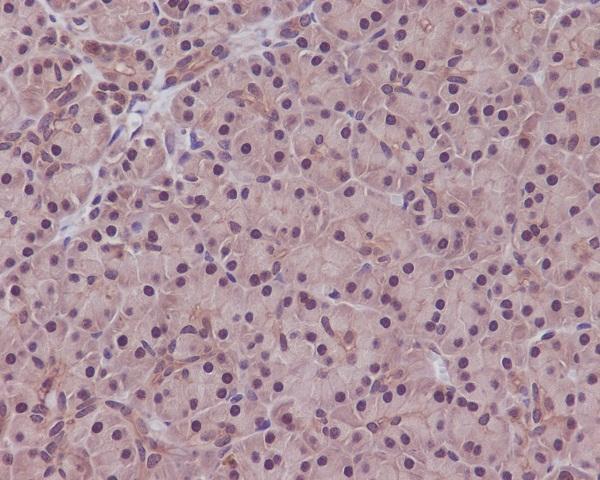
Real IHC data Immunohistochemical analysis of paraffin-embedded human pancreas, using PPP1CA+PPP1CB Antibody(M02801) PPP1CA was detected in paraffin-embedded tissue section. Heat mediated antigen retrieval was performed in citrate buffer (pH6, epitope retrieval solution) for 20 mins. The tissue section was blocked with 10% goat serum. The tissue section was then incubated with 1ug/ml rabbit anti-PPP1CA Antibody (M02801)overnight at 4 Biotinylated goat anti-rabbit IgG was used as secondary antibody and incubated for 30 minutes at 37 The tissue section was developed using Strepavidin-Biotin-Complex (SABC)(Catalog # SA1022) with DAB as the chromogen.
Anti-PPP1CA+PPP1CB Rabbit Monoclonal Antibody
Cat # M02801
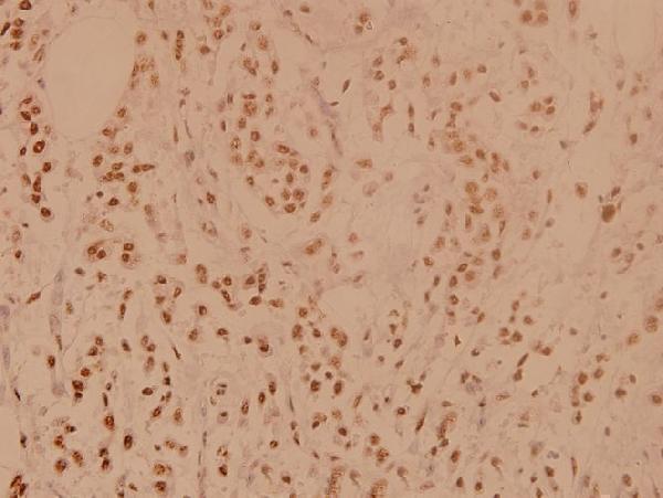
Real IHC data Immunohistochemistry (IHC) analyzes of PP1α (G315) pAb in paraffin-embedded human breast carcinoma tissue at 1:100.
Anti-PP1 Alpha (G315) PPP1CA Antibody
Cat # A02801-1

A02801-1 is shown in paraffin-embedded human breast carcinoma at 1:100 (A02801-1 image caption). M02801 is shown in paraffin-embedded human pancreas with citrate pH 6 retrieval and DAB detection (M02801 image caption).

Which to pick: For tissue IHC, choose A02801-1 for a PPP1CA-labeled antibody with a 1:50–1:200 catalog range; choose M02801 if a rabbit monoclonal that recognizes PPP1CA+PPP1CB suits the experiment (A02801-1 title, dilution and image caption; M02801 title, clone and image caption). For IF/ICC, choose M02801: it lists both applications and a 1:50 IF dilution, though its supplied figure is IHC (M02801 applications, dilution and image caption). Both list Human, Mouse and Rat reactivity, but their pictured IHC examples are human paraffin sections and neither caption reports the fixative (catalog reactivity; A02801-1 and M02801 image captions).

Each figure is that product's own IHC / IF validation image from its datasheet.

References

  1. UniProt Consortium. UniProt entry P62136 (PP1A_HUMAN, Serine/threonine-protein phosphatase PP1-alpha catalytic subunit).
  2. Human Protein Atlas. PPP1CA tissue IHC expression (reliability: Enhanced).
  3. Human Protein Atlas. PPP1CA subcellular location (ICC-IF): Mainly localized to the nucleoplasm, plasma membrane and cytosol. In addition localized to the flagellar centriole, mid piece and end piece..
  4. Human Protein Atlas. PPP1CA antibody validation summary (2 antibodies).
  5. S100A4 targets PPP1CA/IL-17 to inhibit the senescence of sheep endometrial epithelial cells. Frontiers in veterinary science 2024 — PMC11633043.
  6. Nodularin-R Synergistically Enhances Abiraterone Against Castrate- Resistant Prostate Cancer via PPP1CA Inhibition. Journal of cellular and molecular medicine 2024 — PMC11569623.
  7. CircGPRC5A enhances colorectal cancer progress by stabilizing PPP1CA and inducing YAP dephosphorylation. Journal of experimental & clinical cancer research : CR 2023 — PMC10698990.
  8. Comprehensive analysis of PPPCs family reveals the clinical significance of PPP1CA and PPP4C in breast cancer. Bioengineered 2022 — PMC8805822.
  9. PubMed PMID:8392016 — UniProt-cited evidence.
  10. PubMed PMID:8384581 — UniProt-cited evidence.
  11. PubMed PMID:14702039 — UniProt-cited evidence.