This website uses cookies to ensure you get the best experience on our website.


- Table of Contents
Plan PPP1R12A staining in paraffin sections around the cytoplasmic and membranous pattern seen in most cells (HPA tissue IHC). Breast glandular cells stain strongly, while adipocytes are reported as undetected (HPA tissue IHC); start the catalog antibody at 0.5–1 µg/mL (datasheet: PB9737).

Expected localisation, validated protocols, controls and antibodies — the at-a-glance facts below, then the full design guide.
| Expected localisation | Cytoplasmic and membranous (HPA tissue IHC) | |
| Staining pattern | Most cells show cytoplasmic and membranous staining (HPA tissue IHC) | |
| Antigen retrieval | Citrate pH 6 HIER, heat-mediated (datasheet PB9737) | |
| Positive control | Appendix+4 more · see all | |
| Negative control | Adipose tissue |
| Fixation | Keep fixation consistent across paraffin sections. (standard IHC practice; not target-specific) | |
| Caveat | Adipocytes may lack staining despite broad tissue expression (HPA tissue IHC) | |
| Regulation | No stimulus-dependent regulation specified (UniProt) | |
| Isoform / epitope | Five isoforms; confirm antibody epitope coverage (UniProt) |
The catalog antibody’s IHC-P protocol is accompanied by published PPP1R12A staining methods for tumor sections (PMC8210562) and pancreatic tissues (PMC4843125).
| Sample | Paraffin-embedded Human Glioma tissue; fixative not specified (datasheet PB9737) |
| Fixation | Image fixative and duration unreported (datasheet PB9737); verify before use. |
| Sectioning | 4–5 µm sections on charged slides (standard) |
| Deparaffinisation | Xylene, graded ethanol series to water (standard) |
| Antigen retrieval | Heat retrieval: Citrate pH 6, 20 min (datasheet PB9737) |
| Peroxidase block | 3% H2O2, 10 min, room temperature (standard) |
| Blocking | 10% goat serum (datasheet PB9737) |
| Primary antibody | Rabbit anti-PPP1R12A, 0.5-1μg/ml (datasheet PB9737) |
| Primary incubation | Overnight at 4 °C (datasheet PB9737) |
| Detection | Streptavidin-biotin complex (SABC), DAB chromogen (datasheet PB9737) |
| Counterstain | Hematoxylin, blue, dehydrate and mount (standard) |
| Expected result | PPP1R12A-positive staining in lymphoid tissue of appendix (HPA tissue IHC: High). HPA tissue profile: Cytoplasmic and membranous expression in most cells. No signal in the no-primary control. |
PPP1R12A is a cytoplasmic protein associated with stress fibers and actomyosin filaments; it has no transmembrane segment (UniProt O14974). In paraffin-section IHC, expect cytoplasmic staining, sometimes with a membranous appearance, across many cell types (HPA tissue IHC). HPA rates the tissue staining Approved, with medium consistency between antibody staining and RNA expression (HPA tissue IHC).
| Cytoplasmic chromogen in breast or endometrial glandular cells, with some apparent membrane staining. | This fits the reported cytoplasmic and membranous pattern; both named glandular populations stain High (HPA tissue IHC). Judge the signal within cells against adjacent background and a negative reagent control (general IHC practice). |
| Strong, widespread nuclear staining replaces the cytoplasmic pattern. | Treat this as a possible artefact and review the controls and staining conditions: cytoplasm and stress fibers are the UniProt locations (UniProt O14974). HPA reports uncertain nucleolar localization in ICC-IF, which does not establish widespread nuclear IHC staining (HPA subcellular). |
| Adipocytes stain as strongly as the selected positive tissue. | Adipocytes are reported Not detected, so this result warrants a specificity check (HPA tissue IHC). Cross-reactivity or endogenous detection activity is possible; compare a negative reagent control and an independently validated antibody before assigning the signal to PPP1R12A (general IHC practice). |
| Chromogen covers tissue spaces or obscures cell boundaries. | Diffuse background cannot establish a PPP1R12A-positive cell or compartment (general IHC practice). Check the negative reagent control, blocking, washes, and detection reagent; retain the tissue context because HPA reports expression in most cells (HPA tissue IHC). |
| No signal appears in a breast glandular-cell or bronchial epithelial-cell section. | Both populations are reported High, so first check tissue preservation and the run controls (HPA tissue IHC; general IHC practice). A failed run, unsuitable retrieval, or antibody conditions could explain absent signal; HPA supplies no target-specific fixation-sensitivity claim. |
| Cellular location | Cytoplasm and stress fibers are reported locations, including association with actomyosin filaments (UniProt O14974). HPA tissue IHC also describes membranous expression; assess that appearance alongside cytoplasmic staining (HPA tissue IHC). |
| Tissue and cell choice | Breast glandular cells, bronchial respiratory epithelium, and cortical neurons are High; adipocytes are Not detected (HPA tissue IHC). Low tissue specificity means a positive result elsewhere is not automatically unexpected (HPA tissue IHC). |
| Antibody evidence | HPA071956 is Approved for IHC, while HPA041296 has no stated IHC status (HPA antibodies). HPA's overall Approved tissue rating has medium RNA–staining consistency; neither statement validates an unspecified catalog antibody (HPA tissue IHC; HPA antibodies). |
| IF/ICC Q: What location should a separate IF guide assess? | A: Mainly cytosol and actin filaments; HPA marks both Supported. Nucleoli and plasma membrane are additional, Uncertain locations (HPA subcellular). This ICC-IF evidence is a localization comparison, not an IHC protocol or an IHC validation claim. |
| Situation | Likely cause | Next action |
|---|---|---|
| Positive tissue gives no chromogen. | The assay may have failed, or retrieval and antibody conditions may be unsuitable (general IHC practice). HPA lists breast glandular cells as High (HPA tissue IHC). | Check run controls and section integrity, then review the IHC-validated antibody's documented retrieval and dilution conditions (general IHC practice). Do not infer PPP1R12A-specific fixation sensitivity from the HPA tissue score. |
| Adipocytes show strong cellular staining. | This conflicts with the reported Not detected adipocyte result (HPA tissue IHC). Cross-reactivity or endogenous detection activity may contribute (general IHC practice). | Compare a negative reagent control and an HPA High cell population on the same run; investigate detection activity and antibody specificity before scoring adipocytes positive (HPA tissue IHC; general IHC practice). |
| Most nuclei are dark while cytoplasm is weak. | A predominantly nuclear result does not match UniProt's cytoplasmic and stress-fiber locations (UniProt O14974). HPA's additional nucleolar ICC-IF signal is Uncertain (HPA subcellular). | Inspect controls, counterstain, and chromogen distribution; repeat under documented IHC conditions if needed (general IHC practice). Do not use uncertain ICC-IF nucleolar staining to validate broad nuclear IHC. |
| The whole section has hazy chromogen. | Background from detection reagents, inadequate blocking, or insufficient washing can obscure cellular localization (general IHC practice). HPA's broad expression pattern alone cannot identify a diffuse haze as specific staining (HPA tissue IHC). | Review the negative reagent control, blocking, washes, and chromogen development (general IHC practice). Score only discernible cellular signal in the expected context (HPA tissue IHC). |
| Membrane-like staining is the only prominent signal. | HPA describes membranous tissue staining, but plasma-membrane localization is Uncertain in ICC-IF (HPA tissue IHC; HPA subcellular). The two observations do not establish a membrane-only target pattern. | Look for accompanying cytoplasmic staining and compare a High reference population and assay controls (HPA tissue IHC; general IHC practice). Report the observed distribution without claiming confirmed plasma-membrane localization. |
| Signal varies markedly between sections in one run. | Section quality, retrieval, and reagent handling can alter IHC signal (general IHC practice). HPA's Approved rating still notes medium consistency with RNA expression (HPA tissue IHC). | Compare run controls and the same HPA High cell population across sections; review processing and assay records before interpreting variation as biology (HPA tissue IHC; general IHC practice). |



Comprehensive Human Protein Atlas IHC scoring per tissue (reliability: Approved — Medium consistency between antibody staining and RNA expression data.). Rows are taken directly from the HPA tissue chart — click any row's HPA link to view the source.
| Tissue | Cell type | Level | Evidence | Source |
|---|---|---|---|---|
| Appendix | Lymphoid tissue | High | Protein (IHC) | HPA → |
| Breast | Glandular cells | High | Protein (IHC) | HPA → |
| Bronchus | Respiratory epithelial cells | High | Protein (IHC) | HPA → |
| Cerebral cortex | Neuronal cells | High | Protein (IHC) | HPA → |
| Endometrium | Glandular cells | High | Protein (IHC) | HPA → |
| Tissue | Cell type | Level | Evidence | Source |
|---|---|---|---|---|
| Adipose tissue | Adipocytes | Not detected | Protein (IHC) | HPA → |
Troubleshoot PPP1R12A chromogenic IHC in paraffin sections using the PB9737 tissue caption, protein localisation data, and matched staining controls.
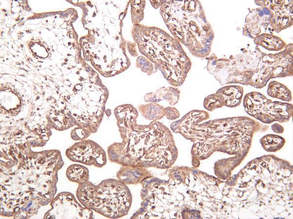
Real IHC images cover human glioma paraffin sections (PB9737 caption) and human placenta (M01743-1 caption); an IF/ICC image covers SiHa cells (PA1681 caption).
PB9737 has an IHC image of human glioma paraffin sections and lists IHC and IF applications (PB9737 caption; catalog applications); M01743-1 has a human placenta IHC image and lists IHC and IF applications (M01743-1 caption; catalog applications). PA1681 has an IF/ICC image of SiHa cells and lists IF and ICC applications (PA1681 caption; catalog applications).
Which to pick: For tissue IHC, choose PB9737 for its human glioma paraffin-section image (PB9737 caption), or M01743-1 for its human placenta IHC image and monoclonal format (M01743-1 caption; catalog); neither caption reports a fixative (PB9737 caption; M01743-1 caption). For IF/ICC, PA1681 has a SiHa cell image but no listed IHC application (PA1681 caption; catalog applications). For work across species, PB9737 lists human, monkey, mouse and rat reactivity, while its pictured tissue IHC result is human (PB9737 catalog reactivity; PB9737 caption).