This website uses cookies to ensure you get the best experience on our website.


- Table of Contents
Plan PPP1R14A chromogenic IHC-P with the catalog antibody at 0.5–1 μg/mL (datasheet A05752-1). Compare high staining in lung alveolar type II cells with undetected staining in adipocytes, and assess cytoplasmic and nuclear signal (HPA tissue IHC).

Expected localisation, validated protocols, controls and antibodies — the at-a-glance facts below, then the full design guide.
| Expected localisation | Cytoplasmic and nuclear tissue staining (HPA tissue IHC) | |
| Staining pattern | Cytoplasmic and nuclear staining across tissues and vessels (HPA tissue IHC) | |
| Antigen retrieval | Citrate pH 6 HIER, heat-mediated (datasheet A05752-1) | |
| Positive control | Adrenal gland+4 more · see all | |
| Negative control | Adipose tissue+4 more · see all |
| Fixation | Keep fixation consistent across sections (standard IHC practice; not target-specific) | |
| Caveat | Staining has medium agreement with RNA data (HPA tissue IHC) | |
| Regulation | Expression regulation unreported (UniProt) | |
| Isoform / epitope | 2 isoforms; epitope coverage unknown (UniProt) |
The catalog antibody protocol (datasheet A05752-1) is accompanied by published PPP1R14A IHC examples in human penile tissue (PMC13097017) and colon tissue (PMC11632118).
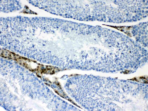
| Sample | Paraffin-embedded mouse testis tissues; fixative not specified (datasheet A05752-1) |
| Fixation | Image fixative and duration unreported (datasheet A05752-1); verify before use. |
| Sectioning | 4–5 µm sections on charged slides (standard) |
| Deparaffinisation | Xylene, graded ethanol series to water (standard) |
| Antigen retrieval | Heat retrieval: Citrate pH 6, 20 min (datasheet A05752-1) |
| Peroxidase block | 3% H2O2, 10 min, room temperature (standard) |
| Blocking | 10% goat serum (datasheet A05752-1) |
| Primary antibody | Rabbit anti-PPP1R14A, 0.5-1μg/ml (datasheet A05752-1) |
| Primary incubation | Overnight at 4 °C (datasheet A05752-1) |
| Detection | Streptavidin-biotin complex (SABC), DAB chromogen (datasheet A05752-1) |
| Counterstain | Hematoxylin, blue, dehydrate and mount (standard) |
| Expected result | PPP1R14A-positive staining in glandular cells of adrenal gland (HPA tissue IHC: High). HPA tissue profile: Cytoplasmic and nuclear expression in several tissues and in blood vessles. No signal in the no-primary control. |
PPP1R14A is annotated as cytoplasmic and has no transmembrane segment (UniProt Q96A00). In paraffin IHC, HPA reports cytoplasmic and nuclear staining across several tissues, including blood vessels; high staining occurs in adrenal glandular cells, brain glial cells and lung alveolar type II cells (HPA tissue IHC). HPA rates the tissue profile Enhanced, with medium agreement between staining and RNA data and external verification pending (HPA tissue IHC).
| Distinct staining in adrenal glandular cells, brain glial cells or lung alveolar type II cells. | These are reported high-staining cell populations (HPA tissue IHC). Score the identified cells and their compartments; a positive area elsewhere in the section does not establish staining in the intended population (general IHC practice). |
| Cytoplasmic staining, with nuclear staining in some cells. | Both compartments occur in the reported tissue profile (HPA tissue IHC). UniProt annotates cytoplasmic localization (UniProt Q96A00); nuclear signal in a tissue section therefore needs evaluation alongside cell identity and controls. |
| Predominantly extracellular or sharply cell-surface staining, or staining across unrelated cell types. | The reported tissue pattern is cytoplasmic and nuclear (HPA tissue IHC), and no transmembrane segment is annotated (UniProt Q96A00). Check for nonspecific staining, cross-reactivity or detection artefact before assigning these patterns to PPP1R14A (general IHC practice). |
| Diffuse chromogen over tissue and spaces between cells, with poor cell boundaries. | This does not resolve the reported positive cell populations (HPA tissue IHC). Background from detection reagents or incomplete blocking can obscure interpretation; compare a negative reagent control and inspect the counterstain (general IHC practice). |
| No signal in a reported high-staining cell population. | A negative section alone cannot distinguish a technical failure from variation in the specimen or antibody performance (general IHC practice). Recheck the identity of the expected cells; HPA reports high staining in specific cell populations, not every cell in their tissues (HPA tissue IHC). |
| Tissue and cell choice | Adrenal glandular cells, brain glial cells and lung alveolar type II cells are reported High; adipocytes in adipose tissue and heart cardiomyocytes are Not detected (HPA tissue IHC). Judge each named cell population, not its whole tissue. |
| Compartment and topology | UniProt annotates cytoplasm and no transmembrane segment (UniProt Q96A00). HPA tissue IHC also reports nuclear staining (HPA tissue IHC); this difference makes cell-resolved assessment useful and does not establish a membrane pattern. |
| Isoforms and processing | UniProt lists two isoforms: isoform 1 in aorta and testis, and isoform 2 in aorta; it annotates no signal peptide or propeptide (UniProt Q96A00). The supplied IHC evidence does not assign staining to either isoform (HPA tissue IHC). |
| Phosphorylation | PPP1R14A has annotated phosphorylation sites, and phosphorylation markedly increases its inhibitory activity (UniProt Q96A00). Total-protein IHC staining does not establish phosphorylation state or inhibitory activity without a separately validated state-specific assay (general IHC practice). |
| Antibody evidence | HPA lists HPA042097 and CAB005044 as IHC Enhanced, and CAB009803 as IHC Supported (HPA antibodies). Apply those ratings to the listed antibodies; the tissue profile still has medium staining–RNA agreement and awaits external verification (HPA tissue IHC). |
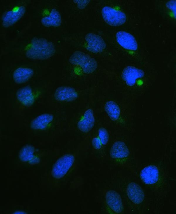
| IF/ICC Q&A: Is nuclear signal plausible? | Yes. HPA ICC-IF reports mainly nucleoplasmic localization, with additional nuclear bodies (HPA subcellular). This cell-image result can inform interpretation, while the paraffin IHC tissue profile reports both cytoplasmic and nuclear staining (HPA tissue IHC). |
| Situation | Likely cause | Next action |
|---|---|---|
| A reported high-staining population appears entirely negative. | The expected cell type may be absent from the section, or the staining run may have failed (general IHC practice); HPA levels describe specific cell populations (HPA tissue IHC). | Confirm cell identity on the counterstain, then review the IHC-validated antibody and run controls. Review retrieval and detection settings as general IHC checks; PPP1R14A-specific fixation sensitivity is unreported in these sources. |
| Chromogen covers the section without clear cell boundaries. | Diffuse background may arise from detection chemistry or nonspecific reagent binding (general IHC practice). It cannot be scored against HPA's cell-specific pattern (HPA tissue IHC). | Compare the negative reagent control, check blocking and washes, and reassess the IHC-validated antibody's working conditions (general IHC practice). Score only staining that can be assigned to intact cells. |
| Staining appears mainly extracellular or confined to cell surfaces. | That distribution departs from the reported cytoplasmic and nuclear tissue pattern (HPA tissue IHC) and the absence of a transmembrane segment (UniProt Q96A00). | Check morphology and counterstain, inspect negative controls, and repeat with an independently validated IHC antibody if available (general IHC practice). Treat the unusual compartment as unresolved until it reproduces. |
| Cells listed as Not detected are strongly stained. | The cell type may be misidentified, or staining may reflect cross-reactivity or endogenous detection activity (general IHC practice). HPA lists adipocytes in adipose tissue and heart cardiomyocytes as Not detected (HPA tissue IHC). | Verify the precise cell type and compare a negative reagent control. If using enzyme-based detection, check the relevant endogenous enzyme block (general IHC practice); avoid judging an entire tissue by one negative cell category. |
| Positive cells show mixed cytoplasmic and nuclear intensity. | Both compartments are reported in tissue IHC (HPA tissue IHC), while HPA ICC-IF emphasizes the nucleoplasm (HPA subcellular); compartment intensity alone does not settle specificity. | Score cytoplasmic and nuclear staining separately in identified cells and compare run controls (general IHC practice). Record the antibody used because HPA validation ratings differ by antibody (HPA antibodies). |
| A faint signal is seen in a reported low-staining population. | HPA reports Low staining in bronchial respiratory epithelial cells and urinary bladder urothelial cells (HPA tissue IHC). Weak signal can be difficult to separate from background (general IHC practice). | Compare cells with nearby background and the negative reagent control, then record intensity with cell identity (general IHC practice). Use a reported high-staining population to check run performance (HPA tissue IHC). |


Comprehensive Human Protein Atlas IHC scoring per tissue (reliability: Enhanced — Medium consistency between antibody staining and RNA expression data. Pending external verification.). Rows are taken directly from the HPA tissue chart — click any row's HPA link to view the source.
| Tissue | Cell type | Level | Evidence | Source |
|---|---|---|---|---|
| Adrenal gland | Glandular cells | High | Protein (IHC) | HPA → |
| Caudate | Glial cells | High | Protein (IHC) | HPA → |
| Cerebellum | Granular cells - cytoplasm/membrane | High | Protein (IHC) | HPA → |
| Cerebral cortex | Glial cells | High | Protein (IHC) | HPA → |
| Hippocampus | Glial cells | High | Protein (IHC) | HPA → |
| Tissue | Cell type | Level | Evidence | Source |
|---|---|---|---|---|
| Adipose tissue | Adipocytes | Not detected | Protein (IHC) | HPA → |
| Appendix | Endocrine cells | Not detected | Protein (IHC) | HPA → |
| Breast | Adipocytes | Not detected | Protein (IHC) | HPA → |
| Cervix | Glandular cells | Not detected | Protein (IHC) | HPA → |
| Duodenum | Endocrine cells | Not detected | Protein (IHC) | HPA → |
Troubleshooting PPP1R14A staining in paraffin sections requires attention to retrieval, compartment, cell type and the limits of antibody validation.
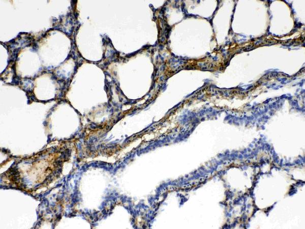
A05752-1 has IHC images from paraffin sections of mouse testis, rat lung, and human intestinal and lung cancers (IHC image captions), plus IF data from U20S cells (IF image caption).
A05752-1 is listed for IHC and IF/ICC, with human, mouse, and rat reactivity (catalog applications/reactivity). Its IHC captions show the paraffin sections listed above, while its IF caption shows U20S cells (catalog image captions).
Which to pick: Choose A05752-1 for paraffin-section chromogenic IHC; its captions document citrate retrieval and DAB detection, with a listed IHC concentration of 0.5–1 μg/ml (IHC image captions; catalog dilution). For IF/ICC, A05752-1 has an IF image from U20S cells and a listed concentration of 2 μg/ml (IF image caption; catalog dilution). It is listed as reactive with human, mouse, and rat; the host is rabbit, clonality is unreported, and the IHC captions do not report the fixative (catalog reactivity/host/clone; IHC image captions).