This website uses cookies to ensure you get the best experience on our website.


- Table of Contents
Plan PPP1R15A paraffin IHC around granular cytoplasmic staining (HPA tissue IHC). The catalog antibody has an IHC dilution range of 2–5 μg/ml (datasheet A02394-2); duodenal glandular cells show high staining and can guide tissue selection (HPA tissue IHC).

Expected localisation, validated protocols, controls and antibodies — the at-a-glance facts below, then the full design guide.
| Expected localisation | Granular cytoplasm (HPA tissue IHC); ER/outer mitochondrial membrane expected (UniProt) | |
| Staining pattern | Granular cytoplasmic staining in several tissues (HPA tissue IHC) | |
| Antigen retrieval | EDTA pH 8.0 HIER, heat-mediated (datasheet A02394-2) | |
| Positive control | Duodenum+4 more · see all | |
| Negative control | Cervix+4 more · see all |
| Fixation | Keep fixation consistent across sections. (standard IHC practice; not target-specific) | |
| Caveat | Antibody staining has medium concordance with RNA expression (HPA tissue IHC) | |
| Regulation | Growth arrest and DNA damage inducible (UniProt) | |
| Isoform / epitope | 2 isoforms; check epitope coverage across both (UniProt) |
The catalog antibody’s IHC-P protocol (datasheet A02394-2) is accompanied by a published chromogenic protocol for human lung sections (PMC8566588).
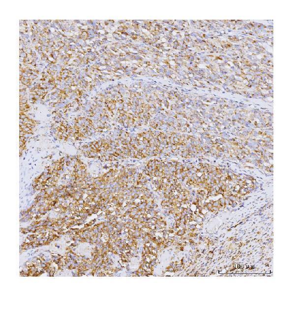
| Sample | Paraffin-embedded human melanom tissue; fixative not specified (datasheet A02394-2) |
| Fixation | Image fixative and duration unreported (datasheet A02394-2); verify before use. |
| Sectioning | 4–5 µm sections on charged slides (standard) |
| Deparaffinisation | Xylene, graded ethanol series to water (standard) |
| Antigen retrieval | Heat retrieval: EDTA pH 8.0 (datasheet A02394-2); 20 min, 95–100 °C (standard) |
| Peroxidase block | 3% H2O2, 10 min, room temperature (standard) |
| Blocking | 10% goat serum (datasheet A02394-2) |
| Primary antibody | Rabbit anti-PPP1R15A, 2-5 μg/ml (datasheet A02394-2) |
| Primary incubation | Overnight at 4 °C (datasheet A02394-2) |
| Detection | HRP-conjugated secondary, DAB chromogen (datasheet A02394-2) |
| Counterstain | Hematoxylin, blue, dehydrate and mount (standard) |
| Expected result | PPP1R15A-positive staining in glandular cells of duodenum (HPA tissue IHC: High). HPA tissue profile: Granular cytoplasmic expression in several tissues. No signal in the no-primary control. |
PPP1R15A should appear as granular cytoplasmic staining in several tissues, especially duodenal and prostatic glandular cells (HPA tissue IHC: High; granular cytoplasmic profile). Its membrane associations are with the endoplasmic reticulum and mitochondrial outer membrane, through an N-terminal amphipathic region without an annotated transmembrane segment (UniProt O75807: subcellular location and topology). Treat tissue predictions as guides: HPA rates the IHC evidence Approved, with medium consistency between staining and RNA expression (HPA tissue IHC: reliability).
| Granular cytoplasmic staining in duodenal or prostatic glandular cells, with the cells identifiable on the counterstained section. | This matches the reported compartment and the two listed High cell populations (HPA tissue IHC: granular cytoplasmic profile; High in duodenal and prostatic glandular cells). Compare intensity within the same staining run; HPA levels are reference observations, not a required score for every specimen (HPA tissue IHC: reported levels; general IHC practice). |
| A dominant nuclear or sharply surface-restricted signal replaces cytoplasmic granules. | This conflicts with HPA's tissue pattern and UniProt's cytoplasmic-facing membrane topology (HPA tissue IHC: granular cytoplasmic profile; UniProt O75807: topology). Review morphology and the detection controls before assigning the signal to PPP1R15A (general IHC practice). |
| Strong staining appears in cardiomyocytes or cervical squamous epithelial cells. | HPA reports PPP1R15A as Not detected in those named cells; the result warrants a specificity check, including cross-reactivity or endogenous detection activity (HPA tissue IHC: cardiomyocytes and cervical squamous epithelial cells Not detected; general IHC practice). It does not prove that every cell in either tissue must be negative (HPA tissue IHC: cell-specific observations). |
| Colour spreads across stroma or cell-free areas and obscures cell boundaries. | This is diffuse background rather than the reported granular cytoplasmic pattern (HPA tissue IHC: granular cytoplasmic profile). Assess the negative detection control and the distribution of precipitate before scoring cells (general chromogenic IHC practice). |
| The expected glandular cells show no signal in a duodenum or prostate control section. | An absent signal in cells listed as High makes the run difficult to interpret (HPA tissue IHC: High in duodenal and prostatic glandular cells). Check control tissue preservation, retrieval, antibody and detection steps together; one negative section cannot establish that PPP1R15A is absent from the specimen (general IHC practice). |
| Compartment and topology (UniProt O75807: subcellular location and topology). | PPP1R15A associates with endoplasmic reticulum and mitochondrial outer membranes through an N-terminal amphipathic region; its annotated regions face the cytoplasm (UniProt O75807: topology). Interpret membrane-associated granularity within the cytoplasm without requiring a visible organelle outline in chromogenic sections (UniProt O75807: location; general IHC practice). |
| Cell-specific reference pattern (HPA tissue IHC: reported cell levels). | Duodenal and prostatic glandular cells are listed High; appendix, breast and colon glandular cells are listed Medium (HPA tissue IHC: cell levels). Cervical squamous epithelial cells and cardiomyocytes are listed Not detected (HPA tissue IHC: cell levels). Use the named cells when comparing sections, rather than treating an entire organ as uniformly positive or negative (HPA tissue IHC: cell-specific entries). |
| Validation scope (HPA antibodies: HPA020240 and CAB018395; HPA tissue IHC: reliability). | Both listed antibodies have Approved IHC status; the tissue summary notes medium consistency with RNA expression (HPA antibodies: IHC Approved; HPA tissue IHC: reliability). Those ratings support comparison with the observed pattern but do not establish that an unexpected signal is target-specific (HPA tissue IHC: validation summary; general IHC practice). |
| Isoforms and epitope uncertainty (UniProt O75807: isoforms). | UniProt lists two isoforms, but the supplied record gives no antibody epitope or isoform-specific staining evidence (UniProt O75807: isoforms; HPA antibodies: supplied fields). Avoid assigning a staining difference to one isoform without independent evidence (general IHC interpretation practice). |
| IF/ICC Q: Should vesicular fluorescence define the paraffin IHC pattern? (HPA subcellular ICC-IF: vesicles, uncertain). | A: No. HPA calls the ICC-IF location vesicles with uncertain confidence, whereas its tissue IHC profile is granular cytoplasmic (HPA subcellular ICC-IF: uncertain; HPA tissue IHC: profile). Interpret each application against its own evidence (general assay interpretation practice). |
| Situation | Likely cause | Next action |
|---|---|---|
| The positive control lacks staining in duodenal or prostatic glandular cells (HPA tissue IHC: High). | A failed staining step or an unsuitable control section is possible; the HPA level alone does not identify which step failed (HPA tissue IHC: High; general IHC practice). | Target-specific fixation effects are not established by the supplied assay evidence. Verify with a matched IHC source before attributing a result to fixation. |
| The negative detection control develops colour (general chromogenic IHC practice). | Detection reagents or endogenous tissue activity may contribute signal independently of primary-antibody binding (general chromogenic IHC practice). | Check the control and address the relevant endogenous activity within the IHC detection workflow before interpreting cell staining (general chromogenic IHC practice). |
| Staining is widespread and diffuse, obscuring cytoplasmic granules (HPA tissue IHC: granular cytoplasmic profile). | Background from antibody concentration, incomplete blocking or washing may obscure the reported pattern (general IHC practice; HPA tissue IHC: profile). | Compare a matched negative control, then review blocking, washing and the catalog antibody's IHC-P dilution guidance (general IHC practice); no dilution value is supplied here (HPA antibodies: supplied fields). |
| A dominant nuclear or cell-surface pattern appears (HPA tissue IHC: granular cytoplasmic profile). | The compartment conflicts with the tissue profile and cytoplasmic-facing topology, so assignment to PPP1R15A is uncertain (HPA tissue IHC: profile; UniProt O75807: topology). | Confirm cellular boundaries with the counterstain and compare the positive and negative detection controls before scoring (general IHC practice). |
| An HPA-listed Not detected cell population stains strongly (HPA tissue IHC: cardiomyocytes or cervical squamous epithelial cells). | Cross-reactivity or endogenous detection activity is possible, while HPA observations do not establish an absolute biological absence (HPA tissue IHC: cell-specific levels; general IHC practice). | Check the named cell type, inspect the negative detection control and seek independent specificity evidence before reporting a new cell-type pattern (general IHC practice; HPA tissue IHC: cell-specific reference). |


Comprehensive Human Protein Atlas IHC scoring per tissue (reliability: Approved — Medium consistency between antibody staining and RNA expression data.). Rows are taken directly from the HPA tissue chart — click any row's HPA link to view the source.
| Tissue | Cell type | Level | Evidence | Source |
|---|---|---|---|---|
| Duodenum | Glandular cells | High | Protein (IHC) | HPA → |
| Prostate | Glandular cells | High | Protein (IHC) | HPA → |
| Appendix | Glandular cells | Medium | Protein (IHC) | HPA → |
| Bone marrow | Hematopoietic cells | Medium | Protein (IHC) | HPA → |
| Breast | Glandular cells | Medium | Protein (IHC) | HPA → |
| Tissue | Cell type | Level | Evidence | Source |
|---|---|---|---|---|
| Cervix | Squamous epithelial cells | Not detected | Protein (IHC) | HPA → |
| Heart muscle | Cardiomyocytes | Not detected | Protein (IHC) | HPA → |
| Liver | Cholangiocytes | Not detected | Protein (IHC) | HPA → |
| Ovary | Follicle cells | Not detected | Protein (IHC) | HPA → |
| Seminal vesicle | Glandular cells | Not detected | Protein (IHC) | HPA → |
Troubleshoot PPP1R15A staining in paraffin sections by checking retrieval, compartment, controls and cell-level scoring against the supplied IHC evidence.
The catalog includes rabbit anti-PPP1R15A antibodies for IHC and IF/ICC in human samples, with rat reactivity listed for both and mouse reactivity for A02394-5 (catalog: applications and reactivity). A02394-2 has a human paraffin-section IHC figure (caption: A02394-2).
A02394-2 has a human paraffin-section IHC image and lists IF/ICC among its applications (caption and catalog: A02394-2). A02394-5 lists IHC and IF/ICC with human, mouse and rat reactivity; no IHC or IF figure is supplied for it (catalog: A02394-5).
Which to pick: Choose A02394-2 for human paraffin-section IHC because its own caption documents EDTA retrieval at pH 8.0 and staining at 2 μg/ml; the fixative is unreported (caption: A02394-2). For IF/ICC, both SKUs list the application, with 5 μg/ml for A02394-2 and 1:50 for A02394-5; neither has a supplied IF image (catalog: A02394-2, A02394-5). For work that includes mouse samples, choose the rabbit polyclonal A02394-5, which lists human, mouse and rat reactivity; its IHC processing is undocumented by an image caption (catalog: A02394-5).