This website uses cookies to ensure you get the best experience on our website.


- Table of Contents
Plan PPP1R1A staining in paraffin sections using the catalog antibody’s IHC protocol. Compare nuclear and cytoplasmic staining in pancreatic endocrine cells with the tissue pattern reported by HPA (HPA tissue IHC).

Expected localisation, validated protocols, controls and antibodies — the at-a-glance facts below, then the full design guide.
| Expected localisation | Nuclear and cytoplasmic staining (HPA tissue IHC) | |
| Staining pattern | Islet endocrine cells: nuclear and cytoplasmic (HPA tissue IHC) | |
| Antigen retrieval | EDTA pH 8.0 HIER, heat-mediated (datasheet M11390) | |
| Positive control | Pancreas | |
| Negative control | Adipose tissue+4 more · see all |
| Fixation | Keep fixation consistent across paraffin sections. (standard IHC practice; not target-specific) | |
| Caveat | Antibody staining and RNA show low consistency (HPA tissue IHC) | |
| Regulation | Expression regulation unreported (UniProt) | |
| Isoform / epitope | No isoforms or signal/propeptide annotated (UniProt) |
The catalog antibody’s IHC-P protocol is accompanied by one published paraffin-section pancreas protocol with fluorescence imaging (PMC3717856).
| Sample | Paraffin-embedded human liver tissue; fixative not specified (datasheet M11390) |
| Fixation | Image fixative and duration unreported (datasheet M11390); verify before use. |
| Sectioning | 4–5 µm sections on charged slides (standard) |
| Deparaffinisation | Xylene, graded ethanol series to water (standard) |
| Antigen retrieval | Heat retrieval: EDTA pH 8.0 (datasheet M11390); 20 min, 95–100 °C (standard) |
| Peroxidase block | 3% H2O2, 10 min, room temperature (standard) |
| Blocking | 10% goat serum (datasheet M11390) |
| Primary antibody | Rabbit monoclonal (clone GEG-16) anti-PPP1R1A, 1:50 recommended; image 2 μg/ml (datasheet M11390) |
| Primary incubation | Overnight at 4 °C (datasheet M11390) |
| Detection | HRP-conjugated secondary, DAB chromogen (datasheet M11390) |
| Counterstain | Hematoxylin, blue, dehydrate and mount (standard) |
| Expected result | PPP1R1A-positive staining in pancreatic endocrine cells of pancreas (HPA tissue IHC: Medium). HPA tissue profile: Nuclear and cytoplasmic expression mainly in islets of Langerhans and renal tubules. No signal in the no-primary control. |
In paraffin-section IHC, expect PPP1R1A in nuclei and cytoplasm, mainly in pancreatic islets and renal tubules (HPA tissue IHC). Pancreatic endocrine cells show medium staining; renal tubular cells show low staining (HPA tissue IHC). HPA rates the tissue staining Approved, while reporting low consistency with RNA expression data (HPA tissue IHC). UniProt annotates no transmembrane segment and gives no subcellular location (UniProt Q13522).
| Discrete nuclear and cytoplasmic staining in pancreatic islet cells, with weaker staining in renal tubules (HPA tissue IHC). | This fits the reported distribution: pancreatic endocrine cells are medium and renal tubular cells are low (HPA tissue IHC). Score the cell population and each compartment separately so weak tubular signal is not mistaken for a failed run (HPA tissue IHC; general IHC practice). |
| Strong, exclusively membrane-bound staining with no discernible nuclear or cytoplasmic signal (HPA tissue IHC comparison). | Flag the compartment mismatch as a possible artefact and review controls and morphology (HPA tissue IHC; general IHC practice). UniProt annotates no transmembrane segment, but that alone cannot rule out every association with a membrane (UniProt Q13522 topology). |
| Prominent signal in adipocytes or other cells listed as not detected by HPA (HPA tissue IHC). | Treat this as possible cross-reactivity or endogenous detection activity; inspect the negative control and compare with the islet pattern (HPA tissue IHC; general IHC practice). A reported 'not detected' result is an IHC observation, not proof that every specimen must be negative (HPA tissue IHC). |
| Diffuse colour across cells and surrounding tissue, obscuring islet boundaries (general IHC practice). | The distribution is difficult to score as PPP1R1A when background lacks the reported cell and compartment pattern (HPA tissue IHC). Check blocking, washes, detection controls and antibody dilution as general IHC troubleshooting steps (general IHC practice). |
| No convincing staining in pancreatic endocrine cells on a run intended to detect PPP1R1A (HPA tissue IHC comparison). | The expected medium positive reference is missing (HPA tissue IHC). First verify tissue identity and assay controls, then review retrieval, dilution and detection as general IHC steps; the supplied sources give no PPP1R1A-specific retrieval or fixation requirement (HPA tissue IHC; general IHC practice). |
| Cell and tissue selection (HPA tissue IHC) | Pancreatic endocrine cells provide the clearest reported positive at medium intensity; renal tubular cells are low, while adipocytes are reported as not detected (HPA tissue IHC). These are useful comparison populations when reading a paraffin section (HPA tissue IHC; general IHC practice). |
| Strength of tissue-pattern evidence (HPA tissue IHC) | The tissue result is Approved, with external characterization supporting antibody staining, but HPA reports low consistency with RNA expression data (HPA tissue IHC). Interpret a discrepant specimen alongside controls and morphology rather than treating RNA abundance as an IHC intensity scale (HPA tissue IHC; general IHC practice). |
| Antibody validation scope (HPA antibodies) | HPA lists HPA041542 and CAB007811 as Approved for IHC; neither has ICC validation in the supplied antibody record (HPA antibodies). Approved is the stated status here; the payload does not assign either antibody an Enhanced IHC rating (HPA antibodies). |
| Topology and localisation evidence (UniProt Q13522; HPA tissue IHC) | UniProt annotates no transmembrane segment and no subcellular location; the nuclear and cytoplasmic expectation comes from observed tissue staining (UniProt Q13522 topology; HPA tissue IHC). Use that observed pattern to judge a slide without claiming UniProt independently confirms either compartment (UniProt Q13522; HPA tissue IHC). |
| Reported modifications (UniProt Q13522) | UniProt lists modified residues, including phosphorylation at Thr35 and Ser43, Ser46 and Ser47 (UniProt Q13522). The supplied records do not establish whether these modifications change IHC staining or whether either HPA antibody distinguishes modified PPP1R1A (UniProt Q13522; HPA antibodies). |
| Situation | Likely cause | Next action |
|---|---|---|
| The pancreatic islet reference has no signal (HPA tissue IHC comparison). | A failed detection step, unsuitable assay setting or an incorrectly identified reference section is possible (general IHC practice). | Confirm endocrine-cell morphology and control performance; review antigen retrieval, antibody dilution and chromogenic detection using the assay's documented settings (general IHC practice). No PPP1R1A-specific retrieval condition is supplied (HPA tissue IHC). |
| The renal tubules appear negative while the islet reference stains (HPA tissue IHC comparison). | Tubular staining is reported as low, compared with medium in pancreatic endocrine cells (HPA tissue IHC). | Inspect tubular cells at an appropriate magnification and compare them with a negative control before calling the run unsuccessful (HPA tissue IHC; general IHC practice). |
| Adipocytes show prominent colour (HPA tissue IHC comparison). | HPA reports adipocytes as not detected; cross-reactivity or endogenous detection activity is possible (HPA tissue IHC; general IHC practice). | Compare a negative control and the positive islet pattern; review detection and blocking steps if colour persists outside the expected cells (HPA tissue IHC; general IHC practice). |
| Signal is diffuse across the section (general IHC practice). | Background from insufficient blocking, washing or an overly concentrated reagent can obscure cellular staining (general IHC practice). | Check negative controls, then review blocking, wash steps and the documented antibody dilution; score only a distinguishable cellular pattern (general IHC practice). |
| Staining is confined to membranes (HPA tissue IHC comparison). | The pattern differs from HPA's nuclear and cytoplasmic description, so artefact or nonspecific staining is possible (HPA tissue IHC; general IHC practice). | Check section morphology and detection controls, then compare the staining with pancreatic endocrine cells on a reference section (HPA tissue IHC; general IHC practice). |
| Can IF/ICC confirm the nuclear and cytoplasmic pattern? | The supplied HPA subcellular record has no main location or ICC-IF images, and the listed antibodies have no ICC validation status (HPA subcellular; HPA antibodies). | Treat IF/ICC localisation as unconfirmed by this payload and use a separately validated IF/ICC guide for that application (HPA subcellular; HPA antibodies). |


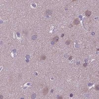


Comprehensive Human Protein Atlas IHC scoring per tissue (reliability: Approved — Low consistency between antibody staining and RNA expression data. External characterization data supports antibody staining.). Rows are taken directly from the HPA tissue chart — click any row's HPA link to view the source.
| Tissue | Cell type | Level | Evidence | Source |
|---|---|---|---|---|
| Pancreas | Pancreatic endocrine cells | Medium | Protein (IHC) | HPA → |
| Tissue | Cell type | Level | Evidence | Source |
|---|---|---|---|---|
| Adipose tissue | Adipocytes | Not detected | Protein (IHC) | HPA → |
| Adrenal gland | Glandular cells | Not detected | Protein (IHC) | HPA → |
| Appendix | Glandular cells | Not detected | Protein (IHC) | HPA → |
| Bone marrow | Hematopoietic cells | Not detected | Protein (IHC) | HPA → |
| Breast | Adipocytes | Not detected | Protein (IHC) | HPA → |
Troubleshoot PPP1R1A staining in paraffin sections using the M11390 liver image as the workflow reference and HPA tissue patterns as context.
M11390 has real IHC data from human paraffin-embedded liver sections (M11390 IHC caption); IF/ICC is listed for human, mouse and rat (M11390 catalog applications and reactivity).
M11390 shows PPP1R1A staining in paraffin-embedded human liver and liver cancer sections (M11390 IHC captions). IF/ICC is listed, with human, mouse and rat reactivity, but no IF image is supplied (M11390 catalog applications, reactivity and image alts).
Which to pick: For tissue IHC, choose rabbit monoclonal M11390: its own caption documents staining in a paraffin-embedded human liver section, with EDTA pH 8.0 retrieval and DAB detection; the fixative is unreported (M11390 catalog; M11390 IHC caption). For IF/ICC or work involving mouse or rat, M11390 is the listed option based on its application and reactivity entries; the supplied images do not demonstrate IF or staining in those species (M11390 catalog applications, reactivity and image alts).