PRAME / Melanoma antigen preferentially expressed in tumors · IHC design guide

Design Immunohistochemistry for PRAME

Plan PRAME IHC in paraffin sections using testis as a positive tissue, with cytoplasmic staining expected in elongated or late spermatids (HPA tissue IHC). The catalog antibody is validated for IHC-P at 1:50–1:100 (datasheet).

Evidence assembled Oct 2026 · For research use; verify linked source records and product datasheet before use
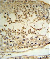
Immunohistochemistry protocol sheet for PRAME (IHC for PRAME): expected localisation Cytoplasmic in testicular spermatids (HPA tissue IHC), antibody A06628-1, validated IHC image, and IHC protocol steps
Printable PRAME IHC protocol sheet — expected localisation Cytoplasmic in testicular spermatids (HPA tissue IHC), antibody A06628-1, controls and protocol steps. Open the full PRAME IHC guide →

PRAME Immunohistochemistry Experimental Design Guide

Expected localisation, validated protocols, controls and antibodies — the at-a-glance facts below, then the full design guide.

Must know before staining
Expected localisation Cytoplasmic in testicular spermatids (HPA tissue IHC)
Staining pattern Cytoplasmic staining in elongated or late spermatids (HPA tissue IHC)
Antigen retrieval Tris-EDTA pH 9.0 HIER, 95–98 °C, 20 min (rule: nuclear antigen)
Positive control ⓘ Testis
Negative control ⓘ Adipose tissue+4 more · see all
Important caveats
Reasons your staining may differ from the expected pattern.
Fixation Keep formalin fixation consistent across paraffin sections (standard IHC practice; not target-specific)
Caveat Listed tissues outside testis show no detected staining (HPA tissue IHC)
Regulation Testis enriched; high in late spermatids (HPA tissue IHC)
Isoform / epitope No annotated isoforms; one 1–509 chain (UniProt)
Section 1

Recommended PRAME IHC & IF Protocols

The catalog antibody’s IHC-P protocol is paired with four published PRAME IHC protocols covering melanocytic lesions and liposarcoma (PMC10813997; PMC4939879; PMC12615063; PMC10591151).

Recommended immunohistochemistry (IHC-P) protocol parameters
SampleFFPE human testis tissue (datasheet A06628-1)
FixationImage formalin-fixed; duration unreported (datasheet A06628-1); verify before use.
Sectioning4–5 µm sections on charged slides (standard)
DeparaffinisationXylene, graded ethanol series to water (standard)
Antigen retrievalHeat-induced epitope retrieval in Tris-EDTA buffer, pH 9.0, 20 min at 95–98 °C (standard rule: nuclear antigen)
Peroxidase block3% H2O2, 10 min, room temperature (standard)
Blocking10% normal serum of the secondary host, 30 min, room temperature (standard)
Primary antibodyRabbit anti-PRAME, 1:50-1:100 (datasheet A06628-1)
Primary incubationOvernight at 4 °C (standard)
DetectionHRP-polymer secondary, DAB chromogen 5–10 min (standard)
CounterstainHematoxylin, blue, dehydrate and mount (standard)
Expected resultPRAME-positive staining in elongated or late spermatids of testis (HPA tissue IHC: High). HPA tissue profile: Cytoplasmic expression in seminiferous ducts in testis. No signal in the no-primary control.
💡Decision noteStart with Tris-EDTA HIER at pH 9.0, 95–98 °C for 20 min (page retrieval specification); the published protocols also describe pH 6 citrate and pH 8 retrieval (PMC4939879; PMC10591151).
Section 2

What Is the Expected PRAME Staining Pattern?

In paraffin-section IHC, expect cytoplasmic PRAME staining in elongated or late spermatids within testicular seminiferous ducts (HPA tissue IHC: Enhanced; High in elongated or late spermatids). PRAME has no annotated transmembrane segment (UniProt P78395 topology). Nuclear and other locations are also recorded (UniProt P78395 subcellular location), so interpret compartment in the context of the assay and tissue.

What am I looking at on my slide?
Strong, cell-associated cytoplasmic staining in elongated or late spermatids of testis.Matches the observed testis IHC pattern (HPA tissue IHC: cytoplasmic expression in seminiferous ducts; High in elongated or late spermatids). Assess both the cell type and compartment.
Predominantly nuclear staining in testicular spermatids, with little of the expected cytoplasmic signal.Does not reproduce the HPA testis IHC pattern (HPA tissue IHC: cytoplasmic expression). Recheck morphology and controls before calling it PRAME; nuclear localization is documented in other contexts (UniProt P78395; HPA subcellular ICC-IF).
Strong staining in testicular cells other than the expected spermatids, or in adipocytes.Compare with cell-specific controls: HPA reports High staining in elongated or late spermatids and no detection in adipocytes (HPA tissue IHC). Unexpected staining warrants a check for antibody cross-reactivity or endogenous detection activity.
Diffuse chromogen across tissue, including regions without clear cell boundaries.This is difficult to assign to PRAME-positive cells. Review blocking, washing, detection reagents and a no-primary control as general IHC checks; the HPA tissue pattern is cell-associated (HPA tissue IHC: seminiferous ducts).
No staining in a testis section containing identifiable elongated or late spermatids.The positive control has failed relative to the reported High signal (HPA tissue IHC: elongated or late spermatids). Check tissue morphology, antibody performance and detection before interpreting other sections as negative.
💡Expected PRAME appearanceA convincing positive is High, cell-associated cytoplasmic staining in elongated or late spermatids of testis (HPA tissue IHC); diffuse deposit or staining in HPA-negative adipocytes is suspicious (HPA tissue IHC: adipocytes Not detected).
How each factor affects the staining
Tissue and cell selectionTestis with identifiable elongated or late spermatids provides a reported positive pattern; adipocytes and adrenal glandular cells are reported Not detected (HPA tissue IHC). Interpret those calls at the named cell level.
IHC versus IF/ICC compartment: what should IF show?For the separate IF/ICC assay, HPA reports mainly nucleoplasmic localization (approved), with additional plasma-membrane localization (supported) (HPA subcellular ICC-IF). That does not replace the cytoplasmic testis IHC reference pattern (HPA tissue IHC).
Antibody validationHPA rates tissue IHC reliability Enhanced and lists IHC Enhanced for HPA045153 and CAB080138 (HPA tissue IHC; HPA antibodies). HPA068562 is listed as ICC Supported, without an IHC rating in the supplied record (HPA antibodies).
Topology and processingUniProt annotates a 509-aa chain, no signal peptide, no annotated transmembrane segment and no propeptide (UniProt P78395 topology and processing). These annotations do not establish an IHC retrieval condition or a staining compartment on their own.
Antigen retrieval and chromogenic detectionThe supplied sources give no PRAME-specific retrieval condition or fixation sensitivity. If optimizing a paraffin-section IHC workflow, evaluate retrieval and detection with positive tissue and appropriate controls as general IHC practice.
Why is my staining missing, weak or wrong?
SituationLikely causeNext action
Testis positive control is blank.The observed High spermatid signal has not been reproduced (HPA tissue IHC); the cause cannot be assigned from a blank slide alone.Confirm elongated or late spermatids are present, then check the IHC-validated antibody, retrieval and detection steps with the same control section; no PRAME-specific retrieval setting is supplied.
Only nuclei stain in testicular spermatids.The compartment differs from the reported cytoplasmic testis IHC pattern (HPA tissue IHC), although PRAME can localize to nuclei (UniProt P78395).Check morphology and the IHC assay controls; report the discrepancy rather than treating a nuclear-only testis result as the established HPA IHC pattern.
Adipocytes or adrenal glandular cells stain strongly.These named cell types are reported Not detected (HPA tissue IHC); cross-reactivity or endogenous detection activity is possible.Run a no-primary control and inspect detection and blocking steps; assess staining in the specific HPA-negative cell type before assigning PRAME positivity.
Brown deposit is widespread or obscures cellular detail.Diffuse signal cannot be matched confidently to the cell-associated testis pattern (HPA tissue IHC); excess background is a general IHC possibility.Inspect no-primary and tissue controls, washing and detection background; score only interpretable cells with preserved morphology.
Results disagree across antibodies.Validation differs by assay: HPA045153 and CAB080138 are IHC Enhanced, whereas HPA068562 is ICC Supported with no supplied IHC rating (HPA antibodies).For paraffin-section IHC, verify the antibody's IHC status and compare the same positive tissue and cell compartment before reconciling results.
IF/ICC shows nucleoplasmic staining while testis IHC looks cytoplasmic.These are different reported assay observations (HPA subcellular ICC-IF: nucleoplasm approved; HPA tissue IHC: cytoplasmic spermatids).Interpret each against its own assay reference and controls; use the separate IF/ICC guide for IF workflow decisions.

Sample controls for PRAME IHC & IF

🧪Run testis first; elongated or late spermatids should stain (HPA: High in elongated or late spermatids). Use adipose tissue as a negative tissue, focusing on adipocytes (HPA: Not detected in adipocytes); on the testis slide, cells outside the listed spermatid population should show only background staining, but their PRAME-negative status is unconfirmed.
Positive control tissue: Testis (Elongated or late spermatids, HPA High)
Negative control tissue: Adipose tissue (HPA Not detected)
ICC-IF cell lines (HPA subcellular resource): HPA ICC-IF images show PRAME in K-562, U2OS, with annotated localisation: Nucleoplasm (approved) (HPA subcellular).
Technical controls: Include a no-primary or secondary-only control, a control immunoglobulin matched to the primary antibody’s host species and isotype for a monoclonal antibody (or nonimmune IgG for a polyclonal antibody), and a PRAME-knockout biological control where available (standard IHC/IF control practice). For testis sections, block and check endogenous peroxidase signal in chromogenic IHC; assess tissue autofluorescence in IF with a no-primary control (standard IHC/IF practice).
⚠️Feasibility: The selected antibody caption reports formalin-fixed, paraffin-embedded human testis stained with peroxidase and DAB, but reports no fixation duration or target-specific fixation window (caption: A06628-1). Retrieval requirements are unreported, and the supplied evidence does not establish whether frozen sections or IF are easier than paraffin IHC (caption: A06628-1; HPA: subcellular ICC-IF images). No testis-specific artefact is documented in the supplied evidence; check peroxidase background or autofluorescence with the corresponding controls (standard IHC/IF practice).

HPA tissue IHC evidence for PRAME

Comprehensive Human Protein Atlas IHC scoring per tissue (reliability: Enhanced — High consistency between antibody staining and RNA expression data.). Rows are taken directly from the HPA tissue chart — click any row's HPA link to view the source.

Positive expression · recommended positive controls

TissueCell typeLevelEvidenceSource
Testis Elongated or late spermatids High Protein (IHC) HPA →

Undetected expression · recommended negative controls

TissueCell typeLevelEvidenceSource
Adipose tissue Adipocytes Not detected Protein (IHC) HPA →
Adrenal gland Glandular cells Not detected Protein (IHC) HPA →
Appendix Glandular cells Not detected Protein (IHC) HPA →
Bone marrow Hematopoietic cells Not detected Protein (IHC) HPA →
Breast Adipocytes Not detected Protein (IHC) HPA →
Section 3

Advanced PRAME IHC Tips

Troubleshoot PRAME staining in paraffin section IHC by checking retrieval, tissue context, cellular compartment, background, and scoring before interpreting a result.

How should I retrieve PRAME in paraffin sections when staining is weak?
Start with Tris-EDTA pH 9.0 HIER at 95–98 °C for 20 min (page retrieval setting). Cool sections consistently, then compare a testis control and a no-primary control on the same staining run to separate poor retrieval from detection background (HPA: high staining in elongated or late spermatids; standard IHC practice). If signal remains weak, adjust heating time in a small series while keeping buffer and detection conditions fixed, and reject conditions that damage morphology (standard IHC practice). Only then assess an alternative retrieval buffer as a fallback, recording its pH and time because a changed staining pattern alone does not establish specificity (standard IHC practice).
Does formalin fixation change PRAME sensitivity in paraffin IHC?
An image caption documents formalin-fixed, paraffin-embedded human testis stained with the C-terminal antibody and peroxidase/DAB detection (A06628-1 tissue-IHC caption). It provides no fixation comparison, so PRAME-specific sensitivity to fixation duration or fixative choice is unknown (A06628-1 tissue-IHC caption). Record fixative, duration, section age, and retrieval conditions for each case; compare differently processed specimens only after staining them alongside the same control section (standard IHC practice). If staining varies, inspect preservation and morphology before attributing the difference to PRAME biology, and use matched processing when assessing intensity across cases (standard IHC practice).
Should PRAME staining be nuclear or cytoplasmic?
Assess nuclear and cytoplasmic staining separately: PRAME is mainly nucleoplasmic in subcellular imaging, while tissue IHC shows cytoplasmic expression in testis seminiferous ducts (HPA: subcellular; HPA: tissue IHC). UniProt also lists chromosome, Golgi, cell membrane, and cytoplasm, with chromatin association and interferon-gamma-responsive Golgi recruitment (UniProt P78395 localisation). Therefore, do not discard a signal solely because it differs from the dominant compartment in another assay; first verify cell identity, morphology, and the no-primary control (HPA: subcellular and tissue IHC; standard IHC practice). Report the fraction of stained cells in each compartment and whether staining follows tissue structure rather than section edges (standard IHC practice).
Could an epitope or PRAME isoform explain discordant staining?
The record lists one 1–509 chain, 0 annotated isoforms, no transmembrane segment, and no annotated glycosylation sites (UniProt P78395 processing, isoforms, topology, glycosylation). The tissue-IHC image uses a C-terminal antibody, but its caption does not define an exact epitope or establish that every intracellular pool is detected equally (A06628-1 tissue-IHC caption). Phosphoserines at positions 9 and 16 are annotated; their effect on this antibody's staining is untested in the supplied evidence (UniProt P78395 modified residues; A06628-1 tissue-IHC caption). When results disagree, compare independently validated epitopes on adjacent sections and check whether retrieval changes signal without compromising morphology (standard IHC practice).
How can IF help assess an ambiguous PRAME IHC pattern?
Use IF as a complementary localisation check and multiplex PRAME with a separately validated marker for elongated or late spermatids in testis (HPA: high staining in elongated or late spermatids; standard IF practice). Choose spectrally separated fluorophores, favoring a far-red channel when tissue autofluorescence obscures shorter wavelengths, and include single-stain and no-primary controls (standard IF practice). PRAME has no annotated transmembrane segment, so access to an intracellular C-terminal epitope may require permeabilisation; compare permeabilised and unpermeabilised samples without assuming a membrane-facing epitope (UniProt P78395 topology; A06628-1 tissue-IHC caption; standard IF practice). Evaluate nuclear and cytoplasmic channels separately when comparing IF with chromogenic IHC (HPA: subcellular and tissue IHC).
What should I check when PRAME DAB staining appears diffuse?
Inspect a no-primary section for nonspecific detection and verify that endogenous peroxidase was blocked before applying the peroxidase-based detection system (A06628-1 tissue-IHC caption; standard IHC practice). If diffuse color persists, titrate the primary antibody and detection reagents, improve washing, and inspect whether the signal follows folds, damaged regions, or section edges (standard IHC practice). Compare testis containing elongated or late spermatids with an appropriate negative tissue control, such as adipose adipocytes, processed in the same run (HPA: high in elongated or late spermatids; HPA: not detected in adipocytes). Preserve counterstain and tissue detail so weak cellular staining can be distinguished from deposited DAB (standard IHC practice).
How should I score heterogeneous PRAME staining in IHC? ⚠ ANSWER MARKED FOR VERIFICATION
Define the target cell population before scoring and record nuclear and cytoplasmic positivity separately because the reported compartments differ by assay and tissue context (HPA: subcellular and tissue IHC). For an H-score, classify intensity as 0–3 and sum each intensity multiplied by its percentage of target cells, yielding 0–300 (standard IHC scoring practice). Also report percent positive target cells or positive-cell density per mm², with the denominator specified as evaluable cells or viable tissue area (standard IHC scoring practice). Exclude folds and necrotic or poorly preserved regions, and compare cases stained under matched processing and detection conditions (standard IHC practice).
How can I distinguish true PRAME staining from artefact?
A convincing result has cellular staining in the expected population, with testis elongated or late spermatids providing a documented positive reference (HPA: high in elongated or late spermatids). Interpret compartment in context: tissue IHC reports cytoplasmic expression in seminiferous ducts, whereas subcellular imaging chiefly reports nucleoplasm with additional plasma membrane localisation (HPA: tissue IHC; HPA: subcellular). Staining confined to section edges, folds, or necrotic areas, or reproduced in a no-primary control, warrants an artefact check rather than a positive score (standard IHC practice). Check the peroxidase block when DAB appears in unexpected cells, and corroborate uncertain findings with cell identity and matched controls (standard IHC practice).
Boster reagents

Best PRAME / Melanoma antigen preferentially expressed in tumors IHC Antibodies

The catalog labels A06628-1 anti-MAPE, leaving PRAME specificity unconfirmed (catalog title). Its IHC image shows formalin-fixed, paraffin-embedded human testis (catalog image caption); no IF image is supplied (catalog IF images).

Real IHC data MAPE Antibody (C-term) IHC analysis in formalin fixed and paraffin embedded human testis tissue followed by peroxidase conjugation of the secondary antibody and DAB staining. This data demonstrates the use of the MAPE Antibody (C-term) for immunohistochemistry. Clinical relevance has not been evaluated.
Anti-MAPE Antibody (C-term)
Cat # A06628-1

A06628-1 is the sole card SKU (catalog SKU). Its own image documents DAB staining of formalin-fixed, paraffin-embedded human testis, and the catalog lists human reactivity and IHC-P (catalog image caption; catalog reactivity; catalog applications).

Which to pick: For tissue IHC, A06628-1 is the sole candidate: it is rabbit polyclonal, lists IHC-P at 1:50–1:100, and has its own human testis IHC image (catalog host; catalog dilution_raw; catalog IHC dilution; catalog image caption). No listed SKU supports an IF/ICC or cross-species recommendation (catalog applications; catalog IF images; catalog reactivity). Confirm target identity before use: A06628-1 is titled anti-MAPE, leaving PRAME specificity unconfirmed (catalog title).

Each figure is that product's own IHC / IF validation image from its datasheet.

References

  1. UniProt Consortium. UniProt entry P78395 (PRAME_HUMAN, Melanoma antigen preferentially expressed in tumors).
  2. Human Protein Atlas. PRAME tissue IHC expression (reliability: Enhanced).
  3. Human Protein Atlas. PRAME subcellular location (ICC-IF): Mainly localized to the nucleoplasm. In addition localized to the plasma membrane..
  4. Human Protein Atlas. PRAME antibody validation summary (3 antibodies).
  5. PRAME Is an Effective Tool for the Diagnosis of Nevus-Associated Cutaneous Melanoma. Cancers 2024 — PMC10813997.
  6. Cancer-testis antigens PRAME and NY-ESO-1 correlate with tumour grade and poor prognosis in myxoid liposarcoma. The journal of pathology. Clinical research 2015 — PMC4939879.
  7. PRAME Immunohistochemical Expression in Recurrent/Traumatized Melanocytic Nevi and the Pitfall of Expression by Reactive Fibroblasts. Dermatology practical & conceptual 2025 — PMC12615063.
  8. PRAME immunohistochemistry compared to traditional FISH testing in spitzoid neoplasms and other difficult to diagnose melanocytic neoplasms. Frontiers in medicine 2023 — PMC10591151.
  9. PubMed PMID:9047241 — UniProt-cited evidence.
  10. PubMed PMID:15461802 — UniProt-cited evidence.
  11. PubMed PMID:14702039 — UniProt-cited evidence.