This website uses cookies to ensure you get the best experience on our website.


- Table of Contents
Real validated PRDM14 Western blot protocols, expected-band and isoform facts, troubleshooting for weak or shifted signal, and recommended anti-PRDM14 WB antibodies. Everything you need to plan the experiment before you commit precious samples.
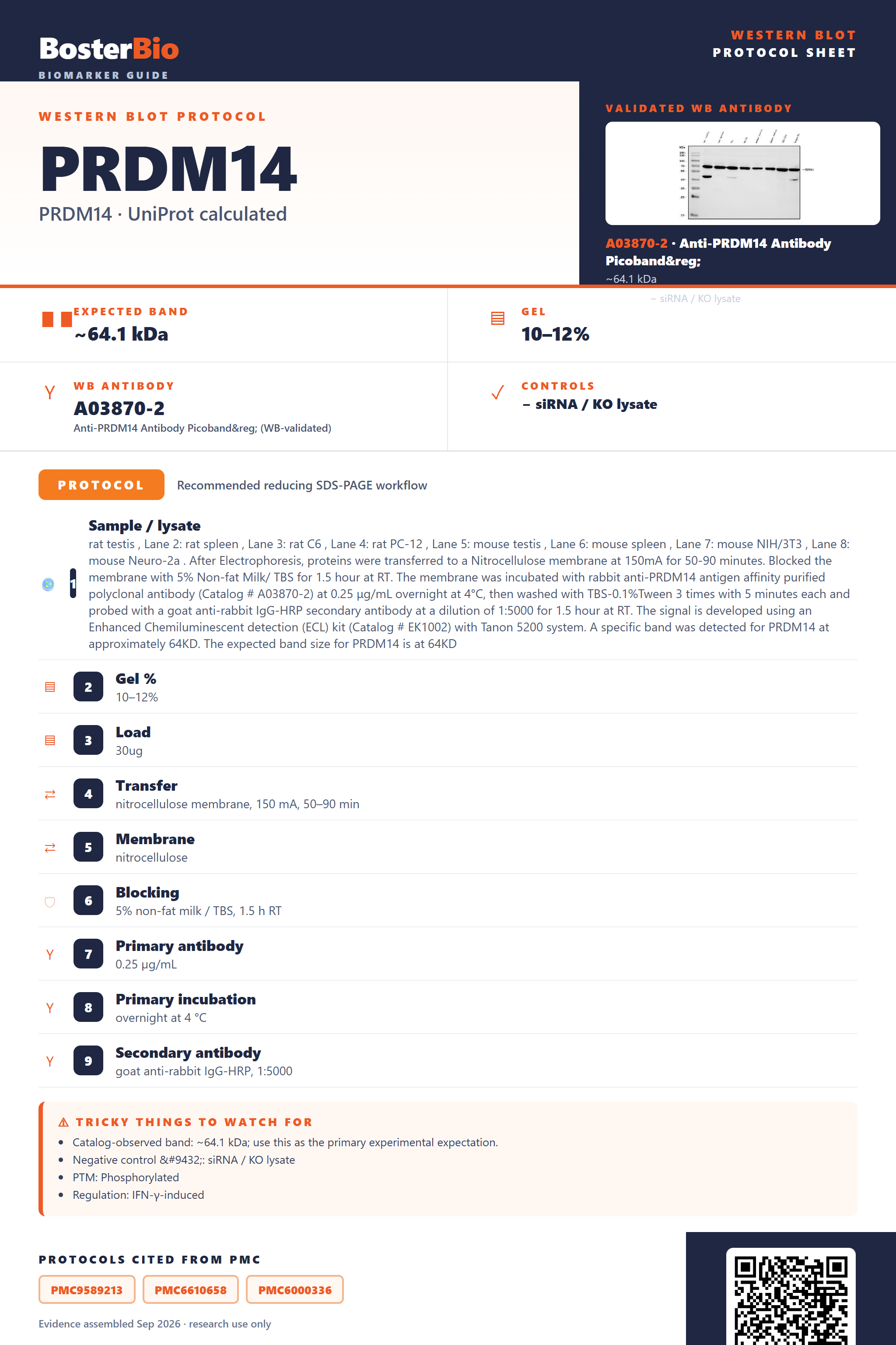
Expected bands, validated protocols, controls and antibodies — the at-a-glance facts below, then the full design guide.
| Expected band | ~64.1 kDa | |
| Gel | 10–12% | |
| Negative control | siRNA / KO lysate |
| PTM | Phosphorylated | |
| Caveat | — | |
| Regulation | IFN-γ-induced | |
| Isoform | 1 isoform(s) |
Literature-validated Western blot parameters for PRDM14 — gel percentage, transfer, blocking, antibody incubation and detection, extracted from published methods.
| Sample / lysate | rat testis , Lane 2: rat spleen , Lane 3: rat C6 , Lane 4: rat PC-12 , Lane 5: mouse testis , Lane 6: mouse spleen , Lane 7: mouse NIH/3T3 , Lane 8: mouse Neuro-2a . After Electrophoresis, proteins were transferred to a Nitrocellulose membrane at 150mA for 50-90 minutes. Blocked the membrane with 5% Non-fat Milk/ TBS for 1.5 hour at RT. The membrane was incubated with rabbit anti-PRDM14 antigen affinity purified polyclonal antibody (Catalog # A03870-2) at 0.25 μg/mL overnight at 4°C, then washed with TBS-0.1%Tween 3 times with 5 minutes each and probed with a goat anti-rabbit IgG-HRP secondary antibody at a dilution of 1:5000 for 1.5 hour at RT. The signal is developed using an Enhanced Chemiluminescent detection (ECL) kit (Catalog # EK1002) with Tanon 5200 system. A specific band was detected for PRDM14 at approximately 64KD. The expected band size for PRDM14 is at 64KD |
| Gel % | 10–12% |
| Load | 30ug |
| Transfer | nitrocellulose membrane, 150 mA, 50–90 min |
| Membrane | nitrocellulose |
| Blocking | 5% non-fat milk / TBS, 1.5 h RT |
| Primary antibody | 0.25 µg/mL |
| Primary incubation | overnight at 4 °C |
| Secondary antibody | goat anti-rabbit IgG-HRP, 1:5000 |
| Wash | TBS-0.1% Tween, 3 × 5 min |
| Detection | ECL |
| Exposure / imaging | Tanon 5200 |
| Observed band | 64 kDa |
PRDM14 is predicted at 64 kDa and, lacking glycosylation, disulfide dimerization, or splice isoforms, should appear as a single clean band near that size.
| single sharp band near 64 kDa | matches the predicted mass of PRDM14, which carries no glycosylation, disulfide linkage, or cleavage to shift it |
| band detected in nuclear or whole-cell lysate but faint in cytoplasmic-only fractions | PRDM14 is annotated as a nuclear protein, so nuclear-inclusive lysis is needed to recover it |
| no additional band at roughly double the mass on non-reducing gels | no disulfide bonds are annotated, so PRDM14 does not form a covalent homodimer |
| no smeared or diffuse band above the main species | the absence of annotated glycosylation sites means there is no heterogeneous carbohydrate modification to broaden the band |
| single band with no doublet | only one isoform of PRDM14 is annotated, so no alternate-splicing band is expected |
| no smaller cleaved species below the main band | no signal peptide or propeptide is annotated, so the full-length translated product is the mature protein |
| Predicted mass (64.1 kDa) | sets the baseline expected migration position on SDS-PAGE |
| Absence of glycosylation sites | prevents a higher or smeared band relative to the predicted mass |
| Absence of disulfide bonds | prevents an approximately doubled-mass band from covalent homodimer formation under non-reducing conditions |
| No signal peptide or propeptide | means the full translated protein is the mature form, with no smaller processed species |
| Single annotated isoform | means no additional bands arise from alternative splicing |
| Zinc-finger and repeat domains with bound metal | can cause folded zinc-finger regions to retain structure in SDS-PAGE, sometimes giving slightly anomalous (often higher) migration than the predicted mass |
| Situation | Likely cause | Next action |
|---|---|---|
| No band in lysate | PRDM14 is a nuclear protein, so lysis conditions that do not adequately solubilize the nuclear fraction can fail to extract it | use a nuclear or whole-cell lysis buffer with sufficient detergent and confirm nuclear protein recovery with a nuclear marker |
| Band higher than expected | the multiple zinc-finger and repeat domains can remain partly folded and bind SDS poorly, causing anomalous migration above the predicted 64 kDa | boil samples longer in fresh reducing sample buffer to fully denature the zinc-finger regions before loading |
| Multiple bands | extra bands likely reflect nonspecific antibody cross-reactivity or proteolytic clipping of the zinc-finger domains, since only one isoform is annotated | validate specificity with a knockdown or knockout control and include fresh protease inhibitors during lysis |
| Weak or no signal | PRDM14 is a low-abundance nuclear transcription factor, so total protein loaded may be insufficient | increase total lysate or nuclear extract loaded and consider a more sensitive detection method |
| Fragments below expected size | since no propeptide or signal peptide cleavage is annotated, smaller fragments indicate proteolytic degradation during sample handling rather than natural processing | keep samples cold, add protease inhibitors immediately at lysis, and minimize freeze-thaw cycles |

Comprehensive Human Protein Atlas IHC scoring per tissue. Rows are taken directly from the HPA tissue chart — click any row's HPA link to view the source.
| Tissue | Cell type | Level | Evidence | Source |
|---|
| Tissue | Cell type | Level | Evidence | Source |
|---|
Deeper troubleshooting and optimisation questions for PRDM14, answered from its protein features.
BosterBio's PRDM14 antibodies are among the best-performing WB antibodies on the market — well cited, thoroughly validated, and orthogonally cross-validated against negative tissues and complementary methods.
For PRDM14 Western blot, we recommend the validated Boster antibody below, selected for demonstrated specificity in an actual WB run rather than predicted reactivity, giving you a documented starting point for your own optimization and controls.
Which to pick: Only one PRDM14 antibody is catalogued here: A03870-2. It includes a real WB validation image (SDS-PAGE, 5-20% gel), so it's the practical default choice — just confirm species reactivity and lysate loading match your experimental setup before use.