PRDX4 / Peroxiredoxin-4 · IHC design guide

Design Immunohistochemistry for PRDX4

Plan PRDX4 chromogenic IHC around cytoplasmic staining, with high signal in pancreatic exocrine glandular cells and testicular Leydig cells (HPA tissue IHC). For paraffin sections, the catalog antibody was demonstrated at 1 μg/ml (datasheet PB9383).

Evidence assembled Oct 2026 · For research use; verify linked source records and product datasheet before use
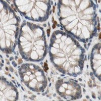
Immunohistochemistry protocol sheet for PRDX4 (IHC for PRDX4): expected localisation Cytoplasmic staining in tissue IHC (HPA tissue IHC), antibody PB9383, validated IHC image, and IHC protocol steps
Printable PRDX4 IHC protocol sheet — expected localisation Cytoplasmic staining in tissue IHC (HPA tissue IHC), antibody PB9383, controls and protocol steps. Open the full PRDX4 IHC guide →

PRDX4 Immunohistochemistry Experimental Design Guide

Expected localisation, validated protocols, controls and antibodies — the at-a-glance facts below, then the full design guide.

Must know before staining
Expected localisation Cytoplasmic staining in tissue IHC (HPA tissue IHC)
Staining pattern Ubiquitous cytoplasmic staining (HPA tissue IHC)
Antigen retrieval Citrate pH 6 HIER, heat-mediated (datasheet PB9383)
Positive control ⓘ Pancreas+4 more · see all
Negative control ⓘ None in HPA (detected in all 45 tissues); use no-primary + isotype controls
Important caveats
Reasons your staining may differ from the expected pattern.
Fixation Keep fixation consistent across samples. (standard IHC practice; not target-specific)
Caveat Staining and RNA have medium consistency (HPA tissue IHC)
Regulation Expression regulation is unannotated (UniProt)
Isoform / epitope No isoforms listed; signal peptide spans 1–37 (UniProt)
Section 1

Recommended PRDX4 IHC & IF Protocols

Compare the catalog antibody’s IHC-P protocol with 3 published PRDX4 IHC protocols (PMC6036164; PMC7114939; PMC13312543).

Recommended immunohistochemistry (IHC-P) protocol parameters
SampleParaffin-embedded mouse Brain tissues; fixative not specified (datasheet PB9383)
FixationImage fixative and duration unreported (datasheet PB9383); verify before use.
Sectioning4–5 µm sections on charged slides (standard)
DeparaffinisationXylene, graded ethanol series to water (standard)
Antigen retrievalHeat retrieval: Citrate pH 6, 20 min (datasheet PB9383)
Peroxidase block3% H2O2, 10 min, room temperature (standard)
Blocking10% goat serum (datasheet PB9383)
Primary antibodyRabbit anti-PRDX4, 0.5-1μg/ml (datasheet PB9383)
Primary incubationOvernight at 4 °C (datasheet PB9383)
DetectionStreptavidin-biotin complex (SABC), DAB chromogen (datasheet PB9383)
CounterstainHematoxylin, blue, dehydrate and mount (standard)
Expected resultPRDX4-positive staining in exocrine glandular cells of pancreas (HPA tissue IHC: High). HPA tissue profile: Ubiquitous cytoplasmic expression. No signal in the no-primary control.
💡Decision noteStart with heat-mediated citrate retrieval at pH 6 (PB9383 datasheet). Try trypsin retrieval if needed (PMC13312543 methods).
Section 2

What Is the Expected PRDX4 Staining Pattern?

PRDX4 should produce predominantly cytoplasmic staining consistent with endoplasmic reticulum localisation; a smaller cytosolic pool is also reported (UniProt Q13162: localisation; HPA subcellular). It has no transmembrane segment (UniProt Q13162: topology). In tissue, expect widespread cytoplasmic staining, with particularly strong signal in pancreatic exocrine glandular cells and testicular Leyig cells (HPA tissue IHC). HPA rates the tissue pattern Enhanced, with medium agreement between staining and RNA data (HPA tissue IHC).

What am I looking at on my slide?
Strong cytoplasmic staining in pancreatic exocrine glandular cells.This matches an HPA High cell population and the reported endoplasmic reticulum and cytosolic localisation (HPA tissue IHC; HPA subcellular). In a chromogenic section, compartment and cell identity are more informative than colour intensity alone (standard IHC practice).
Staining is predominantly nuclear or outlines cell membranes, with little cytoplasmic signal.That distribution conflicts with the reported endoplasmic reticulum and cytosolic locations and absence of a transmembrane segment (HPA subcellular; UniProt Q13162: topology). Treat it as a possible artefact and check control sections before scoring it as PRDX4 (standard IHC practice).
A cell population reported as low stains more strongly than nearby expected positive cells.For example, HPA reports low staining in cerebellar granular-layer cells and salivary glandular cells (HPA tissue IHC). Unexpected dominance may reflect cross-reactivity or endogenous detection activity; low does not mean definitively negative (standard IHC practice; HPA tissue IHC).
Chromogen appears broadly across the section, including areas without clear cellular boundaries.Diffuse deposits obscure the cytoplasmic pattern reported by HPA (HPA tissue IHC). Assess reagent background, incomplete blocking or washing, and, with enzyme detection, endogenous activity using appropriate controls (standard IHC practice).
Pancreatic exocrine glandular cells show no detectable signal.HPA reports High staining in these cells, so a blank result warrants a run-control review before a biological interpretation (HPA tissue IHC). Check that the expected cells are present and that detection and counterstaining worked (standard IHC practice).
💡Expected PRDX4 appearanceCall a section positive when identifiable pancreatic exocrine glandular cells show strong cytoplasmic signal; predominantly nuclear or membrane-only staining is suspect (HPA tissue IHC; HPA subcellular; UniProt Q13162: topology).
How each factor affects the staining
Compartment resolutionPRDX4 is mainly retained in the endoplasmic reticulum, with a small cytoplasmic fraction (UniProt Q13162: localisation). Chromogenic IHC supports a cytoplasmic call but generally cannot prove that each deposit lies within the endoplasmic reticulum (standard IHC practice).
Choice of comparison tissuePancreatic exocrine glandular cells and testicular Leydig cells are reported High; several other listed populations are Medium or Low (HPA tissue IHC). Compare named cell populations within intact tissue rather than treating whole-organ colour as one score (standard IHC practice).
Protein processingUniProt annotates a signal peptide at residues 1–37 and the mature chain at 38–271 (UniProt Q13162: processing). The payload gives no antibody epitope, so these annotations cannot predict this antibody's staining strength or retrieval response.
Confidence in the reference patternThe HPA tissue result is Enhanced, and multiple listed antibodies have Enhanced IHC status; HPA describes only medium agreement with RNA expression (HPA tissue IHC; HPA antibodies). Use the pattern as a reference while retaining section-level controls (standard IHC practice).
Q: Can IF/ICC establish the expected compartment?A: HPA ICC-IF supports endoplasmic reticulum as the main location and cytosol as an additional location (HPA subcellular). Those cell-image observations inform interpretation but do not establish a paraffin-section staining outcome (standard IHC practice).
Why is my staining missing, weak or wrong?
SituationLikely causeNext action
No signal in pancreatic exocrine glandular cells.The observed result differs from HPA's High tissue finding; the immediate cause is undetermined (HPA tissue IHC).Verify cell identity, section integrity, antibody application and detection controls; review the IHC-validated antibody's own instructions for retrieval and dilution (standard IHC practice).
Signal is mainly nuclear or membrane-like.This differs from PRDX4's reported endoplasmic reticulum and cytosolic distribution (HPA subcellular; UniProt Q13162: topology).Inspect the counterstain and staining controls, then repeat with the IHC-validated antibody under its documented conditions before assigning specificity (standard IHC practice).
A low-reference cell population is intensely stained.HPA calls some named populations Low, so strong staining there needs investigation; cross-reactivity or endogenous detection activity are possible explanations (HPA tissue IHC; standard IHC practice).Compare neighbouring expected positive cells and use suitable negative and detection controls; do not equate an HPA Low designation with absence (HPA tissue IHC; standard IHC practice).
The whole section has diffuse chromogen.Non-specific reagent deposition or endogenous enzyme activity can obscure cellular localisation in chromogenic IHC (standard IHC practice).Check blocking, washes and detection-only controls, then score only signal that can be assigned to intact cells (standard IHC practice).
Staining is present but too faint to score confidently.HPA reports a range of tissue staining levels, so weak signal alone does not identify an assay failure (HPA tissue IHC).Check a High reference population in the same run, then review the antibody's documented dilution and retrieval conditions (HPA tissue IHC; standard IHC practice).
The section differs from an HPA ICC-IF image.HPA's ICC-IF localisation and tissue IHC pattern come from different preparations (HPA subcellular; HPA tissue IHC).Judge the paraffin section by identifiable tissue cells, cytoplasmic localisation and IHC controls; use the ICC-IF image only as compartment context (HPA tissue IHC; HPA subcellular; standard IHC practice).

Sample controls for PRDX4 IHC & IF

🧪Start with pancreas: exocrine glandular cells should stain strongly (HPA: High in pancreatic exocrine glandular cells). HPA detects PRDX4 in all 45 scored tissues, so there is no documented negative tissue; use no-primary and isotype controls, and expect cells without specific staining on the positive slide to show only background chromogen without assigning any pancreatic cell type as PRDX4-negative (HPA: no negative tissue rows; standard IHC practice).
Positive control tissue: Pancreas (Exocrine glandular cells, HPA High)
Negative control tissue: None in HPA: PRDX4 is detected in all 45 scored tissues. Use a no-primary (secondary-only) and an isotype control instead.
ICC-IF cell lines (HPA subcellular resource): HPA ICC-IF images show PRDX4 in A-431, U-251MG, U2OS, with annotated localisation: Endoplasmic reticulum (supported) (HPA subcellular).
Technical controls: Run a no-primary, secondary-only control and a rabbit IgG isotype control matched to the primary antibody’s clonality, alongside a PRDX4 knockout specimen or peptide-blocked primary where available (PB9383 caption: rabbit primary; standard IHC practice). Check endogenous peroxidase and biotin background because the documented detection uses a biotinylated secondary, SABC and DAB (PB9383 caption: detection method; standard IHC practice).
⚠️Feasibility: No target-specific fixation window or fixation effect is reported, and the selected PB9383 paraffin-section caption does not state a fixative (PB9383 caption: fixative not stated). That IHC workflow uses heat retrieval in citrate buffer at pH 6 for 20 minutes, but retrieval dependence and frozen-section feasibility are unreported; HPA ICC-IF images support ER and cytosolic localization without establishing that IF is easier (PB9383 caption: retrieval; HPA subcellular: ER and cytosol). In pancreatic sections, assess background from endogenous peroxidase or biotin before interpreting DAB staining (PB9383 caption: SABC/DAB detection; standard IHC practice).

HPA tissue IHC evidence for PRDX4

Comprehensive Human Protein Atlas IHC scoring per tissue (reliability: Enhanced — Medium consistency between antibody staining and RNA expression data.). Rows are taken directly from the HPA tissue chart — click any row's HPA link to view the source.

Positive expression · recommended positive controls

TissueCell typeLevelEvidenceSource
Pancreas Exocrine glandular cells High Protein (IHC) HPA →
Testis Leydig cells High Protein (IHC) HPA →
Adipose tissue Adipocytes Medium Protein (IHC) HPA →
Adrenal gland Glandular cells Medium Protein (IHC) HPA →
Appendix Glandular cells Medium Protein (IHC) HPA →

Undetected expression · recommended negative controls

TissueCell typeLevelEvidenceSource
None in HPA: PRDX4 is detected in all 45 scored tissues. Use a no-primary (secondary-only) and an isotype control instead.
Section 3

Advanced PRDX4 IHC Tips

Troubleshoot PRDX4 staining in paraffin sections by checking retrieval, cell identity, compartment, and detection controls before comparing signal intensity.

How should I adjust retrieval when PRDX4 staining is weak in paraffin sections?
Begin with heat-mediated citrate retrieval at pH 6 for 20 minutes (datasheet PB9383). The reported paraffin-section staining used 1 μg/mL antibody overnight at 4°C, so match those conditions when assessing retrieval (caption PB9383). If staining remains weak, compare longer heating or an alternative retrieval buffer on matched sections while holding antibody and detection conditions constant (standard IHC practice). Evaluate cytoplasmic signal in an appropriate positive-control cell population, given PRDX4’s predominantly endoplasmic-reticulum localisation (UniProt Q13162 localisation; HPA tissue IHC). Reject conditions that improve apparent intensity mainly through tissue damage or no-primary background (standard IHC practice).
Could fixation explain variable PRDX4 staining between paraffin blocks?
No PRDX4-specific fixation sensitivity is supplied, and the PB9383 paraffin caption leaves its fixative unstated (caption PB9383). Document fixative and duration for each block; compare equally processed positive controls before calling weak staining biological (standard IHC practice). Use routine neutral-buffered formalin only as a laboratory baseline, without treating it as an established PRDX4 requirement (standard IHC practice). Keep section thickness, citrate pH 6 retrieval, and 1 μg/mL antibody exposure consistent across comparisons (caption PB9383; standard IHC practice). Add no-primary slides to identify pigment and chromogen background when evaluating apparently negative cell populations (standard IHC practice).
What PRDX4 staining pattern should I expect in tissue sections?
PRDX4 is predominantly retained in the endoplasmic reticulum, with a smaller cytoplasmic fraction (UniProt Q13162 localisation; HPA subcellular). Expect cytoplasmic, potentially reticular DAB staining rather than dominant nuclear or plasma-membrane staining (UniProt Q13162 localisation; standard IHC practice). Use pancreas exocrine glandular cells or testis Leydig cells as high-expression reference populations when suitable sections are available (HPA tissue IHC). HPA also reports medium endothelial staining in cerebral cortex, so identify the stained cell type before comparing brain regions (HPA tissue IHC). Check section morphology and the no-primary control if staining is exclusively nuclear or concentrated at tissue edges (standard IHC practice).
Could PRDX4 processing or an unknown epitope explain a negative stain?
PRDX4 has no annotated isoforms, a 1–37 signal peptide, and a mature 38–271 chain (UniProt Q13162 processing and isoforms). Obtain the antibody’s immunogen or mapped epitope before assigning an epitope-based cause; the supplied PB9383 caption does not specify one (caption PB9383). An epitope restricted to the removed signal peptide would be a concern when assessing mature PRDX4 staining (UniProt Q13162 processing; standard IHC practice). PRDX4 has no transmembrane segment or annotated glycosylation sites, so neither feature explains failed retrieval here (UniProt Q13162 topology and glycosylation). Compare results under citrate pH 6 retrieval with detection conditions held constant (caption PB9383; standard IHC practice).
How can IF help assess PRDX4 localisation alongside cell identity?
Treat IF with PB9383 as exploratory: the supplied antibody evidence describes paraffin-section chromogenic IHC, without an IF validation (caption PB9383). Multiplex PRDX4 with a validated marker for pancreatic exocrine glandular cells or testis Leydig cells, both reported high-signal populations (HPA tissue IHC). Choose spectrally separated fluorophores, favoring a far-red PRDX4 channel if shorter wavelengths show tissue autofluorescence (standard IF practice). If the epitope is inside the ER lumen, permeabilize for compartment access; the PB9383 epitope is unspecified (UniProt Q13162 localisation; caption PB9383; standard IF practice). Include single-stain and secondary-only controls when assessing overlap or diffuse haze (standard IF practice).
How do I distinguish PRDX4 staining from chromogenic background?
The PB9383 paraffin-section example used 10% goat serum blocking, a biotinylated secondary antibody, and DAB detection (caption PB9383). Apply an endogenous peroxidase block before chromogen development and include a no-primary control to assess detection background (standard IHC practice). Because this detection method uses biotin, check whether an avidin–biotin blocking step reduces signal in a matched control section (caption PB9383; standard IHC practice). If background persists, titrate primary antibody below the reported 1 μg/mL condition and keep development time consistent (caption PB9383; standard IHC practice). Interpret staining only where morphology remains intact and signal exceeds the control (standard IHC practice).
How should I quantify PRDX4 staining across tissue samples? ⚠ ANSWER MARKED FOR VERIFICATION
Define the cell population and anatomical region before scoring, since reported PRDX4 intensity differs among cell types and tissues (HPA tissue IHC). For cell-based analysis, record the percentage of positive cells and staining intensity, then calculate an H-score from 0–300 (standard IHC practice). For spatial analysis, report positive-cell density per mm² of viable tissue and document excluded areas (standard IHC practice). Normalize comparisons to the same cell type or viable tissue area, using identical retrieval, imaging, and DAB development settings (standard IHC practice). Keep positive-control and no-primary sections alongside each batch to detect assay drift and background (standard IHC practice).
When should I doubt an apparent PRDX4-positive cell or region?
Favor cytoplasmic or ER-associated staining in morphologically intact cells, consistent with PRDX4 localisation (UniProt Q13162 localisation; HPA subcellular). Check cell identity: HPA reports high staining in pancreatic exocrine glandular cells and testis Leydig cells, but low staining in cerebellar granular-layer cells (HPA tissue IHC). Treat dominant nuclear or membrane-edge staining, section-edge accentuation, and necrotic-region signal cautiously until controls resolve them (UniProt Q13162 localisation; standard IHC practice). Compare suspicious DAB deposits with a no-primary slide and an endogenous peroxidase-blocked slide (standard IHC practice). HPA describes only medium agreement between antibody staining and RNA expression, so corroborate unexpected patterns independently (HPA tissue IHC reliability).
Boster reagents

Best PRDX4 / Peroxiredoxin-4 IHC Antibodies

PB9383 has IHC images from paraffin sections of mouse and rat brain and human tonsil (PB9383 IHC captions), plus an IF image from A549 cells (PB9383 IF caption).

Real IHC data IHC analysis of Peroxiredoxin 4 using anti-Peroxiredoxin 4 antibody (PB9383). Peroxiredoxin 4 was detected in paraffin-embedded section of mouse Brain tissues. Heat mediated antigen retrieval was performed in citrate buffer (pH6, epitope retrieval solution) for 20 mins. The tissue section was blocked with 10% goat serum. The tissue section was then incubated with 1μg/ml rabbit anti-Peroxiredoxin 4 Antibody (PB9383) overnight at 4°C. Biotinylated goat anti-rabbit IgG was used as secondary antibody and incubated for 30 minutes at 37°C. The tissue section was developed using Strepavidin-Biotin-Complex (SABC)(Catalog # SA1022) with DAB as the chromogen.
Anti-Peroxiredoxin 4/PRDX4 Antibody ®
Cat # PB9383

PB9383 is the rendering SKU; its IHC captions show paraffin sections of mouse and rat brain and human tonsil (PB9383 IHC captions). Its IF caption shows A549 cells (PB9383 IF caption), and its catalog lists human, mouse and rat reactivity (PB9383 catalog).

Which to pick: For tissue IHC, choose PB9383: its IHC captions show staining in paraffin sections from all three listed species; the fixative is unreported (PB9383 IHC captions). For IF/ICC, PB9383 has an A549 cell IF image and lists IF and ICC applications (PB9383 IF caption; PB9383 catalog). For cross-species IHC, PB9383 has image evidence in human, mouse and rat tissue, while M05000-1 lists reactivity to those species and IHC use but supplies no IHC image in this payload (PB9383 IHC captions; M05000-1 catalog).

Each figure is that product's own IHC / IF validation image from its datasheet.

References

  1. UniProt Consortium. UniProt entry Q13162 (PRDX4_HUMAN, Peroxiredoxin-4).
  2. Human Protein Atlas. PRDX4 tissue IHC expression (reliability: Enhanced).
  3. Human Protein Atlas. PRDX4 subcellular location (ICC-IF): Mainly localized to the endoplasmic reticulum. In addition localized to the cytosol..
  4. Human Protein Atlas. PRDX4 antibody validation summary (6 antibodies).
  5. The Combination Of Weak Expression Of PRDX4 And Very High MIB-1 Labelling Index Independently Predicts Shorter Disease-free Survival In Stage I Lung Adenocarcinoma. International journal of medical sciences 2018 — PMC6036164.
  6. PRDX4 overexpression is associated with poor prognosis in gastric cancer. Oncology letters 2020 — PMC7114939.
  7. The impact of PRDX4 and the EGFR mutation status on cellular proliferation in lung adenocarcinoma. International journal of medical sciences 2019 — PMC6775271.
  8. PRDX4 expression potentially links redox adaptation to oncogenic signaling and tumor progression in pancreatic ductal adenocarcinoma. Translational oncology 2026 — PMC13312543.
  9. PubMed PMID:9388242 — UniProt-cited evidence.
  10. PubMed PMID:15489334 — UniProt-cited evidence.
  11. PubMed PMID:19892738 — UniProt-cited evidence.