This website uses cookies to ensure you get the best experience on our website.


- Table of Contents
Plan PREX1 IHC-P around cytoplasmic staining in immune and neuronal cells (HPA tissue IHC). This guide covers fixation, antibody use and cell-type controls for paraffin sections (datasheet A03098-1; HPA tissue IHC).

Expected localisation, validated protocols, controls and antibodies — the at-a-glance facts below, then the full design guide.
| Expected localisation | Cytoplasmic staining; membrane staining in Bergmann glia (HPA tissue IHC) | |
| Staining pattern | Cytoplasmic staining in immune and neuronal cells (HPA tissue IHC) | |
| Antigen retrieval | Citrate pH 6 HIER, heat-mediated (datasheet A03098-1) | |
| Positive control | Cerebellum+4 more · see all | |
| Negative control | Adipose tissue+4 more · see all |
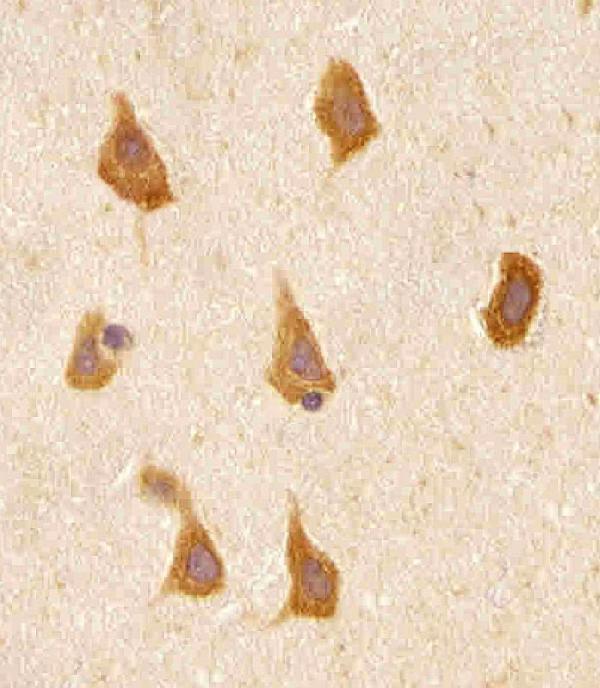
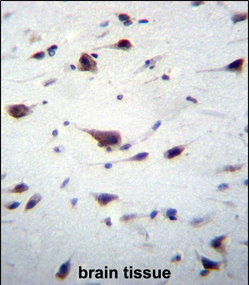
| Fixation | Formalin-fixed paraffin sections were used (selected-SKU IHC image A03098-1); keep fixation consistent (standard IHC practice; not target-specific) | |
| Caveat | Presumed off-target binding may mimic PREX1 staining (HPA tissue IHC) | |
| Regulation | Expression is highest in leukocytes and brain (UniProt) | |
| Isoform / epitope | 3 isoforms; epitope conservation is unknown (UniProt) |
The catalog antibody protocol (datasheet: A03098-1) is followed by published PREX1 IHC protocols for glioblastoma tissue microarrays (PMC5352422) and human melanoma sections (PMC5065759).
| Sample | Formaldehyde-fixed, paraffin-embedded human brain tissue (datasheet A03098-1) |
| Fixation | Image formalin-fixed; duration unreported (datasheet A03098-1); verify before use. |
| Sectioning | 4–5 µm sections on charged slides (standard) |
| Deparaffinisation | Xylene, graded ethanol series to water (standard) |
| Antigen retrieval | Heat retrieval: Citrate pH 6 (datasheet A03098-1); 20 min, 95–100 °C (standard) |
| Peroxidase block | 3% H2O2, 10 min, room temperature (standard) |
| Blocking | 10% normal serum of the secondary host, 30 min, room temperature (standard) |
| Primary antibody | Rabbit anti-PREX1, 1:10-1:50 (datasheet A03098-1) |
| Primary incubation | 1 hours at 37°C (datasheet A03098-1) |
| Detection | HRP-polymer secondary, DAB chromogen 5–10 min (standard) |
| Counterstain | Hematoxylin, blue, dehydrate and mount (standard) |
| Expected result | PREX1-positive staining in bergmann glia - cytoplasm/membrane of cerebellum (HPA tissue IHC: High). HPA tissue profile: Cytoplasmic expression in several tissues, most abundant in immune and neuronal cells. No signal in the no-primary control. |
PREX1 should appear mainly in the cytoplasm, with some plasma membrane association; it has no transmembrane segment (UniProt Q8TCU6 localisation and topology). In paraffin tissue sections, expect prominent staining in lung macrophages and cerebellar Bergmann glia, with weaker or less widespread staining in other listed cell populations (HPA tissue IHC: High in both; overall reliability: Supported). Interpret the cell type and compartment together because HPA reports presumed off-target binding (HPA tissue IHC reliability description).
| Cytoplasmic staining in lung macrophages or cytoplasmic and membrane staining in cerebellar Bergmann glia. | These are the clearest listed positive patterns: each is High in HPA tissue IHC. PREX1 is mainly cytosolic, with some plasma membrane association (UniProt Q8TCU6 localisation). Judge staining within the annotated cells, since HPA reports only medium consistency with RNA and notes presumed off-target binding (HPA tissue IHC reliability description). |
| Predominantly nuclear staining, with little corresponding cytoplasmic signal in the expected positive cells. | A nuclear dominant pattern does not fit the supplied localisation evidence: UniProt places PREX1 mainly in cytosol and partly at the plasma membrane, while HPA tissue IHC describes cytoplasmic expression (UniProt Q8TCU6 localisation; HPA tissue IHC profile). Treat this as a possible artefact and reassess staining controls before assigning it to PREX1 (general IHC practice). |
| Strong staining in an annotated cell type where HPA reports PREX1 as not detected, such as adipocytes in adipose tissue. | That pattern conflicts with the listed cell specific observation (HPA tissue IHC: adipocytes, Not detected). Cross-reactivity or endogenous chromogenic detection activity are possible explanations, rather than evidence that PREX1 is present there (general IHC practice). HPA itself notes presumed off-target binding in its tissue assessment (HPA tissue IHC reliability description). |
| Diffuse chromogen across cells and surrounding tissue, without a discernible cytoplasmic pattern. | A uniform deposit cannot establish cell specific PREX1 staining; the reference pattern is cytoplasmic in several tissues and most abundant in immune and neuronal cells (HPA tissue IHC profile). Consider background from detection reagents or inadequate blocking, and inspect a negative control alongside the section (general IHC practice). |
| No staining in lung macrophages or cerebellar Bergmann glia on an otherwise evaluable section. | Both populations are listed as High positives (HPA tissue IHC: lung macrophages and cerebellar Bergmann glia). First check section quality and the antibody and chromogen detection steps using appropriate controls (general IHC practice). A failed run is one possibility; the HPA pattern alone does not identify its cause or establish a PREX1 specific fixation effect. |
| Cellular distribution | PREX1 is mainly cytosolic, although some is apparently associated with the plasma membrane, and no transmembrane segment is listed (UniProt Q8TCU6 localisation and topology). Thus cytoplasm should anchor interpretation; the membrane component can support a result in the right cells, especially Bergmann glia, whose HPA annotation includes cytoplasm and membrane (HPA tissue IHC: cerebellum). |
| Tissue and cell context | HPA lists High staining in lung macrophages and cerebellar Bergmann glia; Medium staining in bone marrow hematopoietic cells, lymph node germinal center cells, spleen red pulp cells, and late spermatids; and Low staining in tonsil germinal center cells (HPA tissue IHC). Compare like cell types rather than treating every cell in a positive tissue as positive (general IHC practice). |
| Isoform and epitope coverage | UniProt lists 3 PREX1 isoforms, but the supplied record gives no antibody epitope or isoform coverage (UniProt Q8TCU6 isoforms; supplied HPA antibody summary). A staining difference cannot be assigned to a particular isoform from these sources. Interpret the observed compartment and annotated cell type before making any isoform claim (UniProt Q8TCU6 localisation; HPA tissue IHC). |
| Processing and antibody validation | UniProt lists no signal peptide or propeptide and describes a 1–1659 chain (UniProt Q8TCU6 processing). The supplied HPA antibody, HPA001927, has IHC status Supported; the tissue assessment reports medium consistency with RNA and presumed off-target binding (HPA antibody summary; HPA tissue IHC reliability description). These records support cautious pattern comparison, not a claim that every chromogenic signal is specific. |
| Does IF/ICC show the same pattern? | HPA ICC-IF places PREX1 in vesicles (Approved) and cytosol (Supported), with images listed for A-431, U-251MG, and U2OS (HPA subcellular ICC-IF). This supports a cytosolic component across applications, while the vesicle annotation comes from ICC-IF rather than tissue IHC. Evaluate paraffin tissue staining against the tissue IHC cell annotations and UniProt localisation (HPA tissue IHC; UniProt Q8TCU6). |
| Situation | Likely cause | Next action |
|---|---|---|
| A listed High positive cell population has no visible chromogen (HPA tissue IHC: lung macrophages or Bergmann glia). | Section quality, primary antibody application, or detection failure may explain a blank run (general IHC practice); no PREX1 specific fixation sensitivity is supplied. | Review a positive control section, tissue morphology, primary antibody application, and detection reagents; repeat only the step implicated by those checks (general IHC practice). |
| Nuclear staining dominates while the expected cytoplasm is clear. | The compartment conflicts with the mainly cytosolic PREX1 assignment and HPA's cytoplasmic tissue profile (UniProt Q8TCU6 localisation; HPA tissue IHC profile). | Compare an antibody omitted control and a listed positive tissue, then assess whether the nuclear signal follows the detection system rather than the expected cells (general IHC practice; HPA tissue IHC positives). |
| Strong chromogen appears in HPA negative cells, including adipocytes in adipose tissue. | Cross-reactivity or endogenous detection activity is plausible; HPA notes presumed off-target binding (general IHC practice; HPA tissue IHC reliability description). | Check a primary antibody omitted control and appropriate blocking for the chromogenic system; score the annotated cell type separately from nearby positive cells (general IHC practice). |
| A uniform haze obscures boundaries and prevents cell level scoring. | Nonspecific reagent binding or excess chromogen development can obscure a cellular pattern (general IHC practice). | Review blocking, reagent washes, detection timing, and the negative control; accept a result only when cellular staining can be distinguished from background (general IHC practice). |
| Cytoplasm stains clearly, but little membrane accent is visible. | That can still fit PREX1: UniProt describes it as mainly cytosolic, with only some amount apparently associated with plasma membrane (UniProt Q8TCU6 localisation). | Score the cytoplasmic pattern in the correct annotated cells first; use the Bergmann glia membrane annotation as supporting context, not a required feature in every tissue (HPA tissue IHC: cerebellum; general IHC practice). |
| A tissue labelled positive appears mixed, with many unstained neighboring cells. | HPA's levels refer to named cell populations, such as macrophages in lung and germinal center cells in lymph node, rather than every cell in those tissues (HPA tissue IHC positives). | Locate and score the annotated population against its neighboring cells and confirm that the counterstain preserves cell identification (HPA tissue IHC positives; general IHC practice). |


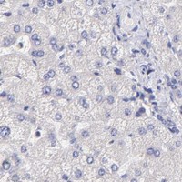
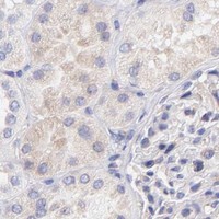


Comprehensive Human Protein Atlas IHC scoring per tissue (reliability: Supported — Medium consistency between antibody staining and RNA expression data. Presumed off target binding observed and disregarded. Antibody staining in cells/structures not annotated, view images.). Rows are taken directly from the HPA tissue chart — click any row's HPA link to view the source.
| Tissue | Cell type | Level | Evidence | Source |
|---|---|---|---|---|
| Cerebellum | Bergmann glia - cytoplasm/membrane | High | Protein (IHC) | HPA → |
| Lung | Macrophages | High | Protein (IHC) | HPA → |
| Bone marrow | Hematopoietic cells | Medium | Protein (IHC) | HPA → |
| Lymph node | Germinal center cells | Medium | Protein (IHC) | HPA → |
| Spleen | Cells in red pulp | Medium | Protein (IHC) | HPA → |
| Tissue | Cell type | Level | Evidence | Source |
|---|---|---|---|---|
| Adipose tissue | Adipocytes | Not detected | Protein (IHC) | HPA → |
| Adrenal gland | Glandular cells | Not detected | Protein (IHC) | HPA → |
| Appendix | Glandular cells | Not detected | Protein (IHC) | HPA → |
| Breast | Adipocytes | Not detected | Protein (IHC) | HPA → |
| Bronchus | Respiratory epithelial cells | Not detected | Protein (IHC) | HPA → |
Troubleshoot PREX1 staining in paraffin sections by checking retrieval, cell type, compartment, and controls before comparing signal across samples.
A03098-1 has real IHC-P images from human brain paraffin sections (catalog image captions). No IF/ICC data are supplied (catalog).
A03098-1 is listed for human IHC-P (catalog applications and reactivity). Its IHC images show PREX1 staining in paraffin-embedded human brain sections, with paraformaldehyde fixation in one caption and formalin fixation in another (catalog image captions).
Which to pick: For human tissue IHC-P, choose A03098-1: this rabbit polyclonal antibody has a listed dilution of 1:10–1:50 (catalog), and its own images document human brain paraffin sections (catalog image captions). For IF/ICC, A03098-1 has no listed application, dilution, or image (catalog). For other species, A03098-1 has documented reactivity only with human samples (catalog).