This website uses cookies to ensure you get the best experience on our website.


- Table of Contents
Plan PRKAB2 paraffin-section IHC around cytoplasmic staining in respiratory epithelial cells, glandular cells and hepatocytes (HPA tissue IHC). Use consistent fixation, score cytoplasmic signal and start with the IHC-validated antibody at 0.5–1 μg/mL (datasheet PB9739).

Expected localisation, validated protocols, controls and antibodies — the at-a-glance facts below, then the full design guide.
| Expected localisation | General cytoplasmic staining (HPA tissue IHC) | |
| Staining pattern | Cytoplasm in respiratory epithelium, glands and hepatocytes (HPA tissue IHC) | |
| Antigen retrieval | EDTA pH 8.0 HIER, heat-mediated (datasheet PB9739) | |
| Positive control | Bronchus+4 more · see all | |
| Negative control | None in HPA (detected in all 45 tissues); use no-primary + isotype controls |
| Fixation | Keep fixation consistent across sections (standard IHC practice; not target-specific) | |
| Caveat | Staining and RNA show only medium consistency (HPA tissue IHC) | |
| Regulation | Expression regulation not annotated (UniProt) | |
| Isoform / epitope | 2 isoforms; confirm epitope coverage (UniProt) |
The catalog antibody protocol (datasheet: PB9739) and one published PRKAB2 IHC protocol (PMC13232994) provide starting conditions for paraffin sections.
| Sample | Paraffin-embedded mouse intestine tissue; fixative not specified (datasheet PB9739) |
| Fixation | Image fixative and duration unreported (datasheet PB9739); verify before use. |
| Sectioning | 4–5 µm sections on charged slides (standard) |
| Deparaffinisation | Xylene, graded ethanol series to water (standard) |
| Antigen retrieval | Heat retrieval: EDTA pH 8.0 (datasheet PB9739); 20 min, 95–100 °C (standard) |
| Peroxidase block | 3% H2O2, 10 min, room temperature (standard) |
| Blocking | 10% goat serum (datasheet PB9739) |
| Primary antibody | Rabbit anti-PRKAB2, 0.5-1μg/ml (datasheet PB9739) |
| Primary incubation | Overnight at 4 °C (datasheet PB9739) |
| Detection | Streptavidin-biotin complex (SABC), DAB chromogen (datasheet PB9739) |
| Counterstain | Hematoxylin, blue, dehydrate and mount (standard) |
| Expected result | PRKAB2-positive staining in respiratory epithelial cells of bronchus (HPA tissue IHC: High). HPA tissue profile: General cytoplasmic expression. No signal in the no-primary control. |
In paraffin IHC, PRKAB2 has a general cytoplasmic pattern, with high staining reported in respiratory, glandular, exocrine glandular, and liver cells (HPA tissue IHC: Enhanced; medium consistency with RNA). A continuous cell-surface rim is unexpected because PRKAB2 has no annotated transmembrane segment (UniProt O43741 topology). ICC-IF has a different reported emphasis: mainly nucleoplasmic, with additional cytosolic localization (HPA subcellular ICC-IF: approved).
| Cytoplasmic staining in hepatocytes, duodenal glandular cells, or bronchial respiratory epithelial cells. | This matches the general cytoplasmic IHC profile and cell types scored High (HPA tissue IHC). Judge signal within identifiable cells; HPA's Enhanced rating carries a medium-consistency caveat relative to RNA data (HPA tissue IHC). |
| A crisp cell-surface rim dominates, with little intracellular staining. | Treat this as a possible staining artefact: the rim conflicts with the general cytoplasmic IHC profile (HPA tissue IHC) and the absence of a transmembrane segment (UniProt O43741 topology). Recheck the control section before assigning PRKAB2 localization. |
| Strong staining appears chiefly in an unexpected cell population rather than the documented high-staining cells. | Consider antibody cross-reactivity or endogenous chromogenic detection activity (general IHC practice). HPA reports high staining in specified cell populations, not every cell in those tissues (HPA tissue IHC); morphology and detection controls matter. |
| Color spreads across stroma, empty areas, or many cell types without clear intracellular boundaries. | Diffuse background cannot establish PRKAB2 expression (general IHC practice). Compare the background with the specific cytoplasmic pattern in documented high-staining cells (HPA tissue IHC), then assess blocking, washing, and detection controls. |
| No cellular signal appears in a section containing well-preserved hepatocytes or pancreatic exocrine glandular cells. | A failed stain is plausible because both cell populations were scored High (HPA tissue IHC). Check tissue quality and the IHC workflow before calling the specimen PRKAB2-negative (general IHC practice); HPA lists no validated negative tissue here (HPA tissue IHC). |
| Evidence strength and tissue choice | The tissue IHC profile is Enhanced, with medium consistency between staining and RNA (HPA tissue IHC). Use an HPA High cell population as a practical positive comparator; skeletal muscle and tongue are RNA-enhanced tissues, not documented High IHC examples in this payload (HPA tissue IHC). |
| IHC and ICC-IF compartment calls | Paraffin tissue IHC is described as generally cytoplasmic (HPA tissue IHC). ICC-IF places PRKAB2 mainly in nucleoplasm and additionally in cytosol (HPA subcellular ICC-IF: approved). Interpret each application against its own observation; this guide gives no ICC-IF protocol. |
| Topology and processing | PRKAB2 has no annotated transmembrane segment or signal peptide, and its annotated chain spans residues 1–272 (UniProt O43741 topology and processing). These annotations support scrutiny of a membrane-rim pattern; they do not identify an antibody epitope or establish how processing affects IHC. |
| Isoforms and modification sites | UniProt lists two isoforms and modified residues, including phosphorylation sites at 39, 40, 69, 95, and 108 (UniProt O43741 isoforms and modified residues). Epitope coverage is not supplied, so neither isoform-specific staining nor modification-dependent signal can be predicted from this record. |
| Situation | Likely cause | Next action |
|---|---|---|
| The documented positive cells show no chromogenic signal. | The staining run or section may have failed; high PRKAB2 staining is reported in hepatocytes and pancreatic exocrine glandular cells (HPA tissue IHC; general IHC practice). | Confirm recognizable positive cells are present, then review retrieval, primary antibody use, detection reagents, and counterstain visibility (general IHC practice). Do not infer target-specific retrieval sensitivity from HPA. |
| Background obscures cell boundaries throughout the section. | Incomplete blocking, washing, or detection control can produce diffuse color (general IHC practice). HPA's expected tissue pattern is general cytoplasmic expression (HPA tissue IHC). | Inspect a detection-only control and review blocking, washes, and reagent handling (general IHC practice). Score only intracellular signal that remains distinguishable from background. |
| A strong rim outlines cells, but their cytoplasm is faint. | A membrane-dominant result conflicts with the reported cytoplasmic IHC pattern and lack of a transmembrane segment (HPA tissue IHC; UniProt O43741 topology). | Compare the rim with a documented High-cell control and the detection-only control; review morphology and staining deposits before interpreting it as PRKAB2 (HPA tissue IHC; general IHC practice). |
| Signal is concentrated in cells other than the documented High populations. | Cross-reactivity or endogenous detection activity is possible (general IHC practice); HPA High calls specify particular cells within each tissue (HPA tissue IHC). | Verify cell identity by morphology and compare detection controls and an HPA High-cell section (HPA tissue IHC; general IHC practice). Avoid declaring an unlisted population positive from color alone. |
| The paraffin section is nuclear-only, while an ICC-IF image also looks nuclear. | ICC-IF reports mainly nucleoplasmic PRKAB2, but tissue IHC reports a general cytoplasmic pattern (HPA subcellular ICC-IF: approved; HPA tissue IHC). The assays have distinct observations. | Evaluate the paraffin result against tissue IHC controls and morphology; review retrieval and detection workflow if cytoplasmic signal is absent (HPA tissue IHC; general IHC practice). Do not use ICC-IF localization as an IHC acceptance rule. |
| Low staining is mistaken for a validated negative control. | HPA lists low staining in several cell populations but supplies no negative tissue in this record (HPA tissue IHC). Low expression is an uncertain absence-of-signal benchmark. | Label low-staining cells as low comparators only; include a documented High population and an appropriate detection control when judging assay performance (HPA tissue IHC; general IHC practice). |


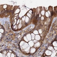
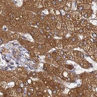


Comprehensive Human Protein Atlas IHC scoring per tissue (reliability: Enhanced — Medium consistency between antibody staining and RNA expression data.). Rows are taken directly from the HPA tissue chart — click any row's HPA link to view the source.
| Tissue | Cell type | Level | Evidence | Source |
|---|---|---|---|---|
| Bronchus | Respiratory epithelial cells | High | Protein (IHC) | HPA → |
| Duodenum | Glandular cells | High | Protein (IHC) | HPA → |
| Epididymis | Glandular cells | High | Protein (IHC) | HPA → |
| Fallopian tube | Glandular cells | High | Protein (IHC) | HPA → |
| Gallbladder | Glandular cells | High | Protein (IHC) | HPA → |
| Tissue | Cell type | Level | Evidence | Source |
|---|---|---|---|---|
| None in HPA: PRKAB2 is detected in all 45 scored tissues. Use a no-primary (secondary-only) and an isotype control instead. | ||||
Troubleshoot PRKAB2 staining in paraffin sections by checking retrieval, cellular distribution, detection background and scoring against tissue controls.
PB9739 has real paraffin-section IHC images from mouse and rat intestine and human lung cancer tissue, plus an IF/ICC image from A431 cells (catalog image captions).
PB9739 is listed for human, mouse and rat IHC and for human IF/ICC; its captions show paraffin-section IHC in mouse and rat intestine and human lung cancer tissue, and IF/ICC in A431 cells (catalog applications/reactivity; image captions). M05077 is listed for human frozen-section IHC and ICC, with no supplied IHC or IF figure (catalog reactivity/dilution_raw; image captions).
Which to pick: Choose PB9739 for paraffin-section tissue IHC: its own captions document EDTA pH 8 retrieval and 1 μg/ml primary antibody; the fixative is unreported (PB9739 IHC image captions). Choose PB9739 for IF/ICC based on its A431 image at 2 μg/ml; M05077 is a human-reactive mouse monoclonal clone 6G1 listed for ICC and frozen-section IHC without a supplied figure (PB9739 IF image caption; M05077 catalog clone/reactivity/dilution_raw/image captions). For cross-species IHC, PB9739 is listed for human, mouse and rat, while M05077 is listed for human only (catalog reactivity).