This website uses cookies to ensure you get the best experience on our website.


- Table of Contents
Plan chromogenic PRKCE IHC in paraffin sections around cytoplasmic and membranous staining, with high signal in cerebral cortical neurons and kidney distal tubules (HPA tissue IHC). The catalog antibody lists an IHC concentration of 0.5–1 μg/ml (datasheet: PB9320); compare stained cells with documented negative tissue populations (HPA tissue IHC).

Expected localisation, validated protocols, controls and antibodies — the at-a-glance facts below, then the full design guide.
| Expected localisation | Cytoplasmic and membranous tissue staining (HPA tissue IHC) | |
| Staining pattern | High in cortical neurons and kidney distal tubules (HPA tissue IHC) | |
| Antigen retrieval | EDTA pH 8.0 HIER, heat-mediated (datasheet PB9320) | |
| Positive control | Cerebral cortex+3 more · see all | |
| Negative control | Adipose tissue+4 more · see all |
| Fixation | Keep paraffin-section fixation consistent (standard IHC practice; not target-specific); Matched tissue-IHC evidence does not establish the fixation claim. Validate the specimen-specific method before use. (selected-SKU IHC image PB9320) | |
| Caveat | HGF can shift epithelial PRKCE to the plasma membrane (UniProt) | |
| Regulation | HGF-dependent membrane redistribution (UniProt) | |
| Isoform / epitope | No listed isoforms or cleavage; no extracellular domain (UniProt) |
Compare the catalog antibody's IHC-P protocol (datasheet: PB9320) with three published PRKCE IHC protocols (PMC7533283; PMC9293992; PMC5520703).
| Sample | Tissue sections; selected-image fixative not specified (standard IHC workflow) |
| Fixation | Image fixative and duration unreported (datasheet PB9320); verify before use. |
| Sectioning | 4–5 µm sections on charged slides (standard) |
| Deparaffinisation | Xylene, graded ethanol series to water (standard) |
| Antigen retrieval | Heat retrieval: EDTA pH 8.0 (datasheet PB9320); 20 min, 95–100 °C (standard) |
| Peroxidase block | 3% H2O2, 10 min, room temperature (standard) |
| Blocking | 10% goat serum (datasheet PB9320) |
| Primary antibody | Rabbit anti-PRKCE, 0.5-1μg/ml (datasheet PB9320) |
| Primary incubation | Overnight at 4 °C (datasheet PB9320) |
| Detection | HRP-conjugated secondary, DAB chromogen (datasheet PB9320) |
| Counterstain | Hematoxylin, blue, dehydrate and mount (standard) |
| Expected result | PRKCE-positive staining in neuronal cells of cerebral cortex (HPA tissue IHC: High). HPA tissue profile: Cytoplasmic and membranous expression in few tissues, most abundant expression in CNS. No signal in the no-primary control. |
PRKCE is expected mainly in cytoplasmic and membranous compartments on tissue IHC, with the strongest listed staining in cerebral cortical neurons and kidney distal tubules (HPA: tissue IHC). HPA rates its tissue staining reliability Enhanced (HPA: reliability). A peripheral signal can fit its membrane localisation, but PRKCE has no transmembrane segment; a purely surface-restricted pattern needs scrutiny (UniProt Q02156: topology and subcellular location).
| Cortical neurons or kidney distal tubules show strong cytoplasmic staining, with some membrane-associated signal (HPA: High; tissue IHC). | This matches the reported cell types and compartments. Compare intensity within the named cells, since adjacent unstained cells do not by themselves contradict the cell-specific HPA observations (HPA: tissue IHC). |
| A section shows mainly nuclear staining, with little cytoplasmic signal (HPA: cytoplasmic and membranous tissue profile). | Treat this as a localisation mismatch and investigate artefact. UniProt also lists the nucleus and reports stimulus-associated nuclear translocation by similarity, so nuclear staining alone cannot establish either an error or activation (UniProt Q02156: subcellular location). |
| Strong staining appears in appendix glandular cells or bone marrow hematopoietic cells (HPA: Not detected in those cells). | This conflicts with the listed HPA observations and warrants a specificity check. Cross-reactivity or endogenous detection activity are possibilities, not proven causes; assess a control lacking primary antibody (HPA: tissue IHC; general IHC practice). |
| Chromogen covers tissue broadly, including cells expected to be unstained (HPA: cell-specific tissue IHC). | Diffuse deposit obscures the reported cell pattern. Check primary-free and detection controls, blocking, washes, and chromogen development before assigning the signal to PRKCE (general IHC practice; HPA: tissue IHC). |
| No signal is visible in cerebral cortical neurons or kidney distal tubules (HPA: High in both cell types). | A failed positive control is plausible. Review tissue identity, antibody suitability, retrieval, detection and counterstain before interpreting a study section as PRKCE-negative (HPA: tissue IHC; general IHC practice). |
| Cell-specific reference pattern (HPA: tissue IHC) | Cerebral cortical neurons and kidney distal tubules are High; caudate neurons and hippocampal synapses are Medium. Use the named cells when comparing sections, because HPA levels do not describe every cell in each tissue (HPA: tissue IHC). |
| Lower and undetected reference cells (HPA: tissue IHC) | Cerebellar granular-layer cells are Low; appendix glandular cells and bone marrow hematopoietic cells are Not detected. These are cell-level observations, not claims that entire tissue sections lack PRKCE (HPA: tissue IHC). |
| Antibody evidence (HPA: antibody validation) | The tissue profile is rated Enhanced. HPA lists IHC Enhanced for HPA044496 and CAB001948, while HPA008236 has ICC Supported with no IHC status listed. Match validation to the assay being interpreted (HPA: antibodies; reliability). |
| Compartment and stimulus (UniProt Q02156: subcellular location) | UniProt lists cytoplasm, cytoskeleton, cell membrane, perinuclear region and nucleus. It reports HGF-associated plasma-membrane translocation in stimulated epithelial cells; its Golgi and PMA-related nuclear notes are by similarity. The observed context matters (UniProt Q02156). |
| Topology (UniProt Q02156: topology) | PRKCE has no transmembrane segment, signal peptide or propeptide and is listed as one chain spanning residues 1–737. Membrane-associated staining is compatible with its localisation, but topology alone cannot locate the antibody epitope or predict staining after retrieval (UniProt Q02156). |
| IF/ICC Q&A: Is its pattern the same? (HPA: subcellular ICC-IF) | HPA supports cytosol and intermediate filaments, with additional plasma membrane and vesicle localisation in ICC-IF. These cultured-cell observations help assess compartments; they do not prescribe an IF protocol or replace tissue IHC controls (HPA: subcellular ICC-IF; general IHC practice). |
| Situation | Likely cause | Next action |
|---|---|---|
| Cortical neurons or distal tubules are blank (HPA: High in these cells). | The positive control may have failed; the image alone cannot identify which workflow step failed (HPA: tissue IHC; general IHC practice). | Confirm the named cells are present, then check antibody IHC validation, retrieval, primary incubation and detection with a known-positive section (HPA: antibodies and tissue IHC; general IHC practice). |
| Staining is mostly nuclear (HPA: cytoplasmic and membranous tissue profile). | Localisation is discordant with the tissue profile; UniProt also allows context-dependent nuclear localisation (UniProt Q02156: subcellular location). | Compare with a validated positive section and primary-free control; record stimulation context before interpreting nuclear signal as PRKCE (HPA: tissue IHC; UniProt Q02156; general IHC practice). |
| Appendix glandular cells stain strongly (HPA: Not detected). | The result conflicts with HPA's cell-specific observation; nonspecific binding or endogenous detection activity may contribute (HPA: tissue IHC; general IHC practice). | Inspect a primary-free control, review blocking and detection, and compare the same run with a named positive cell population (HPA: tissue IHC; general IHC practice). |
| Deposit is diffuse across cells and extracellular areas (general IHC practice). | Background can arise from nonspecific detection or excessive chromogen development; appearance alone cannot assign a cause (general IHC practice). | Review primary-free and detection controls, blocking, washes and development time; score cells only after background is resolved (general IHC practice). |
| A cell reported Low looks as strong as a High reference cell (HPA: tissue IHC). | Run conditions or image display may distort relative intensity; HPA categories are observations rather than a quantitative assay scale (HPA: tissue IHC; general IHC practice). | Compare matched sections stained and viewed under the same conditions, then describe intensity and cell type rather than forcing a numerical threshold (HPA: tissue IHC; general IHC practice). |
| ICC-IF shows filaments while tissue IHC looks broadly cytoplasmic (HPA: subcellular ICC-IF; tissue IHC). | HPA reports supported intermediate filaments in ICC-IF and a cytoplasmic tissue profile; these are different preparations (HPA: subcellular ICC-IF; tissue IHC). | Interpret each assay against its own HPA reference and validation status; use tissue positive and detection controls for the IHC call (HPA: antibodies and tissue IHC; general IHC practice). |




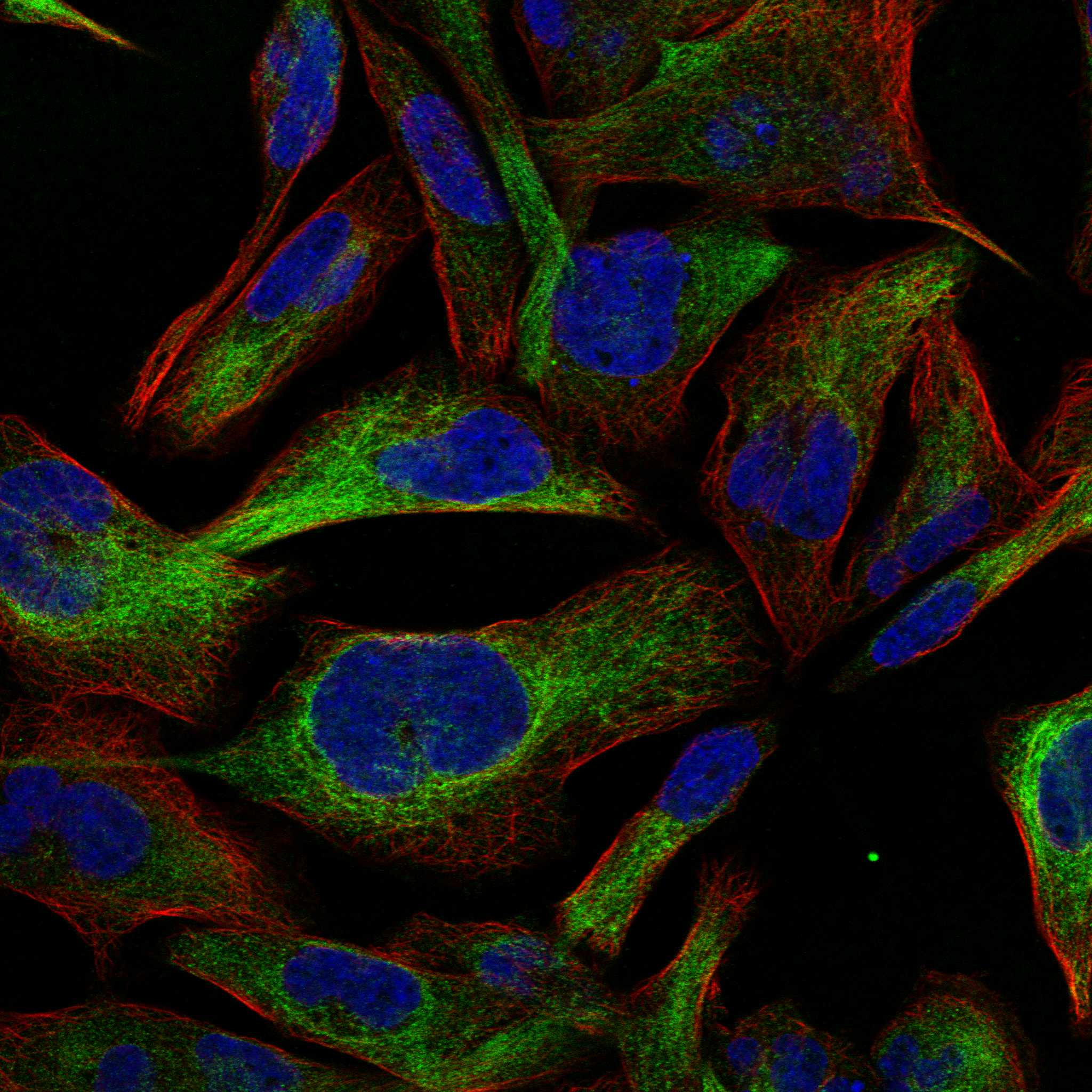
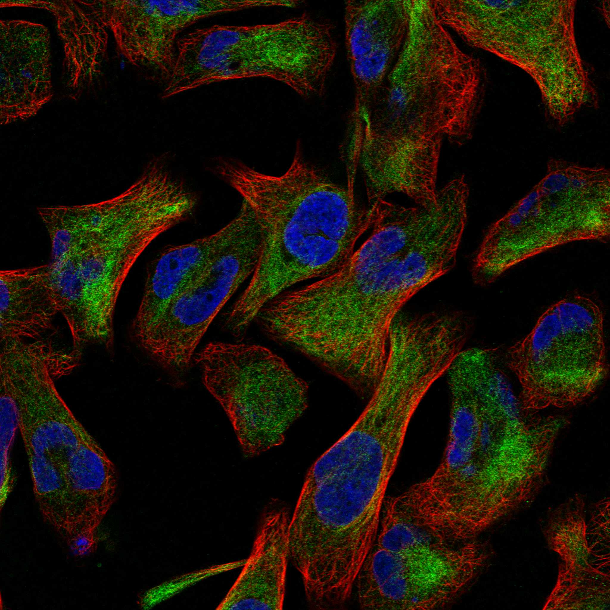
Comprehensive Human Protein Atlas IHC scoring per tissue (reliability: Enhanced — High consistency between antibody staining and RNA expression data. External characterization data supports antibody staining.). Rows are taken directly from the HPA tissue chart — click any row's HPA link to view the source.
| Tissue | Cell type | Level | Evidence | Source |
|---|---|---|---|---|
| Cerebral cortex | Neuronal cells | High | Protein (IHC) | HPA → |
| Kidney | Distal tubules | High | Protein (IHC) | HPA → |
| Caudate | Neuronal cells | Medium | Protein (IHC) | HPA → |
| Hippocampus | Synapses | Medium | Protein (IHC) | HPA → |
| Tissue | Cell type | Level | Evidence | Source |
|---|---|---|---|---|
| Adipose tissue | Adipocytes | Not detected | Protein (IHC) | HPA → |
| Appendix | Glandular cells | Not detected | Protein (IHC) | HPA → |
| Bone marrow | Hematopoietic cells | Not detected | Protein (IHC) | HPA → |
| Breast | Adipocytes | Not detected | Protein (IHC) | HPA → |
| Bronchus | Respiratory epithelial cells | Not detected | Protein (IHC) | HPA → |
Troubleshoot PRKCE staining in paraffin sections by comparing compartment-specific signal with matched controls and documented processing conditions.
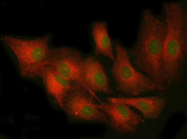
Anti-PRKCE antibodies have IHC images from mouse and rat intestine and paraffin sections of human brain and stomach, plus IF data from A549 cells (PB9320 IHC captions; A01151-1 IF caption).
PB9320 is listed for IHC in human, mouse and rat, with IHC images from mouse and rat intestine and paraffin sections of human brain and stomach (PB9320 applications/reactivity; PB9320 IHC captions). A01151-1 is listed for IF/ICC in human, mouse and rat, with an IF image from A549 cells (A01151-1 applications/reactivity; A01151-1 IF caption).
Which to pick: Choose PB9320 for chromogenic tissue IHC: it is a rabbit polyclonal listed for IHC, and its captions show paraffin-section staining; the fixative is unreported (PB9320 dilution_raw; PB9320 applications; PB9320 IHC captions). Choose A01151-1 for IF/ICC: it is a rabbit polyclonal listed for those applications, with an A549 cell IF image (A01151-1 dilution_raw; A01151-1 applications; A01151-1 IF caption). For cross-species work, both list human, mouse and rat reactivity; choose according to the application and confirm performance in the intended sample (PB9320 reactivity/applications; A01151-1 reactivity/applications).