PRKG1 / cGMP-dependent protein kinase 1 · IHC design guide

Design Immunohistochemistry for PRKG1

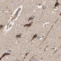
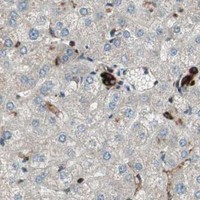
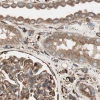
Plan PRKG1 staining in paraffin sections using the catalog antibody's 2–5 μg/mL IHC range (datasheet A01708-3). Compare cytoplasmic staining in adipocytes and glandular cells with low staining in salivary gland glandular cells (HPA tissue IHC).

Evidence assembled Oct 2026 · For research use; verify linked source records and product datasheet before use
Immunohistochemistry protocol sheet for PRKG1 (IHC for PRKG1): expected localisation Cytoplasmic tissue staining (HPA tissue IHC), antibody A01708-3, validated IHC image, and IHC protocol steps
Printable PRKG1 IHC protocol sheet — expected localisation Cytoplasmic tissue staining (HPA tissue IHC), antibody A01708-3, controls and protocol steps. Open the full PRKG1 IHC guide →

PRKG1 Immunohistochemistry Experimental Design Guide

Expected localisation, validated protocols, controls and antibodies — the at-a-glance facts below, then the full design guide.

Must know before staining
Expected localisation Cytoplasmic tissue staining (HPA tissue IHC)
Staining pattern Adipocytes and glandular cells: cytoplasmic staining (HPA tissue IHC)
Antigen retrieval EDTA pH 8.0 HIER, heat-mediated (datasheet A01708-3)
Positive control ⓘ Adipose tissue+4 more · see all
Negative control ⓘ None in HPA (detected in all 45 tissues); use no-primary + isotype controls
Important caveats
Reasons your staining may differ from the expected pattern.
Fixation Keep fixation consistent across paraffin samples (standard IHC practice; not target-specific)
Caveat Staining and RNA expression show medium consistency (HPA tissue IHC)
Regulation Blood-vessel RNA is tissue enhanced (HPA RNA specificity)
Isoform / epitope Three isoforms; confirm antibody epitope coverage (UniProt)
Section 1

Recommended PRKG1 IHC & IF Protocols

The catalog antibody’s IHC-P protocol is accompanied by three published PRKG1 IHC protocols for carotid artery, ligament, and aorta (PMC13446705; PMC7454216; PMC8131144).

Recommended immunohistochemistry (IHC-P) protocol parameters
SampleParaffin-embedded mouse lung tissue; fixative not specified (datasheet A01708-3)
FixationImage fixative and duration unreported (datasheet A01708-3); verify before use.
Sectioning4–5 µm sections on charged slides (standard)
DeparaffinisationXylene, graded ethanol series to water (standard)
Antigen retrievalHeat retrieval: EDTA pH 8.0 (datasheet A01708-3); 20 min, 95–100 °C (standard)
Peroxidase block3% H2O2, 10 min, room temperature (standard)
Blocking10% goat serum (datasheet A01708-3)
Primary antibodyRabbit anti-PRKG1, 2-5μg/ml (datasheet A01708-3)
Primary incubationOvernight at 4 °C (datasheet A01708-3)
DetectionStreptavidin-biotin complex (SABC), DAB chromogen (datasheet A01708-3)
CounterstainHematoxylin, blue, dehydrate and mount (standard)
Expected resultPRKG1-positive staining in adipocytes of adipose tissue (HPA tissue IHC: Medium). HPA tissue profile: General cytoplasmic expression. No signal in the no-primary control.
💡Decision noteStart with heat-mediated EDTA pH 8.0 retrieval (datasheet: A01708-3); consider citrate retrieval when adapting the published carotid artery or ligament protocols (PMC13446705; PMC7454216).
Section 2

What Is the Expected PRKG1 Staining Pattern?

PRKG1 should appear mainly in the cytoplasm in tissue IHC (HPA tissue IHC: general cytoplasmic expression; UniProt Q13976: cytoplasm). HPA reports medium staining in adipocytes, glandular cells, respiratory epithelial cells, glial cells and cerebellar granular-layer cells (HPA tissue IHC). Its tissue IHC profile has Enhanced reliability, with medium consistency between staining and RNA data (HPA tissue IHC). PRKG1 has no transmembrane segment (UniProt Q13976: topology).

What am I looking at on my slide?
Cytoplasmic staining in adipocytes or respiratory epithelial cells, at roughly medium intensity (HPA tissue IHC).This matches two observed tissue and cell patterns (HPA tissue IHC). Score the relevant cells and their compartment; medium is an HPA category, not a required intensity for every specimen (HPA tissue IHC; general IHC practice).
Predominantly nuclear staining, with little cytoplasmic signal.This conflicts with the reported general cytoplasmic tissue pattern (HPA tissue IHC). Check whether counterstain, nonspecific binding or detection background explains the apparent nuclear signal before interpreting it as PRKG1 (general IHC practice).
Strong staining dominates urothelial cells while expected cytoplasmic staining is weak elsewhere.HPA reports low staining in urinary-bladder urothelial cells (HPA tissue IHC). The mismatch warrants checking cross-reactivity and endogenous detection activity; it does not prove either cause (general IHC practice).
Broad haze covers tissue, extracellular spaces or areas without identifiable cells.A haze that obscures cell boundaries cannot establish the cytoplasmic pattern reported for PRKG1 (HPA tissue IHC). Evaluate reagent-only controls, blocking and chromogen development for background (general IHC practice).
No staining in a section containing identifiable adipocytes.HPA reports medium adipocyte staining, so absence is a useful troubleshooting signal (HPA tissue IHC). First verify tissue preservation, antibody application and detection controls; one blank section does not establish true PRKG1 absence (general IHC practice).
💡Expected PRKG1 appearanceCall a result consistent with PRKG1 when identifiable HPA-positive cells show chiefly cytoplasmic staining around the reported medium level; isolated dominant nuclear signal or tissue-wide haze is suspect (HPA tissue IHC: cytoplasmic profile and medium examples; general IHC practice).
How each factor affects the staining
Tissue and cell selectionHPA reports medium staining in adipocytes, adrenal and breast glandular cells, bronchial respiratory epithelial cells, caudate glial cells and cerebellar granular-layer cells (HPA tissue IHC). These are observed examples for comparison, not guarantees for every section (general IHC practice).
Lower-staining comparatorsSalivary glandular, urinary-bladder urothelial and vaginal squamous epithelial cells are listed as low (HPA tissue IHC). Treat them as lower-signal comparators, not confirmed PRKG1-negative controls (HPA tissue IHC).
Validation and RNA contextThe tissue profile is Enhanced but has medium staining–RNA consistency; blood vessel is RNA tissue enhanced (HPA tissue IHC). RNA enrichment helps choose a region to inspect, but does not itself establish a specific IHC staining level there (HPA tissue IHC).
Antibody-specific evidenceCAB009629 is IHC Enhanced, whereas HPA007699 is IHC Approved (HPA antibodies). Record which antibody produced the slide; a profile-level reliability label does not assign the same validation status to both reagents (HPA tissue IHC; HPA antibodies).
Could IF/ICC help assess localisation?Yes: HPA reports approved vesicle and cytosol localisation in ICC-IF, with images from HEK293, U-251MG and U2OS (HPA subcellular). That supports a localisation comparison, while tissue IHC remains the source for tissue staining expectations (HPA tissue IHC; HPA subcellular).
Molecular contextPRKG1 has three reported isoforms, no signal peptide and no transmembrane segment (UniProt Q13976: isoforms, processing and topology). The supplied record gives no antibody epitope, so it cannot establish isoform coverage or an epitope-specific retrieval choice.
Why is my staining missing, weak or wrong?
SituationLikely causeNext action
Cytoplasmic signal is absent in an adipocyte-containing section.Antibody application, retrieval or detection may have failed; HPA reports medium adipocyte staining (HPA tissue IHC; general IHC practice).Confirm the cells are present, inspect a valid positive control, and check the antibody and detection steps (general IHC practice). If adjusting retrieval, document the condition; PRKG1-specific retrieval sensitivity is unreported here.
Nuclei appear darker than cytoplasm.Counterstain or nonspecific signal may dominate; the expected tissue pattern is generally cytoplasmic (HPA tissue IHC; general IHC practice).Review a section without primary antibody and assess counterstain and chromogen development before scoring nuclear staining as PRKG1 (general IHC practice).
Most structures show diffuse chromogen.Detection background or excess staining can obscure cell boundaries (general IHC practice).Compare a no-primary control, check endogenous enzyme blocking where applicable, and optimise antibody concentration and development time using the actual assay reagents (general IHC practice).
Strong signal appears mainly in a cell group HPA lists as low.A tissue-pattern mismatch raises possible nonspecific binding or endogenous detection activity; HPA lists urothelial and vaginal squamous epithelial cells as low (HPA tissue IHC; general IHC practice).Check cell identity and compartment, compare an HPA medium-staining example, and inspect a no-primary control before assigning the signal to PRKG1 (HPA tissue IHC; general IHC practice).
Two antibodies give different tissue patterns.Their HPA IHC validation statuses differ: CAB009629 is Enhanced and HPA007699 is Approved (HPA antibodies). The statuses alone do not identify why a particular section disagrees.Record the antibody ID and compare the same tissue compartments and controls across staining runs; give greater weight to a reproducible cytoplasmic pattern in reported cells (HPA antibodies; HPA tissue IHC; general IHC practice).
An IF/ICC image shows puncta, but tissue IHC looks broadly cytoplasmic.HPA reports vesicles and cytosol in ICC-IF and general cytoplasmic expression in tissue IHC (HPA subcellular; HPA tissue IHC).Interpret each image in its own assay context. Use the tissue IHC profile to score the paraffin section, and use the IF/ICC result only as a localisation cross-check (HPA tissue IHC; HPA subcellular).

Sample controls for PRKG1 IHC & IF

🧪Run adipose tissue first and expect staining in adipocytes (HPA: Medium in adipocytes). HPA detects PRKG1 in all 45 scored tissues, so there is no supported negative tissue; no-primary and isotype controls carry the negative comparison, while cells without specific staining on the positive slide should show only background (HPA: no negative tissue listed).
Positive control tissue: Adipose tissue (Adipocytes, HPA Medium)
Negative control tissue: None in HPA: PRKG1 is detected in all 45 scored tissues. Use a no-primary (secondary-only) and an isotype control instead.
ICC-IF cell lines (HPA subcellular resource): HPA ICC-IF images show PRKG1 in HEK293, U-251MG, U2OS, with annotated localisation: Vesicles (approved), Cytosol (approved) (HPA subcellular).
Technical controls: Include a no-primary, secondary-only control; an isotype control matched to the rabbit primary antibody’s clonality where known; and PRKG1 knockout material or a peptide-block control if the immunizing peptide is available (selected-SKU caption: rabbit primary antibody). For chromogenic adipose sections, quench endogenous peroxidase and assess endogenous biotin because the reported detection uses biotinylated secondary antibody, streptavidin–biotin complex and DAB (selected-SKU caption: SABC/DAB).
⚠️Feasibility: A target-specific fixation window or fixation effect is unreported, and the exact selected-SKU paraffin-section caption does not state a fixative (selected-SKU caption: fixative not stated). The reported mouse lung IHC used heat retrieval in EDTA at pH 8.0, but the caption does not establish that retrieval is essential (selected-SKU caption: EDTA pH 8.0). Frozen-section performance is unreported; ICC-IF images exist for HEK293, U-251MG and U2OS with approved vesicle and cytosol localization, so IF is feasible to assess separately, while adipose sections need careful scoring around lipid vacuoles (HPA: subcellular ICC-IF images; HPA: Medium in adipocytes).

HPA tissue IHC evidence for PRKG1

Comprehensive Human Protein Atlas IHC scoring per tissue (reliability: Enhanced — Medium consistency between antibody staining and RNA expression data.). Rows are taken directly from the HPA tissue chart — click any row's HPA link to view the source.

Positive expression · recommended positive controls

TissueCell typeLevelEvidenceSource
Adipose tissue Adipocytes Medium Protein (IHC) HPA →
Adrenal gland Glandular cells Medium Protein (IHC) HPA →
Appendix Lymphoid tissue Medium Protein (IHC) HPA →
Bone marrow Hematopoietic cells Medium Protein (IHC) HPA →
Breast Glandular cells Medium Protein (IHC) HPA →

Undetected expression · recommended negative controls

TissueCell typeLevelEvidenceSource
None in HPA: PRKG1 is detected in all 45 scored tissues. Use a no-primary (secondary-only) and an isotype control instead.
Section 3

Advanced PRKG1 IHC Tips

These questions address PRKG1 staining in paraffin sections by chromogenic IHC, with one question on planning a separate IF experiment.

Which antigen retrieval should I use when PRKG1 staining is weak?
Start with heat-mediated antigen retrieval in EDTA pH 8.0 for paraffin-section PRKG1 IHC (datasheet A01708-3). The selected image used this retrieval before incubation with 2 μg/mL catalog antibody overnight at 4°C, so keep those conditions together for the initial comparison (datasheet A01708-3). If staining remains weak, vary heating duration on adjacent sections while holding antibody concentration and detection constant; excessive retrieval can damage morphology and increase background (standard IHC practice). If that fails, test another retrieval buffer as a documented fallback, with a no-primary control and the same positive tissue in each run (standard IHC practice).
Could fixation explain weak or uneven PRKG1 staining?
The selected PRKG1 image shows a paraffin-embedded mouse lung section, but its fixative and fixation duration are unreported (datasheet A01708-3). Target-specific fixation sensitivity is therefore unknown; neither the tissue staining profile nor PRKG1 sequence features establish how fixation affects this epitope (HPA tissue IHC; UniProt Q13976). Record the fixative, duration, tissue thickness, and time before processing, then compare adjacent specimens processed under controlled conditions using the same retrieval and detection settings (standard IHC practice). Assess morphology alongside staining: uneven preservation can complicate interpretation, while a positive control processed with each batch helps identify a failed staining run (standard IHC practice).
Where should convincing PRKG1 staining appear in a paraffin section?
Expect predominantly cytoplasmic staining: PRKG1 is annotated in the cytoplasm, and tissue IHC reports general cytoplasmic expression (UniProt Q13976; HPA tissue IHC). A vesicular component is plausible because subcellular imaging places PRKG1 in vesicles and cytosol (HPA subcellular). UniProt also reports colocalisation with TRPC7 at the plasma membrane, but PRKG1 has no transmembrane segment, so membrane-adjacent staining alone needs corroboration (UniProt Q13976). Compare the cellular pattern with a no-primary section, inspect morphology, and require reproducibility in the same cell population before assigning a membrane or nuclear pattern to PRKG1 (standard IHC practice).
Can this IHC stain distinguish PRKG1 isoforms or modified epitopes?
PRKG1 has 3 annotated isoforms—alpha, beta, and isoform 3—so an isoform assignment depends on where the catalog antibody binds (UniProt Q13976). The supplied image establishes staining under its stated conditions, but provides no epitope map or isoform selectivity for this antibody (datasheet A01708-3). Check whether the documented immunogen lies in a shared or isoform-specific sequence before describing a tissue pattern as alpha or beta; treat an undisclosed epitope as unresolved (standard IHC interpretation). PRKG1 includes phosphorylation sites at residues 59 and 515, but ordinary total-protein staining cannot establish phosphorylation state without validated modification-specific evidence (UniProt Q13976; standard IHC interpretation).
How should I plan a separate PRKG1 multiplex IF experiment?
Use chromogenic IHC to establish the cell populations and staining pattern before interpreting a separate multiplex IF experiment (standard IHC/IF practice). Pair PRKG1 with a validated marker for the expected cell type, such as a smooth muscle marker when examining a vascular compartment; PRKG1 participates in smooth muscle relaxation, and its RNA profile is tissue-enhanced in blood vessels (UniProt Q13976; HPA tissue IHC). Select spectrally separated fluorophores after checking tissue autofluorescence and single-color controls, especially where background may resemble weak cytosolic signal (standard IF practice). Because PRKG1 is cytosolic and vesicular with no transmembrane segment, choose permeabilisation that admits antibody to intracellular epitopes and preserves morphology; epitope accessibility still requires validation (HPA subcellular; UniProt Q13976; standard IF practice).
How do I reduce diffuse or vessel-associated DAB background?
First compare the stained section with no-primary and secondary-only controls to locate background introduced by the detection system (standard IHC practice). The selected workflow used a 10% goat serum block, biotinylated secondary antibody, avidin-biotin complex detection, and DAB; test endogenous biotin and enzyme-related signal with appropriate controls when using that system (datasheet A01708-3; standard IHC practice). Optimise the primary concentration around the documented 2 μg/mL condition and keep washes, development time, and section handling consistent across comparisons (datasheet A01708-3; standard IHC practice). Judge residual diffuse colour against expected cytoplasmic staining and tissue morphology, rather than counting every brown deposit as PRKG1 (HPA tissue IHC; standard IHC interpretation).
What should I score when comparing PRKG1 IHC across sections? ⚠ ANSWER MARKED FOR VERIFICATION
Define the anatomical compartment and cell population before scoring because PRKG1 tissue IHC shows general cytoplasmic expression across multiple cell types (HPA tissue IHC). For cellular staining, report the percentage of positive cells and intensity categories, or calculate an H-score from intensity-weighted percentages on a 0–300 scale (standard IHC practice). If measuring positive-cell density, express it per mm² of viable, evaluable tissue and exclude folds, necrosis, and blank space using the same rules for every section (standard IHC practice). Normalise each comparison to the relevant cell population or tissue area, and use consistent retrieval, exposure-free brightfield imaging, and DAB development so technical variation does not dominate the result (standard IHC practice).
How can I distinguish true PRKG1 signal from staining artefact?
A credible pattern is reproducible cytoplasmic staining in intact cells, potentially with vesicular signal; membrane-adjacent staining needs independent support, and a dominant nuclear-only pattern warrants scrutiny (UniProt Q13976; HPA tissue IHC; HPA subcellular). Check that the stained cells match the compartment being studied, rather than inferring identity from brown colour alone; blood vessels are RNA-enriched, while the selected antibody image demonstrates staining in mouse lung (HPA tissue IHC; datasheet A01708-3). Edge accentuation, damaged or necrotic regions, and widespread signal in no-primary controls favor artefact (standard IHC practice). Review endogenous enzyme and avidin-biotin controls where applicable, then confirm the pattern on independently processed sections before interpreting differences as biological (datasheet A01708-3; standard IHC practice).
Boster reagents

Best PRKG1 / cGMP-dependent protein kinase 1 IHC Antibodies

The catalog antibody has paraffin-section IHC images from mouse and rat lung and human breast cancer (IHC image captions), plus cellular IF data from U20S cells (IF image caption).

Real IHC data IHC analysis of CGKI/PRKG1 using anti-CGKI/PRKG1 antibody (A01708-3). CGKI/PRKG1 was detected in paraffin-embedded section of mouse lung tissue. Heat mediated antigen retrieval was performed in EDTA buffer (pH8.0, epitope retrieval solution). The tissue section was blocked with 10% goat serum. The tissue section was then incubated with 2μg/ml rabbit anti-CGKI/PRKG1 Antibody (A01708-3) overnight at 4°C. Biotinylated goat anti-rabbit IgG was used as secondary antibody and incubated for 30 minutes at 37°C. The tissue section was developed using Strepavidin-Biotin-Complex (SABC) (Catalog # SA1022) with DAB as the chromogen.
Anti-cGKI/PRKG1 Antibody ®
Cat # A01708-3

A01708-3 is the sole listed SKU; its IHC captions show paraffin sections of mouse and rat lung and human breast cancer (IHC image captions). Its IF caption shows U20S cells, and the catalog lists IHC, IF, and ICC applications (IF image caption; catalog: applications).

Which to pick: Choose A01708-3 for paraffin-section IHC; its mouse lung caption documents EDTA retrieval at pH 8.0 and primary antibody at 2 μg/ml (A01708-3 IHC image caption). Choose the same SKU for cellular IF/ICC, using 5 μg/ml as the catalog starting concentration (catalog: applications and IF dilution). It lists human, mouse, and rat reactivity, while clonality and the tissue fixative are unreported (catalog: reactivity and clone; IHC image captions).

Each figure is that product's own IHC / IF validation image from its datasheet.

References

  1. UniProt Consortium. UniProt entry Q13976 (KGP1_HUMAN, cGMP-dependent protein kinase 1).
  2. Human Protein Atlas. PRKG1 tissue IHC expression (reliability: Enhanced).
  3. Human Protein Atlas. PRKG1 subcellular location (ICC-IF): Localized to vesicles and cytosol..
  4. Human Protein Atlas. PRKG1 antibody validation summary (2 antibodies).
  5. Vitamin B12 combined with methylene blue alleviates experimental septic shock and is associated with modulation of KCNMB1 and the cGMP-PRKG pathway. PloS one 2026 — PMC13446705.
  6. Upregulation of cGMP-dependent Protein Kinase (PRKG1) in the Development of Adolescent Idiopathic Scoliosis. Orthopaedic surgery 2020 — PMC7454216.
  7. cGMP-dependent protein kinase type I is implicated in the regulation of the timing and quality of sleep and wakefulness. PloS one 2009 — PMC2617781.
  8. Network Pharmacology Prediction and Pharmacological Verification Mechanism of Yeju Jiangya Decoction on Hypertension. Evidence-based complementary and alternative medicine : eCAM 2021 — PMC8131144.
  9. PubMed PMID:2792381 — UniProt-cited evidence.
  10. PubMed PMID:8613202 — UniProt-cited evidence.
  11. PubMed PMID:9192852 — UniProt-cited evidence.