This website uses cookies to ensure you get the best experience on our website.


- Table of Contents
Plan PRMT8 chromogenic IHC on paraffin sections using neuronal staining in caudate or cerebral cortex as a tissue reference (HPA tissue IHC). Start the IHC-validated antibody at 2–5 μg/mL (datasheet A07980-1), and interpret staining in light of the multi-gene targeting caution (HPA tissue IHC).

Expected localisation, validated protocols, controls and antibodies — the at-a-glance facts below, then the full design guide.
| Expected localisation | Neuronal nuclei (HPA tissue IHC); cell membrane (UniProt) | |
| Staining pattern | Nuclear staining in neuronal cells (HPA tissue IHC) | |
| Antigen retrieval | EDTA pH 8.0 HIER, heat-mediated (datasheet A07980-1) | |
| Positive control | Testis+2 more · see all | |
| Negative control | Adipose tissue+4 more · see all |
| Fixation | Keep fixation consistent (standard IHC practice; not target-specific); Matched tissue-IHC evidence does not establish the fixation claim. Validate the specimen-specific method before use. (selected-SKU IHC image A07980-1) | |
| Caveat | Antibody may recognize proteins from more than one gene (HPA tissue IHC) | |
| Regulation | Brain-enriched RNA (HPA tissue data) | |
| Isoform / epitope | 2 isoforms; epitope differences are unspecified (UniProt) |
The catalog antibody has a datasheet IHC-P protocol (datasheet: A07980-1). One published PRMT8 brain IHC protocol provides a second option (PMC7890232 methods).
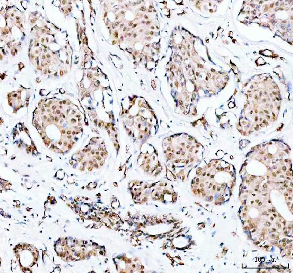
| Sample | Paraffin-embedded human breast cancer tissue; fixative not specified (datasheet A07980-1) |
| Fixation | Image fixative and duration unreported (datasheet A07980-1); verify before use. |
| Sectioning | 4–5 µm sections on charged slides (standard) |
| Deparaffinisation | Xylene, graded ethanol series to water (standard) |
| Antigen retrieval | Heat retrieval: EDTA pH 8.0 (datasheet A07980-1); 20 min, 95–100 °C (standard) |
| Peroxidase block | 3% H2O2, 10 min, room temperature (standard) |
| Blocking | 10% goat serum (datasheet A07980-1) |
| Primary antibody | Rabbit anti-PRMT8, 2-5μg/ml (datasheet A07980-1) |
| Primary incubation | Overnight at 4 °C (datasheet A07980-1) |
| Detection | HRP-conjugated secondary, DAB chromogen (datasheet A07980-1) |
| Counterstain | Hematoxylin, blue, dehydrate and mount (standard) |
| Expected result | PRMT8-positive staining in leydig cells of testis (HPA tissue IHC: High). HPA tissue profile: Nuclear expression in neuronal cells. No signal in the no-primary control. |
In paraffin section IHC, expect predominantly nuclear staining in neuronal cells, with medium staining reported in caudate and cerebral cortex (HPA: tissue IHC). Interpret that pattern cautiously: HPA rates the tissue result Enhanced but flags recognition of proteins from more than one gene and a subcellular conflict with limited external data (HPA: reliability description). UniProt places PRMT8 at the cell membrane despite reporting no transmembrane segment (UniProt Q9NR22: subcellular location and topology).
| Distinct neuronal nuclei stain in caudate or cerebral cortex, with relatively little staining in surrounding cells. | This fits the reported medium neuronal signal and nuclear profile in brain sections (HPA: caudate, cerebral cortex and tissue profile). Score the relevant cell population rather than the whole field. Nuclear staining is an observed IHC pattern, while its disagreement with UniProt's membrane assignment remains unresolved (UniProt Q9NR22: subcellular location; HPA: reliability description). |
| Signal is confined to cell borders, or broad nuclear and cytoplasmic staining obscures individual neuronal nuclei. | A membrane-only result differs from HPA's nuclear IHC profile but could relate to UniProt's membrane localization; compartment alone cannot settle specificity (HPA: tissue profile; UniProt Q9NR22: subcellular location). Broad staining that also appears in a negative control favors staining artifact. Compare matched controls before assigning either pattern to PRMT8 (standard IHC practice). |
| Strong staining appears in an unexpected cell population, including many cells outside the reported neuronal pattern. | Consider antibody cross-reactivity or endogenous chromogen-generating activity, using an appropriate detection control to distinguish them (standard IHC practice). This concern is material because HPA cautions that its tissue staining can represent protein from more than one gene (HPA: reliability description). High Leydig cell staining is itself reported, so it should be assessed separately rather than dismissed solely for being outside brain (HPA: testis). |
| Diffuse color covers tissue and background, with little separation between nuclei and the surrounding section. | The slide does not permit a reliable compartment or cell type call. Background can arise from detection chemistry, inadequate blocking or antibody concentration (standard IHC practice). Check a matched detection control and examine whether the reported neuronal pattern remains visible above background (HPA: tissue profile). Background alone provides no evidence that PRMT8 has changed location. |
| No neuronal signal is visible in caudate or cerebral cortex while the section otherwise appears intact. | Treat this as an unresolved assay result: HPA reports medium neuronal staining in both tissues, but that observation does not guarantee detection with every antibody or run (HPA: caudate and cerebral cortex). Confirm the tissue region and antibody identity, then review the catalog antibody's IHC-P instructions and run controls (standard IHC practice). Do not infer biological absence from one blank slide. |
| Compartment assignment | HPA describes nuclear neuronal staining, whereas UniProt assigns PRMT8 to the cell membrane and lists no transmembrane segment (HPA: tissue profile; UniProt Q9NR22: subcellular location and topology). Record the observed compartment and the discrepancy; neither source alone resolves whether a particular slide's nuclear or membrane signal is target-specific. |
| Tissue and cell choice | Caudate and cerebral cortex provide reported medium neuronal signal; hippocampal neurons and cerebellar Purkinje cells are reported low (HPA: tissue IHC). Adipocytes in adipose tissue and several listed non-neuronal populations are reported undetected (HPA: tissue IHC). These are reference patterns for selecting and interpreting controls, rather than universal guarantees for individual specimens. |
| Testis exception | HPA reports high Leydig cell staining despite brain-enriched RNA in HPA and brain-specific tissue annotation in UniProt (HPA: testis and RNA specificity; UniProt Q9NR22: tissue specificity). HPA also warns that its tissue antibody can recognize protein from more than one gene (HPA: reliability description). Interpret Leydig cell staining with that uncertainty attached. |
| Antibody validation | HPA039747 has Enhanced IHC status, while HPA069769 has Approved IHC status (HPA: antibody validation). Enhanced reflects reproduction by independent antibodies or orthogonal data, yet HPA's tissue profile still carries its cross-gene and localization cautions (HPA: validation definition and reliability description). Record which antibody produced the slide before comparing its pattern with the reference. |
| Isoforms and epitope coverage | UniProt lists two PRMT8 isoforms and a mature chain spanning residues 2–394 (UniProt Q9NR22: isoforms and processing). The supplied record does not map either antibody's epitope to an isoform. If two antibodies disagree, check their documented immunogens or epitopes before attributing the difference to isoform expression (standard IHC interpretation practice). |
| IF/ICC evidence | Q: Can this IHC localization be used as a confirmed IF/ICC pattern? A: HPA supplies no main IF/ICC location and lists no cell lines with ICC-IF images (HPA: subcellular record). Treat nuclear IHC and UniProt membrane localization as separate evidence until an IF/ICC experiment establishes its own pattern (HPA: tissue profile; UniProt Q9NR22: subcellular location). |
| Situation | Likely cause | Next action |
|---|---|---|
| Brain positive-control section has no visible neuronal staining. | The sampled region may lack the expected cells, or an IHC detection step may have failed; HPA reports medium signal specifically in caudate and cortical neurons (HPA: tissue IHC; standard IHC practice). | Verify region and cell morphology. Check the antibody's documented IHC-P dilution and retrieval conditions, then review detection controls before changing conditions (standard IHC practice). |
| All cells show nuclear color, including populations expected to be unstained. | A broadly reactive primary antibody or detection background may be obscuring the cell-restricted pattern (standard IHC practice). HPA explicitly cautions that its tissue signal can include protein from another gene (HPA: reliability description). | Compare a matched primary-omission control and HPA-listed undetected cell populations; assess whether neuronal nuclei remain selectively stained (standard IHC practice; HPA: tissue IHC). |
| Brown signal appears on the control slide without primary antibody. | Endogenous enzyme activity or nonspecific detection-reagent binding is possible in chromogenic IHC (standard IHC practice). This control result cannot establish PRMT8 localization. | Apply detection-system-appropriate endogenous-activity quenching and blocking, then repeat the matched control before scoring the test section (standard IHC practice). |
| Only membrane staining appears in neurons. | UniProt assigns membrane localization, while HPA's observed tissue IHC pattern is nuclear; the supplied evidence does not explain the conflict (UniProt Q9NR22: subcellular location; HPA: tissue profile and reliability description). | Record the compartment, inspect controls and compare an independently validated antibody where available. Report the discrepancy without treating compartment alone as proof of specificity (standard IHC practice). |
| Leydig cells stain strongly while the brain section is weak. | High Leydig cell and medium caudate and cortical neuronal staining match HPA's observations, although HPA flags cross-gene recognition and UniProt calls PRMT8 brain-specific (HPA: testis, brain and reliability description; UniProt Q9NR22: tissue specificity). | Score each cell population separately and retain the specificity caveat. Use antibody comparison or an orthogonal control before treating testis signal as definitive PRMT8 (standard IHC practice). |
| The slide is uniformly pale or strongly colored, preventing neuronal scoring. | Primary antibody concentration, retrieval or chromogen development may be poorly matched to the assay (standard IHC practice). No supplied source establishes PRMT8-specific fixation or retrieval sensitivity. | Start with the catalog antibody's IHC-P instructions; adjust one general IHC condition at a time with positive and detection controls, then reassess neuronal contrast (standard IHC practice; HPA: brain tissue IHC). |
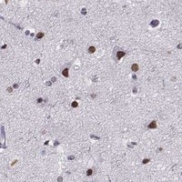


Comprehensive Human Protein Atlas IHC scoring per tissue (reliability: Enhanced — Caution, targets protein from more than one gene. Subcellular location is contradicted by limited external data.). Rows are taken directly from the HPA tissue chart — click any row's HPA link to view the source.
| Tissue | Cell type | Level | Evidence | Source |
|---|---|---|---|---|
| Testis | Leydig cells | High | Protein (IHC) | HPA → |
| Caudate | Neuronal cells | Medium | Protein (IHC) | HPA → |
| Cerebral cortex | Neuronal cells | Medium | Protein (IHC) | HPA → |
| Tissue | Cell type | Level | Evidence | Source |
|---|---|---|---|---|
| Adipose tissue | Adipocytes | Not detected | Protein (IHC) | HPA → |
| Adrenal gland | Glandular cells | Not detected | Protein (IHC) | HPA → |
| Appendix | Glandular cells | Not detected | Protein (IHC) | HPA → |
| Bone marrow | Hematopoietic cells | Not detected | Protein (IHC) | HPA → |
| Breast | Adipocytes | Not detected | Protein (IHC) | HPA → |
These questions focus on interpreting PRMT8 staining in paraffin sections using chromogenic IHC, with one entry on the separate IF/ICC application.
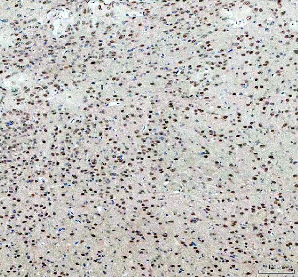
A07980-1 has IHC images from paraffin sections of human breast cancer and brain, and mouse and rat brain (catalog image captions). No IF data are supplied (catalog applications and images).
A07980-1 is listed for IHC in human, mouse and rat (catalog applications and reactivity). Its IHC captions document paraffin sections of human breast cancer and brain, mouse brain and rat brain (catalog image captions).
Which to pick: Choose A07980-1 for tissue IHC, including work across human, mouse and rat, because IHC is listed for all three species and its own captions document paraffin sections from each (catalog applications, reactivity and image captions). Start at 2 μg/ml with EDTA retrieval at pH 8.0 if reproducing the pictured IHC conditions; the fixative is unreported (A07980-1 image captions). No IF/ICC option is validated in this payload, and A07980-1 has no reported clone (catalog applications, IF images and clone field).