This website uses cookies to ensure you get the best experience on our website.


- Table of Contents
Plan chromogenic IHC for PRTN3 in paraffin sections with the catalog antibody at 2–5 μg/ml (datasheet A03863-3). Score cytoplasmic staining in immune cell subsets, especially bone marrow hematopoietic cells and splenic red pulp cells (HPA tissue IHC).

Expected localisation, validated protocols, controls and antibodies — the at-a-glance facts below, then the full design guide.
| Expected localisation | Cytoplasmic in immune cells (HPA tissue IHC) | |
| Staining pattern | Cytoplasmic immune cell subsets, most abundant in marrow and spleen (HPA tissue IHC) | |
| Antigen retrieval | EDTA pH 8.0 HIER, heat-mediated (datasheet A03863-3) | |
| Positive control | Bone marrow+1 more · see all | |
| Negative control | Adipose tissue+4 more · see all |
| Fixation | Keep fixation consistent across sections (standard IHC practice; not target-specific) | |
| Caveat | Secreted PRTN3 can separate protein from RNA location (HPA tissue IHC) | |
| Regulation | TNF or LPS stimulation prompts secretion (UniProt) | |
| Isoform / epitope | No listed isoforms; map epitopes to mature chain 28–248 (UniProt) |
The catalog antibody protocol uses EDTA pH 8.0 heat retrieval (datasheet A03863-3). The published IHC protocols below cover vulvar, gastric, and colorectal cancer specimens (PMC7793473; PMC11912076; PMC10835931).
| Sample | Paraffin-embedded human lung adenocarcinoma tissue; fixative not specified (datasheet A03863-3) |
| Fixation | Image fixative and duration unreported (datasheet A03863-3); verify before use. |
| Sectioning | 4–5 µm sections on charged slides (standard) |
| Deparaffinisation | Xylene, graded ethanol series to water (standard) |
| Antigen retrieval | Heat retrieval: EDTA pH 8.0 (datasheet A03863-3); 20 min, 95–100 °C (standard) |
| Peroxidase block | 3% H2O2, 10 min, room temperature (standard) |
| Blocking | 10% goat serum (datasheet A03863-3) |
| Primary antibody | Rabbit anti-PRTN3, 2-5 μg/ml (datasheet A03863-3) |
| Primary incubation | Overnight at 4 °C (datasheet A03863-3) |
| Detection | HRP-conjugated secondary, DAB chromogen (datasheet A03863-3) |
| Counterstain | Hematoxylin, blue, dehydrate and mount (standard) |
| Expected result | PRTN3-positive staining in hematopoietic cells of bone marrow (HPA tissue IHC: High). HPA tissue profile: Cytoplasmic expression in subsets of immune cells in several tissues, most abundant in bone marrow and spleen. No signal in the no-primary control. |
PRTN3 staining should be concentrated in subsets of immune cells, especially hematopoietic cells in bone marrow and cells in splenic red pulp (HPA: High; tissue IHC reliability Enhanced). Its expected pattern is cytoplasmic, consistent with predominant localization to neutrophil azurophil granules (HPA: cytoplasmic tissue expression; UniProt P24158: cytoplasmic granule). PRTN3 has no transmembrane segment, although secreted protein can be tethered to the neutrophil surface (UniProt P24158: topology and CD177 interaction).
| Strong cytoplasmic staining in a subset of bone marrow hematopoietic cells, with staining also seen in splenic red pulp cells (HPA: High in both sites). | This fits the tissue IHC pattern; assess the stained cells individually rather than treating either whole tissue as uniformly positive (HPA: cytoplasmic expression in subsets of immune cells). Granular cytoplasmic staining is biologically plausible because PRTN3 predominates in neutrophil azurophil granules (UniProt P24158: subcellular location). |
| Predominantly nuclear staining, with little cytoplasmic staining, in otherwise plausible positive cells. | A nuclear-dominant pattern does not match the reported localization (HPA: cytoplasmic tissue expression; UniProt P24158: cytoplasmic granule). Treat it as an unconfirmed staining pattern and check controls and detection conditions before scoring those cells as PRTN3 positive. |
| Strong staining of adipocytes or respiratory epithelial cells across a section. | These are poor matches for the reported cell distribution: adipocytes in adipose tissue and respiratory epithelial cells in bronchus were not detected (HPA: tissue IHC). Consider cross-reactivity or endogenous chromogenic detection activity; confirm whether nearby immune cells account for any localized signal. |
| Color deposits spread across cells and surrounding tissue without a clear cellular pattern. | Diffuse background cannot establish PRTN3 expression. It obscures the subset-restricted cytoplasmic pattern used for interpretation (HPA: tissue IHC). Compare with detection controls and inspect whether deposits track tissue edges or broad areas rather than individual cells. |
| No staining in bone marrow hematopoietic cells while the section otherwise appears interpretable. | This conflicts with a reported high-staining population (HPA: High in bone marrow hematopoietic cells). First check whether that population is present on the tested section, then assess the antibody and detection workflow; a blank slide alone cannot establish absent PRTN3. |
| Choice of IHC antibody | HPA005938 has IHC Enhanced validation; CAB017558 has IHC Supported validation (HPA: antibody records). HPA notes medium consistency between staining and RNA data and disregarded presumed off-target binding (HPA: reliability description), so use cell identity and compartment alongside stain intensity. |
| Granule storage and release | PRTN3 predominates in neutrophil azurophil granules and can be secreted after stimulation or during transmigration (UniProt P24158: subcellular location). Interpret focal extracellular or peripheral color cautiously; its presence does not by itself identify the producing cell. |
| Surface-associated signal | Secreted PRTN3 can bind CD177 at the neutrophil surface despite having no transmembrane segment (UniProt P24158: topology and CD177 interaction). A peripheral signal can therefore be plausible when cell identity and cytoplasmic staining also support the call. |
| Maturation and epitope interpretation | The annotated precursor has a signal peptide, propeptides and a mature myeloblastin chain (UniProt P24158: processing). No antibody epitope is supplied here, so this record cannot predict which processed form a given antibody recognizes. |
| IF/ICC Q: Should fluorescence be exclusively granular? | No. HPA reports approved cytosolic localization with additional vesicle localization in ICC-IF (HPA: subcellular summary); UniProt describes predominant neutrophil granule localization (UniProt P24158: subcellular location). Interpret fluorescence in its own cellular context rather than requiring every positive cell to show resolvable puncta. |
| Situation | Likely cause | Next action |
|---|---|---|
| Bone marrow positive control is blank. | The expected high-staining hematopoietic population may be absent from the examined area, or staining or detection may have failed (HPA: High in bone marrow hematopoietic cells). | Locate hematopoietic cells on the counterstained section, then review the IHC-validated antibody, antigen retrieval and detection steps as general workflow checks; do not infer a PRTN3-specific fixation effect. |
| The whole section has diffuse chromogenic haze. | Nonspecific background or endogenous detection activity may obscure the expected subset-restricted cytoplasmic signal (HPA: tissue IHC pattern). | Compare detection-only controls, review blocking and washing, and score only cells with a distinct interpretable pattern; these are general chromogenic IHC checks. |
| Many nuclei are strongly stained. | Nuclear-dominant signal conflicts with the reported cytoplasmic tissue pattern and granule location (HPA: tissue IHC; UniProt P24158: subcellular location). | Check a known-positive region, inspect counterstain and chromogen separately, and reconsider specificity before assigning nuclear staining to PRTN3. |
| Adipocytes or bronchial respiratory epithelial cells stain strongly. | Those cell types are listed as Not detected (HPA: adipose tissue and bronchus IHC); cross-reactivity or endogenous activity is possible. | Use appropriate detection controls and compare nearby immune cells; do not count a broadly stained negative cell type as confirmation of PRTN3. |
| Only splenic red pulp shows signal, with uncertain cell identity. | HPA reports high staining in red pulp cells but does not identify every stained cell as a neutrophil (HPA: spleen tissue IHC). | Use the counterstained morphology to identify individual cells and report the observed location conservatively; avoid assigning neutrophil identity from PRTN3 color alone. |
| Peripheral staining appears beside cytoplasmic signal. | Secreted PRTN3 can be tethered to neutrophil membranes by CD177 (UniProt P24158: secretion and CD177 interaction). | Check whether the signal outlines identifiable immune cells and whether a cytoplasmic subset remains visible; flag isolated extracellular deposits as uncertain rather than scoring them as cells. |
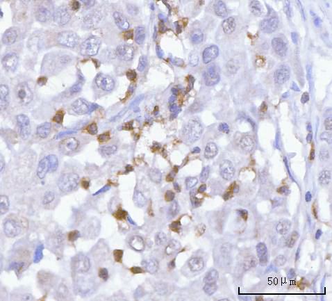
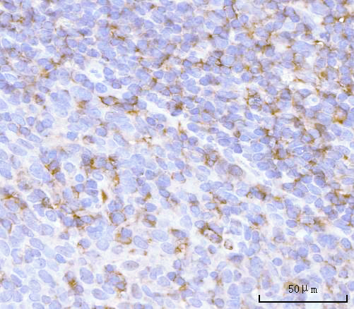


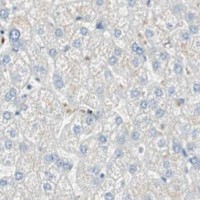


Comprehensive Human Protein Atlas IHC scoring per tissue (reliability: Enhanced — Medium consistency between antibody staining and RNA expression data. At least one protein variant secreted, tissue location of RNA and protein might differ and correlation is complex. Presumed off target binding observed and disregarded. Antibody staining in cells/structures not annotated, view images.). Rows are taken directly from the HPA tissue chart — click any row's HPA link to view the source.
| Tissue | Cell type | Level | Evidence | Source |
|---|---|---|---|---|
| Bone marrow | Hematopoietic cells | High | Protein (IHC) | HPA → |
| Spleen | Cells in red pulp | High | Protein (IHC) | HPA → |
| Tissue | Cell type | Level | Evidence | Source |
|---|---|---|---|---|
| Adipose tissue | Adipocytes | Not detected | Protein (IHC) | HPA → |
| Adrenal gland | Glandular cells | Not detected | Protein (IHC) | HPA → |
| Appendix | Glandular cells | Not detected | Protein (IHC) | HPA → |
| Breast | Adipocytes | Not detected | Protein (IHC) | HPA → |
| Bronchus | Respiratory epithelial cells | Not detected | Protein (IHC) | HPA → |
Troubleshoot chromogenic PRTN3 staining in paraffin sections using the catalog antibody’s tissue image and the expected distribution in immune cells.
Two human-reactive anti-PRTN3 antibodies list IHC use (catalog applications/reactivity). A03863-3 has human paraffin-section IHC images and catalog-listed IF/ICC use (A03863-3 image captions; catalog applications).
A03863-3 has IHC images from paraffin sections of human lung adenocarcinoma and tonsil, and is listed for IF/ICC without a supplied IF image (A03863-3 image captions; catalog applications and IF image alts). M03863-2 is listed for human IHC, with no supplied IHC image (M03863-2 catalog applications/reactivity and IHC image alts).
Which to pick: For tissue IHC, choose A03863-3: its own captions document human paraffin sections, EDTA retrieval at pH 8.0, and 2 μg/ml primary antibody; the fixative is unreported (A03863-3 IHC image captions). For IF/ICC, choose A03863-3 at the catalog-listed 5 μg/ml; M03863-2 is a rabbit monoclonal listed for human IHC but has no supplied IHC image (A03863-3 catalog applications/dilution; M03863-2 catalog clone/applications/reactivity and IHC image alts). Neither SKU has listed cross-species reactivity (catalog reactivity).