This website uses cookies to ensure you get the best experience on our website.
- Table of Contents
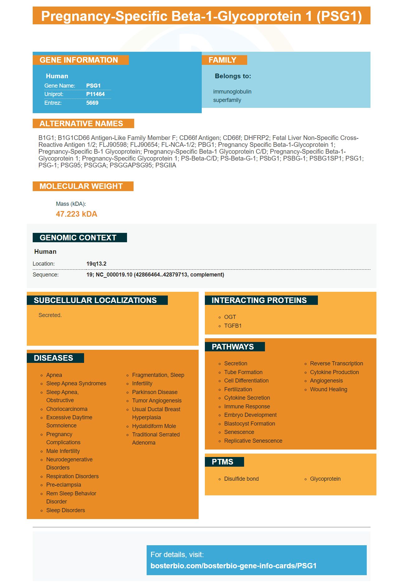
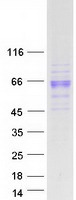
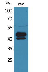
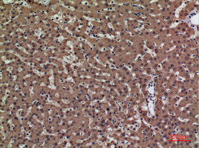
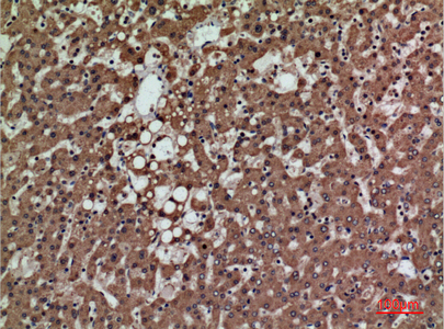
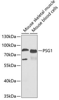
Facts about Pregnancy-specific beta-1-glycoprotein 1.
Human | |
---|---|
Gene Name: | PSG1 |
Uniprot: | P11464 |
Entrez: | 5669 |
Belongs to: |
---|
immunoglobulin superfamily |
B1G1; B1G1CD66 antigen-like family member F; CD66f antigen; CD66f; DHFRP2; Fetal liver non-specific cross-reactive antigen 1/2; FLJ90598; FLJ90654; FL-NCA-1/2; PBG1; pregnancy specific beta-1-glycoprotein 1; pregnancy-specific B-1 glycoprotein; Pregnancy-specific beta-1 glycoprotein C/D; pregnancy-specific beta-1-glycoprotein 1; Pregnancy-specific glycoprotein 1; PS-beta-C/D; PS-beta-G-1; PSbG1; PSBG-1; PSBG1SP1; PSG1; PSG-1; PSG95; PSGGA; PSGGAPSG95; PSGIIA
Mass (kDA):
47.223 kDA
Human | |
---|---|
Location: | 19q13.2 |
Sequence: | 19; NC_000019.10 (42866464..42879713, complement) |
Secreted.
Boster Bio's PSG1 Marker antibody detects recombinant Psg21/His. This antiserum has cross-reactivity with human TGFB1 as well as CD31 endothelial markers. This biomarker is versatile and can be used in immunohistochemistry as well as chemiluminescence.
The PSG1 antiserum detects recombinant Psg-21/His on Western Blets and produces a pattern in mouse placental tissue section staining. It failed to detect PSG endogenously in mouse serum, or placental lymph lysates. The anti-human PSG1 antibody, however, detected endogenous PSG within human placental sera and lysates.
The PSG1 genes are conserved in all mammals. It was isolated from many tissues. The gene encoding Psg21-V5/His has two distinct regions, the c-terminus and the apical domain. These regions are involved with cell proliferation and differentiation regulation. These regions were identified by the Psg antiserum.
The Psg gene is highly expressed in giant cells at E8 to E11. Its mRNA is abundant throughout the cytoplasm. It is expressed at high levels within trophoblast, placental and ectoplacental cells. The mRNA is pooled from six placental fetuses in each litter.
It is not known what role Psg has. Previous studies have shown that Psg is involved in maternal immune regulation. Immunochemical studies in mice have revealed that Psg proteins interact with various immune cells, indicating that the gene has multiple roles in the maternal immune system. The association of Psg and maternal vasculature could indicate that Psg plays a role in angiogenesis or vasoactivity.
Further research showed that anti–Psg1 detected recombinant Psg21 within the labyrinthine portion of the placenta. The presence CD9 was detected in both maternal and foetus tissues. It also stained spongiotrophoblasts and glycogen cells, but with significantly less intensity than the former.
Tumors may not be able to express PSGs despite this. Expression in normal GI tract tissues could suppress PSG expression. PSGs cannot be proven to be tumor markers, but their presence in biopsied tissue might indicate the progression of the disease or a prognosis.
Polyclonal antibodies contain a pool containing immunoglobulin molecules. These antibodies bind to several different epitopes of the target antigen. Polyclonal antibodies are often derived frequently from a variety o animals such as sheep, rabbits, and goats. Monoclonal antibodies only bind to a single epitope.
Positive controls are included in the detection of recombinant Psg21 on Western blots. These antibodies can identify the target protein and confirm antibody activity. To determine whether staining has been nonspecific, negative controls can be used. If this is true, the PSG1 Antiserum is ineffective.
Incomplete or undetectable bands on the blots is often the result of an improper transfer of the antigen. Inconsistencies in the gel's gel will result in uneven spots. To avoid uneven agitation and to identify bands, it is important to incubate the sample. Furthermore, washing is important to remove any contaminant antibodies.
PSG1 functions in the body are conserved between humans, mice, and other mammals. Its two domains regulate TGF-1b1, which allows for the modulation of TGF-1b1's expression. This effect may have important physiological functions in the maternal-fetal interface, including the establishment of a tolerogenic immune environment and the control of immune responses.
The study showed that removing cell surface glycosaminoglycans completely inhibits the binding of PSG1. The removal of GAGs could be achieved either through enzyme treatment or chemical manipulation, or competition with heparin. In vitro tests showed that PSG1 didn't bind to cells without chondroitin or heparan sulfate. However, binding was restored when cells were transfected with syndecans. Preeclampsia may therefore be treated with PSG1.
PSG17 also binds to the CD9 and cyclooxygenase-2 tetraspanin CD9 receptors and induces TGF beta1 secretion from a macrophage cell-line. Both PSG17 as well as PSG19 modify the receptor-binding domains with N-linked carbohydrate. CHO cells can detect the terminal sialic Acid in PSG17.
ALK1 and ALK5 control the mitogenic and paracrine reactions of ECs towards TGF-b. They are required for accessory receptor endoglin. The endoglin binds TGFb and enhances ALK1 cytostatic response and inhibits ALK5. They are also crucial for the development of blood vessel.
Moreover, the TGF-bs are implicated in a number of processes crucial for pregnancy. They regulate trophoblast invasion and proliferation, fetal semiallograft tolerability, and angiogenesis. Their roles in pregnancy will likely be further explored by future research. These antibodies can detect human TGFb in sandwich immunoassays.
PSG1 is highly reactive to human TGFB1. This antibody recognizes betaglycan, endoglin and Tgfbeta-beta-beta III beta-synucleins. These proteins are able to function in different contexts and can cause disparate responses to TGFbeta. This antibody is highly cross-reactive to human TGFB1 in different tissues.
The most studied member of TGF-b's family is the human TGFB1 protein. The precursor, pre-TGFB1, undergoes two proteolytic nucleavages. Proprotein convertase and proprotein (proprotein), cleaves in the rough reticulum the first signal protein. The mature TGF-1b1 fragment that remains is called LAP (latency-associated proteins). These domains are still connected by noncovalent links, which allows them both to associate with each others.
TGFb plays a dual purpose in tumor progression. A better prognosis is associated with higher levels of TGFb found in cancer cells. TGF-b regulates cell cycle, inhibits cMyc in the late G1 phase, promotes VEGF expression at ECs, and regulates cell cycle. TGFb is also associated VEGF, PDGF, bFGF changes.
Boster Bio's antiserum PSG1 is highly reactive with CD31 endothelial marker markers. Human PSG is not detected by PSG1 antiserum. PSG is a protein produced by blood endothelial cell. It is also found inside the placenta. It is produced by recombinant virus protein expression system.
The uterus expresses a wide range of human PSG1. One study showed that embryo transfer to CD9 deficient females resulted was comparable in terms of pregnancy rates. Data suggest that Psg does not play a significant role in the successful implantation and maintenance of human pregnancy. A surrogate Psg null mutation would be a useful surrogate for human embryos. It would also provide important information on Psg's role in pregnancy.
Anti-PSG1 antibody detects Psg presence in the decidua endothelial cell lining at the early stages. The fetal Psg gene does not exist, which suggests that Psg is secreted by fetal tissues and is associated with the maternal blood vessels. Psg is part of the immunoglobulin Superfamily. Eleven human PSG genes are located on chromosome 19q13.2 and seventeen mouse Psg family genes are on chromosome seven.
Sequential immunofluorescence assays are used to determine the Psg and CD31 antibodies. The procedure for immunofluorescence is similar to that of immunohistochemistry, but fluorescent secondary antibodies are used instead of the ABC system. After Psg staining, CD9 staining was performed with the anti-CD31 antibody at a dilution of 1:20. The anti-CD9 immunostaining is performed with the same washing and blocking steps.
Boster Bio's PSG1 antibody is highly reactive with CD31 endothelial cells markers
Boster Bio PSG1 has been used in an earlier study to determine its expression in trophoblast giant and spongiotrophoblast cells. PCR primers were used in order to amplify the N-domains of Psg genes from mouse mice. To minimize non-specific amplification, the primer concentration was optimized. 300 pmol was the optimal concentration for primers. The resulting amplicons could be subcloned into NovaBlue Singles or pSTblue-1 competent cells. Diagnostic PCR screening was used to confirm the existence of specific clones.
PMID: 3260773 by Streydio C., et al. The human pregnancy-specific beta 1-glycoprotein (PS beta G) and the carcinoembryonic antigen (CEA)-related proteins are members of the same multigene family.
PMID: 3180995 by Chan W.-Y., et al. Characterization of cDNA encoding human pregnancy-specific beta 1- glycoprotein from placenta and extraplacental tissues and their comparison with carcinoembryonic antigen.