PSIP1 / PC4 and SFRS1-interacting protein · IHC design guide

Design Immunohistochemistry for PSIP1

Plan chromogenic PSIP1 IHC in paraffin sections using the expected nuclear pattern (HPA tissue IHC). This guide covers tissue selection, fixation, detection, and interpretation of cell type and isoform differences (HPA tissue IHC; UniProt).

Evidence assembled Oct 2026 · For research use; verify linked source records and product datasheet before use
Immunohistochemistry protocol sheet for PSIP1 (IHC for PSIP1): expected localisation Nuclear (HPA tissue IHC), antibody A01960-2, validated IHC image, and IHC protocol steps
Printable PSIP1 IHC protocol sheet — expected localisation Nuclear (HPA tissue IHC), antibody A01960-2, controls and protocol steps. Open the full PSIP1 IHC guide →

PSIP1 Immunohistochemistry Experimental Design Guide

Expected localisation, validated protocols, controls and antibodies — the at-a-glance facts below, then the full design guide.

Must know before staining
Expected localisation Nuclear (HPA tissue IHC)
Staining pattern General nuclear staining across tissue cells (HPA tissue IHC)
Antigen retrieval EDTA pH 8.0 HIER, heat-mediated (datasheet A01960-2)
Positive control ⓘ Appendix+4 more · see all
Negative control ⓘ None in HPA (detected in all 45 tissues); use no-primary + isotype controls
Important caveats
Reasons your staining may differ from the expected pattern.
Fixation Keep fixation consistent across sections (standard IHC practice; not target-specific). Selected-image fixative and duration unreported (datasheet A01960-2); verify before use.
Caveat Brain cell patterns differ: HPA glia versus UniProt neurons (HPA tissue IHC; UniProt)
Regulation Higher in fetal than adult brain (UniProt)
Isoform / epitope 3 isoforms; epitope coverage is unspecified (UniProt)
Section 1

Recommended PSIP1 IHC & IF Protocols

The catalog antibody’s IHC-P protocol uses EDTA pH 8.0 retrieval (datasheet A01960-2). A published cervical biopsy protocol provides a citrate-based comparison (PMC3946365).

Recommended immunohistochemistry (IHC-P) protocol parameters
SampleParaffin-embedded human breast cancer tissue; fixative not specified (datasheet A01960-2)
FixationImage fixative and duration unreported (datasheet A01960-2); verify before use.
Sectioning4–5 µm sections on charged slides (standard)
DeparaffinisationXylene, graded ethanol series to water (standard)
Antigen retrievalHeat retrieval: EDTA pH 8.0 (datasheet A01960-2); 20 min, 95–100 °C (standard)
Peroxidase block3% H2O2, 10 min, room temperature (standard)
Blocking10% goat serum (datasheet A01960-2)
Primary antibodyRabbit anti-PSIP1, 2-5 μg/ml (datasheet A01960-2)
Primary incubationOvernight at 4 °C (datasheet A01960-2)
DetectionHRP-conjugated secondary, DAB chromogen (datasheet A01960-2)
CounterstainHematoxylin, blue, dehydrate and mount (standard)
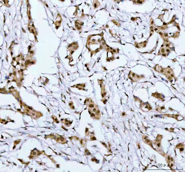
Expected resultPSIP1-positive staining in endocrine cells of appendix (HPA tissue IHC: High). HPA tissue profile: General nuclear expression. No signal in the no-primary control.
💡Decision noteStart with EDTA pH 8.0 for the catalog antibody (datasheet A01960-2). Consider citrate pH 6.0 when adapting the published method (PMC3946365).
Section 2

What Is the Expected PSIP1 Staining Pattern?

PSIP1 is nuclear and remains associated with chromatin throughout the cell cycle (UniProt O75475). In paraffin section IHC, expect predominantly nuclear staining across tissues, with high staining reported in selected cell populations; HPA rates its tissue pattern Enhanced for consistency with RNA expression (HPA tissue IHC). PSIP1 has no transmembrane segment (UniProt O75475 topology). Interpret the nucleus within each identified cell, since staining intensity and cell distribution vary by tissue (HPA tissue IHC).

What am I looking at on my slide?
Distinct nuclear chromogen in glandular cells of endometrium or squamous epithelial cells of esophagus (HPA tissue IHC).This matches HPA's High staining in those cell populations and PSIP1's nuclear location (HPA tissue IHC; UniProt O75475). Compare nuclei with nearby background before scoring intensity; a High HPA category does not require every cell in a section to stain equally (HPA tissue IHC).
Predominantly cytoplasmic or membrane staining, with little nuclear signal.That compartment conflicts with the reported nuclear pattern and nucleoplasmic ICC-IF location (UniProt O75475; HPA subcellular). Consider nonspecific detection or a technical artefact; repeat with a known positive tissue and detection controls before treating the pattern as PSIP1.
Strong staining in a cell population outside the reported High populations, while expected nuclei are faint (HPA tissue IHC).Cross-reactivity or endogenous detection activity is possible under standard IHC practice. Identify the cells and check controls before calling them positive. HPA reports general nuclear expression, so an unlisted population alone is insufficient evidence of a false positive (HPA tissue IHC).
Diffuse chromogen over cells, stroma and empty areas, obscuring nuclear boundaries.Treat this as background under standard IHC practice: it cannot establish PSIP1 localization. Inspect the reagent omission control and adjust blocking, washing or detection conditions; preserve enough counterstain to identify nuclei and the cell populations being scored.
No nuclear signal in a section containing HPA High cell populations (HPA tissue IHC).First suspect an assay or specimen problem under standard IHC practice, then confirm cell identity and the expected population. HPA High describes observed staining, not a guarantee for every preparation (HPA tissue IHC); a single negative section cannot establish absence of PSIP1.
💡Expected PSIP1 appearanceCall positive when identifiable nuclei stain clearly, especially in HPA High cell populations, against low surrounding background; dominant cytoplasmic staining or chromogen outside cells is suspect (HPA tissue IHC; UniProt O75475; standard IHC practice).
How each factor affects the staining
Nuclear residencePSIP1 remains chromatin-associated throughout the cell cycle (UniProt O75475). Use nuclear boundaries and a readable counterstain to judge localization; extranuclear color by itself does not match the reported target location (UniProt O75475).
Tissue and cell contextHPA describes general nuclear expression and low tissue specificity, with High staining in specified endocrine, glandular, squamous epithelial, glial and cerebellar granular-layer cells (HPA tissue IHC). Score identified cell populations rather than treating the whole section as uniformly positive.
Brain cell interpretationHPA reports High staining in glial cells of caudate and cerebral cortex (HPA tissue IHC). UniProt says PSIP1 is expressed in neurons but not astrocytes and describes restricted adult brain expression (UniProt O75475). These statements differ in scope; identify the glial population before using either account to reject a result.
Antibody evidenceHPA lists IHC Supported for HPA019697 and IHC Enhanced for CAB013718; it lists ICC Enhanced for HPA019697 (HPA antibodies). Those ratings apply to the named antibodies and applications. They do not establish the performance of an unspecified catalog antibody in this section.
Isoforms and epitopeUniProt lists three PSIP1 isoforms and a PWWP domain at residues 1–64 (UniProt O75475). The supplied sources do not locate the IHC antibody epitope or establish isoform-specific staining. Do not interpret a staining difference as an isoform difference without antibody-specific evidence.
IF/ICC Q&AQ: What localization should IF/ICC show? A: Nucleoplasmic staining is the HPA ICC-IF result; its image set includes A-431, U-251MG and U2OS (HPA subcellular). This supports a localization check, not an IF/ICC protocol or an IHC intensity benchmark.
Why is my staining missing, weak or wrong?
SituationLikely causeNext action
Known HPA High cells show no nuclear chromogen (HPA tissue IHC).Target-specific fixation effects are not established by the supplied assay evidence. Verify with a matched IHC source before attributing a result to fixation.Confirm morphology and cell identity, then check a positive control and the staining run. Review the antibody's IHC-P instructions and retrieval conditions as general workflow checks; do not infer a PSIP1-specific retrieval requirement.
Cytoplasm or cell borders stain more strongly than nuclei.This conflicts with nuclear PSIP1 and nucleoplasmic ICC-IF localization (UniProt O75475; HPA subcellular). Nonspecific binding or detection background is possible under standard IHC practice.Inspect a reagent omission control, reduce background through validated blocking and washing adjustments, and reassess the same cell populations for a distinct nuclear signal.
Unexpected cell types appear strongly positive.Cross-reactivity or endogenous detection activity is possible under standard IHC practice; HPA also reports general nuclear expression, so an unlisted cell type is not automatically negative (HPA tissue IHC).Verify cell identity and localization, compare the reported HPA populations, and use appropriate detection controls before assigning the signal to PSIP1.
Diffuse color hides nuclei throughout the section.Broad background can obscure the compartment needed to judge this nuclear target (UniProt O75475; standard IHC practice). The supplied sources do not identify a PSIP1-specific cause.Check the reagent omission control, washing and detection settings; repeat with readable counterstain and background so nuclear boundaries can be assessed.
Brain glial staining seems inconsistent with the UniProt neuron description.HPA reports High glial staining in caudate and cerebral cortex, whereas UniProt states expression in neurons but not astrocytes (HPA tissue IHC; UniProt O75475). 'Glial cells' does not identify an astrocyte subtype.Record the anatomical region and cell morphology, and avoid equating all glia with astrocytes. Treat the source difference as unresolved for that cell subtype without further identification.
Nuclear staining appears widespread but varies among cells.HPA describes general nuclear expression and low tissue specificity while assigning High staining to particular populations (HPA tissue IHC). Uneven intensity alone does not prove an assay failure.Score nuclei within identified populations, document intensity and background, and compare like cells across sections before interpreting variation as a biological difference.

Sample controls for PSIP1 IHC & IF

🧪Run appendix first and score its endocrine cells, which show High PSIP1 staining (HPA: High in appendix endocrine cells). HPA detects PSIP1 in all 45 scored tissues, so there is no supported negative tissue; use no-primary and isotype controls, and regard counterstain-only cells on the positive slide as internal background references only after validating their identity, without assuming another appendix cell type lacks PSIP1 (HPA: no negative rows; detected in all 45 tissues).
Positive control tissue: Appendix (Endocrine cells, HPA High)
Negative control tissue: None in HPA: PSIP1 is detected in all 45 scored tissues. Use a no-primary (secondary-only) and an isotype control instead.
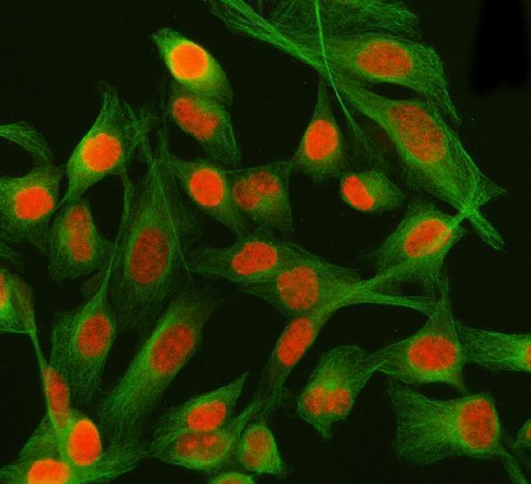
ICC-IF cell lines (HPA subcellular resource): HPA ICC-IF images show PSIP1 in A-431, U-251MG, U2OS, U2OS, siRNA 1 (10x), U2OS, siRNA 1 (40x), U2OS, scrambled (10x), U2OS, scrambled (40x), with annotated localisation: Nucleoplasm (enhanced) (HPA subcellular).
Technical controls: Include a no-primary, secondary-only control, a concentration-matched rabbit IgG isotype control, and PSIP1 knockout material as a biological negative (selected-SKU caption: rabbit primary; standard IHC practice). For appendix sections developed with HRP/DAB, quench endogenous peroxidase and inspect control sections for residual signal (selected-SKU caption: HRP/DAB; standard IHC practice).
⚠️Feasibility: No target-specific fixation window or fixation effect is reported, and the selected A01960-2 paraffin-section caption does not state the fixative (selected-SKU caption: fixative unreported). The caption reports heat retrieval in EDTA at pH 8.0, but does not establish that PSIP1 staining requires that retrieval condition (selected-SKU caption: EDTA pH 8.0). Frozen-section practicality is unreported; ICC-IF images exist in U2OS, but the supplied evidence does not show that IF is easier than paraffin IHC, and endogenous peroxidase in appendix inflammatory cells can complicate HRP/DAB interpretation (HPA: U2OS ICC-IF images; selected-SKU caption: HRP/DAB; standard IHC practice).

HPA tissue IHC evidence for PSIP1

Comprehensive Human Protein Atlas IHC scoring per tissue (reliability: Enhanced — High consistency between antibody staining and RNA expression data.). Rows are taken directly from the HPA tissue chart — click any row's HPA link to view the source.

Positive expression · recommended positive controls

TissueCell typeLevelEvidenceSource
Appendix Endocrine cells High Protein (IHC) HPA →
Caudate Glial cells High Protein (IHC) HPA →
Cerebellum Cells in granular layer High Protein (IHC) HPA →
Cerebral cortex Glial cells High Protein (IHC) HPA →
Colon Endocrine cells High Protein (IHC) HPA →

Undetected expression · recommended negative controls

TissueCell typeLevelEvidenceSource
None in HPA: PSIP1 is detected in all 45 scored tissues. Use a no-primary (secondary-only) and an isotype control instead.
Section 3

Advanced PSIP1 IHC Tips

Troubleshoot PSIP1 staining in paraffin sections by checking retrieval, nuclear localisation, antibody specificity and cell type before comparing chromogenic signals.

What retrieval should I try first when PSIP1 nuclear staining is weak?
Start with heat-mediated retrieval in EDTA, pH 8.0 (datasheet A01960-2). The catalog antibody detected PSIP1 in a paraffin-embedded section after this retrieval, followed by 2 μg/ml primary antibody overnight at 4°C (datasheet A01960-2). Compare retrieval conditions on adjacent sections while holding detection and imaging conditions constant (standard IHC practice). If staining remains weak, test an alternative retrieval condition as a fallback and compare nuclear signal with tissue damage (standard IHC practice). Judge improvement by nuclear staining and preserved morphology, since PSIP1 is nuclear and remains chromatin-associated throughout the cell cycle (UniProt O75475).
Could fixation explain weak PSIP1 staining in paraffin sections?
PSIP1-specific fixation sensitivity is unknown because the selected tissue caption reports a paraffin-embedded section but does not state its fixative (datasheet A01960-2). Record each specimen’s actual fixative and fixation history before comparing staining across blocks (standard IHC practice). First reproduce the documented EDTA, pH 8.0 retrieval and 2 μg/ml overnight primary incubation on a suitable control section (datasheet A01960-2). Then compare sections with known processing histories under the same staining conditions, assessing nuclear signal alongside morphology (standard IHC practice; UniProt O75475 localisation). Do not assign a PSIP1-specific fixation effect from those comparisons without a controlled fixation experiment (standard IHC practice).
How should I assess cytoplasmic or uneven PSIP1 staining?
Expect predominantly nuclear staining: PSIP1 remains chromatin-associated throughout the cell cycle, and the HPA tissue profile reports general nuclear expression (UniProt O75475; HPA tissue IHC). HPA subcellular images further localise the signal to the nucleoplasm (HPA subcellular). Compare DAB deposition with a nuclear counterstain across intact cells, including the EDTA, pH 8.0 retrieved control section (standard IHC practice; datasheet A01960-2). Treat diffuse cytoplasmic colour, section-edge staining and signal over damaged areas as reasons to review background and morphology (standard IHC practice). If a non-nuclear pattern persists, seek independent antibody or orthogonal evidence before assigning it to PSIP1 (standard IHC practice).
Can this antibody distinguish PSIP1 isoforms or modified epitopes in tissue?
PSIP1 has 3 annotated isoforms and a PWWP domain at residues 1–64 (UniProt O75475). The supplied catalog caption documents tissue staining but does not identify the antibody epitope or establish isoform selectivity (datasheet A01960-2). UniProt lists modified residues, including phosphoserines at 102, 105 and 106; their effects on this antibody’s tissue staining are unknown (UniProt O75475; datasheet A01960-2). Use the documented EDTA, pH 8.0 retrieval as the starting condition when comparing specimens (datasheet A01960-2). For isoform-specific or modification-specific conclusions, obtain epitope mapping and independent validation before interpreting differences in DAB intensity (standard IHC practice).
How can IF help check a questionable PSIP1 IHC pattern?
Use IF as an independent localisation check, with a nuclear counterstain and a marker for the cell type under study (standard IF practice). This matters in brain: UniProt reports neuronal expression but not astrocyte expression, whereas HPA tissue IHC lists high staining in glial cells in several regions (UniProt O75475; HPA tissue IHC). Choose fluorophores whose signal can be separated from tissue autofluorescence, and include single-channel and no-primary controls (standard IF practice). PSIP1 lacks a transmembrane segment and is nuclear, so permeabilisation must allow antibody access to the nuclear epitope (UniProt O75475; standard IF practice). Compare IF localisation with IHC nuclei, without treating the IHC caption’s 2 μg/ml condition as an IF-validated dilution (datasheet A01960-2).
What should I check when PSIP1 DAB staining looks widespread?
First separate plausible widespread nuclear staining from unwanted colour: PSIP1 is widely expressed, and HPA describes general nuclear tissue staining (UniProt O75475; HPA tissue IHC). Run a no-primary control and review endogenous peroxidase blocking, reagent carryover and DAB development on adjacent sections (standard IHC practice). The selected caption used 10% goat serum block and an HRP-linked secondary incubated for 30 minutes at 37°C (datasheet A01960-2). Compare intact nuclei with acellular deposits and section edges under the same development conditions (standard IHC practice). If background rises with primary concentration, titrate around the documented 2 μg/ml condition while preserving a positive control (datasheet A01960-2; standard IHC practice).
How should I quantify PSIP1 staining across paraffin sections? ⚠ ANSWER MARKED FOR VERIFICATION
Define the scored compartment as the nucleus before measuring PSIP1, consistent with UniProt and HPA localisation (UniProt O75475; HPA tissue IHC). For a cell-based readout, record the percentage of positive nuclei and staining intensity, then calculate an H-score with the same thresholds across sections (standard IHC practice). If cell density is the question, report positive nuclei per mm² of viable tissue and record the total nuclei or tissue area used for normalisation (standard IHC practice). Keep retrieval at EDTA, pH 8.0 and use matched staining batches when comparing samples (datasheet A01960-2; standard IHC practice). Exclude folds, necrosis and edges by predefined rules (standard IHC practice).
When is a PSIP1-positive IHC pattern convincing rather than artefactual?
A convincing result places signal in intact nuclei, matching PSIP1’s chromatin association and HPA’s general nuclear pattern (UniProt O75475; HPA tissue IHC). Check whether the positive cells fit the sampled tissue and any cell-type markers; UniProt’s neuronal account and HPA’s reported glial staining warrant particular care in brain (UniProt O75475; HPA tissue IHC). Review adjacent morphology for section-edge effects and necrosis, and use a no-primary control to assess endogenous enzyme or detection-system colour (standard IHC practice). The documented paraffin-section example used EDTA, pH 8.0 retrieval and DAB detection (datasheet A01960-2). For an unexpected compartment or cell population, require reproducibility and independent evidence before calling it PSIP1 (standard IHC practice).
Boster reagents

Best PSIP1 / PC4 and SFRS1-interacting protein IHC Antibodies

A01960-2 has pictured IHC in human paraffin sections and IF in HELA cells (catalog image captions). Both SKUs list human IHC/IF; A01960-2 also lists mouse and rat reactivity (catalog).

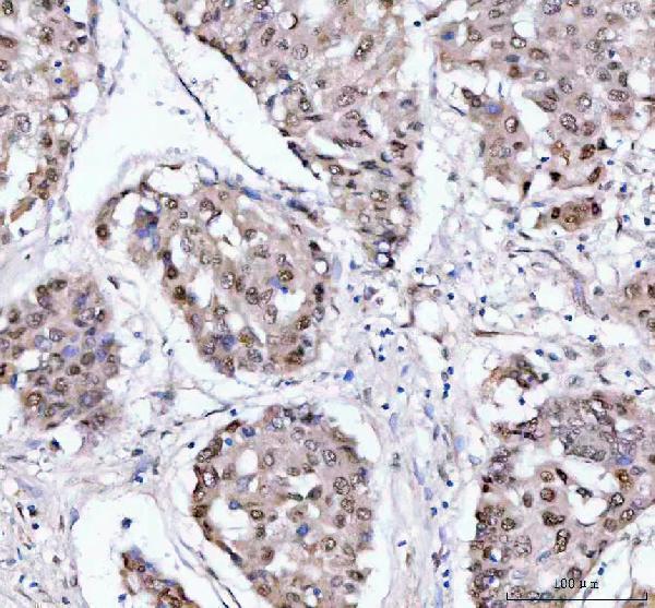
Real IHC data IHC analysis of PSIP1 using anti-PSIP1 antibody (A01960-2). PSIP1 was detected in a paraffin-embedded section of human breast cancer tissue. Heat mediated antigen retrieval was performed in EDTA buffer (pH 8.0, epitope retrieval solution). The tissue section was blocked with 10% goat serum. The tissue section was then incubated with 2 μg/ml rabbit anti-PSIP1 Antibody (A01960-2) overnight at 4°C. Peroxidase Conjugated Goat Anti-rabbit IgG was used as secondary antibody and incubated for 30 minutes at 37°C. The tissue section was developed using HRP Conjugated Rabbit IgG Super Vision Assay Kit (Catalog # SV0002) with DAB as the chromogen.
Anti-PSIP1 Antibody ®
Cat # A01960-2

A01960-2 lists human, mouse and rat reactivity and has IHC images from human breast cancer, liver cancer, ovarian cancer and spleen paraffin sections, plus IF in HELA cells (catalog reactivity and image captions). M01960 lists human IHC and IF/ICC applications but has no sample images in the payload (catalog applications and image captions).

Which to pick: Choose A01960-2 for tissue IHC: its breast cancer paraffin-section caption reports EDTA retrieval at pH 8.0 and 2 μg/ml primary antibody; the fixative is unreported (A01960-2 IHC caption). Choose A01960-2 for IF/ICC when pictured cell data matter: its HELA IF caption uses 5 μg/ml, while M01960 is a human-reactive rabbit monoclonal with listed IF/ICC use but no IF image (A01960-2 IF caption; M01960 catalog). For mouse or rat samples, choose A01960-2 based on its listed reactivity; M01960 lists human reactivity only (catalog reactivity).

Each figure is that product's own IHC / IF validation image from its datasheet.

References

  1. UniProt Consortium. UniProt entry O75475 (PSIP1_HUMAN, PC4 and SFRS1-interacting protein).
  2. Human Protein Atlas. PSIP1 tissue IHC expression (reliability: Enhanced).
  3. Human Protein Atlas. PSIP1 subcellular location (ICC-IF): Localized to the nucleoplasm..
  4. Human Protein Atlas. PSIP1 antibody validation summary (2 antibodies).
  5. A preliminary bioinformatic screen to identify SRI SMC2 PSIP1 TLE4 and MSX1 as potential diagnostic and prognostic markers of osteoarthritis. Scientific reports 2025 — PMC12368170.
  6. Oncogenic human papillomaviruses activate the tumor-associated lens epithelial-derived growth factor (LEDGF) gene. PLoS pathogens 2014 — PMC3946365.
  7. Absence of Ledgf in mouse brain affects the Kmt2a/b and polycomb balance, synaptic transmission and motor function. Acta neuropathologica communications 2026 — PMC12910792.
  8. Integration of transcriptomics, proteomics, and microRNA analyses reveals novel microRNA regulation of targets in the mammalian inner ear. PloS one 2011 — PMC3071727.
  9. PubMed PMID:9822615 — UniProt-cited evidence.
  10. PubMed PMID:10623627 — UniProt-cited evidence.
  11. PubMed PMID:10721720 — UniProt-cited evidence.