This website uses cookies to ensure you get the best experience on our website.


- Table of Contents
Plan PSMB5 paraffin IHC around nuclear staining that varies across tissues (HPA tissue IHC). Use the catalog antibody’s documented IHC conditions (datasheet A03418-1) and include controls because staining has low consistency with RNA expression (HPA tissue IHC).

Expected localisation, validated protocols, controls and antibodies — the at-a-glance facts below, then the full design guide.
| Expected localisation | Nuclear staining in tissues (HPA tissue IHC); cytoplasm also annotated (UniProt) | |
| Staining pattern | Nuclear staining in several tissues at varied intensity (HPA tissue IHC) | |
| Antigen retrieval | EDTA pH 8.0 HIER, heat-mediated (datasheet A03418-1) | |
| Positive control | Cerebellum+4 more · see all | |
| Negative control | Adipose tissue+4 more · see all |
| Fixation | Keep fixation consistent across samples (standard IHC practice; not target-specific) | |
| Caveat | Staining has low consistency with RNA expression (HPA tissue IHC) | |
| Regulation | No specific expression regulator reported (UniProt) | |
| Isoform / epitope | 3 isoforms; propeptide 1–59 is removed; check epitope (UniProt) |
Compare the catalog antibody’s IHC-P protocol (datasheet A03418-1) with published PSMB5 staining of piglet brain (PMC10925635) and HCC tissues (PMC9623531).
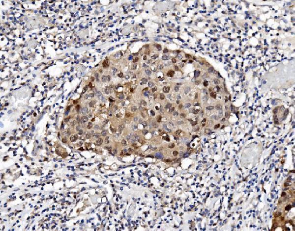
| Sample | Paraffin-embedded human breast cancer tissue; fixative not specified (datasheet A03418-1) |
| Fixation | Image fixative and duration unreported (datasheet A03418-1); verify before use. |
| Sectioning | 4–5 µm sections on charged slides (standard) |
| Deparaffinisation | Xylene, graded ethanol series to water (standard) |
| Antigen retrieval | Heat retrieval: EDTA pH 8.0 (datasheet A03418-1); 20 min, 95–100 °C (standard) |
| Peroxidase block | 3% H2O2, 10 min, room temperature (standard) |
| Blocking | 10% goat serum (datasheet A03418-1) |
| Primary antibody | Rabbit anti-PSMB5, 2-5μg/ml (datasheet A03418-1) |
| Primary incubation | Overnight at 4 °C (datasheet A03418-1) |
| Detection | Streptavidin-biotin complex (SABC), DAB chromogen (datasheet A03418-1) |
| Counterstain | Hematoxylin, blue, dehydrate and mount (standard) |
| Expected result | PSMB5-positive staining in cells in granular layer of cerebellum (HPA tissue IHC: High). HPA tissue profile: Nuclear expression in several tissues with various levels of intensity. No signal in the no-primary control. |
In paraffin section IHC, expect PSMB5 mainly in nuclei, with intensity varying by tissue and cell type (HPA: nuclear expression in several tissues). Granular layer cells in cerebellum and Sertoli cells in testis show high staining (HPA: High in both). Cytoplasmic staining is biologically plausible (UniProt P28074: cytoplasm and nucleus; no transmembrane segment). Interpret the pattern cautiously: HPA rates tissue IHC Approved but reports low consistency with RNA expression and pending external verification (HPA: reliability description).
| Strong nuclear staining in cerebellar granular layer cells or testicular Sertoli cells. | This fits the reported high staining in those specific cells (HPA: High in cerebellar granular layer cells; High in Sertoli cells). Compare the expected cell population, compartment and nearby unstained areas before scoring a section positive (general IHC practice). |
| Predominant staining at cell borders, in extracellular material or as an isolated membrane rim. | This conflicts with the reported nuclear tissue pattern and intracellular localization (HPA: nuclear expression; UniProt P28074: cytoplasm and nucleus, no transmembrane segment). Treat a strong border or extracellular pattern as a possible artefact; cytoplasmic signal alone is not automatically wrong (UniProt P28074: cytoplasm). |
| Strong staining in adipocytes or bone marrow hematopoietic cells while expected positive cells stain weakly. | These cell populations are reported as not detected (HPA: adipocytes and bone marrow hematopoietic cells). Consider antibody cross-reactivity or endogenous chromogenic activity, and compare an appropriate negative detection control (general IHC practice). An HPA result for one cell population does not make every cell in that tissue negative. |
| Diffuse color across many cells and tissue spaces, with little nuclear contrast. | A widespread, poorly localized deposit is hard to reconcile with the reported nuclear tissue profile (HPA: nuclear expression in several tissues). Excess detection background, inadequate blocking or insufficient washing can produce this appearance (general IHC practice); evaluate it against a control lacking primary antibody. |
| No signal in cerebellar granular layer cells or testicular Sertoli cells. | Both are reported high staining sites, so a blank result there warrants a technical check (HPA: High in both cell populations). Check section integrity, retrieval, antibody dilution and detection controls as general IHC practice. HPA's Approved tissue rating still carries a low RNA concordance caveat and awaits external verification (HPA: reliability description). |
| Tissue and cell selection | Use the named cell population when judging signal: granular layer cells and Sertoli cells are High, whereas adipocytes are Not detected (HPA: tissue IHC). Low tissue RNA specificity does not predict equal staining in every cell (HPA: low tissue specificity). |
| Location and proteasome biology | UniProt places PSMB5 in cytoplasm and nucleus and describes nuclear import following AKIRIN2 interaction (UniProt P28074: subcellular location). HPA tissue images are summarized as nuclear, so score nuclear and cytoplasmic compartments separately (HPA: tissue IHC profile). |
| Antibody evidence | One listed antibody is IHC Approved; its ICC evidence is Supported (HPA: HPA049518). HPA also reports low consistency between tissue staining and RNA expression, pending external verification (HPA: reliability description). Confirm an unexpected pattern independently before interpreting it as biology (general IHC practice). |
| Processing and isoforms | The precursor has a 1–59 propeptide, a 60–263 mature chain and three annotated isoforms (UniProt P28074: processing; isoforms). Antibody epitope coverage is unspecified here, so these annotations alone cannot explain a weak stain or an isoform-specific pattern. |
| IF/ICC Q&A: should its pattern match IHC exactly? | No exact match is established: ICC-IF is mainly nucleoplasmic, with additional centrosome, sperm mid-piece and principal-piece localization (HPA: subcellular ICC-IF). Tissue IHC is summarized as nuclear (HPA: tissue IHC profile); assess each application against its own evidence. |
| Situation | Likely cause | Next action |
|---|---|---|
| Known-positive cells are blank. | The reported high-signal cells may have been missed, or a routine IHC step may have failed (HPA: High in cerebellar granular layer cells and Sertoli cells; general IHC practice). | Verify cell identity and section quality, then review retrieval, antibody dilution and detection controls (general IHC practice). No PSMB5-specific retrieval condition or fixation sensitivity is established by the supplied sources. |
| Color is strongest at tissue edges or in empty spaces. | A deposit outside cells does not fit the intracellular locations reported for PSMB5 (UniProt P28074: cytoplasm and nucleus). Uneven reagent coverage or chromogen deposition is a general IHC possibility. | Check reagent coverage, washes and the primary-antibody-omission control; reassess staining within intact cells before assigning a PSMB5 score (general IHC practice). |
| Unexpected cells stain more strongly than expected positive cells. | Cross-reactivity or endogenous detection activity is possible (general IHC practice), especially when the unexpected population is reported Not detected, such as adipocytes (HPA: adipocytes Not detected). | Compare a control lacking primary antibody and, if needed, an independently validated antibody (general IHC practice). Record the exact cell type rather than calling the whole tissue positive or negative. |
| Nuclear and cytoplasmic signals disagree between samples. | Both compartments are annotated, while HPA summarizes tissue IHC as nuclear; compartment balance need not be identical in every observation (UniProt P28074: cytoplasm and nucleus; HPA: nuclear tissue profile). | Score nuclear and cytoplasmic staining separately and compare the same cell type across sections (general IHC practice). Review strong border-only staining as atypical (UniProt P28074: no transmembrane segment). |
| Most of the section has diffuse chromogenic background. | Blocking, washing or endogenous detection activity can obscure localized staining (general IHC practice). HPA's nuclear profile cannot by itself identify which workflow step caused background (HPA: tissue IHC profile). | Inspect the primary-antibody-omission control, then review blocking, washes, detection reagents and chromogen development (general IHC practice). Judge positivity only where cellular boundaries and nuclear contrast remain interpretable. |
| IHC and IF/ICC show different additional structures. | ICC-IF lists centrosome and sperm-piece locations in addition to nucleoplasm, whereas the tissue IHC summary emphasizes nuclei (HPA: subcellular ICC-IF; tissue IHC profile). | Compare each result with evidence for its own application and cell type; do not use an ICC-IF feature alone to score a paraffin section positive (HPA: subcellular ICC-IF; tissue IHC profile). |


Comprehensive Human Protein Atlas IHC scoring per tissue (reliability: Approved — Low consistency between antibody staining and RNA expression data. Pending external verification.). Rows are taken directly from the HPA tissue chart — click any row's HPA link to view the source.
| Tissue | Cell type | Level | Evidence | Source |
|---|---|---|---|---|
| Cerebellum | Cells in granular layer | High | Protein (IHC) | HPA → |
| Testis | Sertoli cells | High | Protein (IHC) | HPA → |
| Adrenal gland | Glandular cells | Medium | Protein (IHC) | HPA → |
| Caudate | Neuronal cells | Medium | Protein (IHC) | HPA → |
| Cerebral cortex | Neuronal cells | Medium | Protein (IHC) | HPA → |
| Tissue | Cell type | Level | Evidence | Source |
|---|---|---|---|---|
| Adipose tissue | Adipocytes | Not detected | Protein (IHC) | HPA → |
| Appendix | Glandular cells | Not detected | Protein (IHC) | HPA → |
| Bone marrow | Hematopoietic cells | Not detected | Protein (IHC) | HPA → |
| Bronchus | Basal cells | Not detected | Protein (IHC) | HPA → |
| Cervix | Glandular cells | Not detected | Protein (IHC) | HPA → |
Troubleshoot PSMB5 staining in paraffin sections by checking retrieval, controls, compartment-specific signal, and reproducible scoring (datasheet A03418-1; UniProt P28074 localisation).
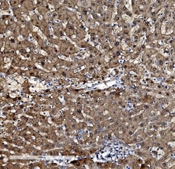
A03418-1 has real IHC data from human paraffin sections and IF data from MCF-7 cells (catalog image captions). Its listed reactivity is human, mouse and rat (catalog: reactivity).
A03418-1 is listed for IHC and IF/ICC (catalog: applications). Its images show IHC in human breast cancer, liver cancer, lymphoma and ovarian serous adenocarcinoma paraffin sections, plus IF in MCF-7 cells (catalog image captions).
Which to pick: For tissue IHC, choose A03418-1: its own IHC captions show staining in human paraffin sections; the fixative is unreported (catalog IHC image captions). For IF/ICC, the same SKU has an MCF-7 IF image and lists both applications (catalog IF image caption; catalog: applications). It lists human, mouse and rat reactivity, but the supplied IHC and IF images demonstrate human samples only (catalog: reactivity; catalog image captions).