PSMB9 / Proteasome subunit beta type-9 · IHC design guide

Design Immunohistochemistry for PSMB9

Plan PSMB9 chromogenic IHC in paraffin sections using the catalog antibody at 2–5 μg/ml (datasheet: A02867-2). Compare nuclear and cytoplasmic staining across cell types, with leukocytes as a strong reference population (HPA tissue IHC).

Evidence assembled Oct 2026 · For research use; verify linked source records and product datasheet before use
Immunohistochemistry protocol sheet for PSMB9 (IHC for PSMB9): expected localisation Nuclear and cytoplasmic tissue staining (HPA tissue IHC), antibody A02867-2, validated IHC image, and IHC protocol steps
Printable PSMB9 IHC protocol sheet — expected localisation Nuclear and cytoplasmic tissue staining (HPA tissue IHC), antibody A02867-2, controls and protocol steps. Open the full PSMB9 IHC guide →

PSMB9 Immunohistochemistry Experimental Design Guide

Expected localisation, validated protocols, controls and antibodies — the at-a-glance facts below, then the full design guide.

Must know before staining
Expected localisation Nuclear and cytoplasmic tissue staining (HPA tissue IHC)
Staining pattern Leukocytes show the strongest nuclear and cytoplasmic staining (HPA tissue IHC)
Antigen retrieval EDTA pH 8.0 HIER, heat-mediated (datasheet A02867-2)
Positive control ⓘ Appendix+4 more · see all
Negative control ⓘ Adipose tissue+4 more · see all
Important caveats
Reasons your staining may differ from the expected pattern.
Fixation Keep fixation conditions consistent across sections. (standard IHC practice; not target-specific)
Caveat Presumed off-target staining has been observed (HPA tissue IHC)
Regulation Lymphoid tissue enhanced at RNA level (HPA tissue RNA)
Isoform / epitope 2 isoforms; propeptide 1–20 is removed; epitope unknown (UniProt)
Section 1

Recommended PSMB9 IHC & IF Protocols

The catalog antibody protocol is accompanied by published PSMB9 chromogenic IHC protocols for bladder tissue (PMC11863778) and knee cartilage (PMC13101164).

Recommended immunohistochemistry (IHC-P) protocol parameters
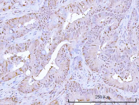
SampleParaffin-embedded human rectum adenocarcinoma tissue; fixative not specified (datasheet A02867-2)
FixationImage fixative and duration unreported (datasheet A02867-2); verify before use.
Sectioning4–5 µm sections on charged slides (standard)
DeparaffinisationXylene, graded ethanol series to water (standard)
Antigen retrievalHeat retrieval: EDTA pH 8.0 (datasheet A02867-2); 20 min, 95–100 °C (standard)
Peroxidase block3% H2O2, 10 min, room temperature (standard)
Blocking10% goat serum (datasheet A02867-2)
Primary antibodyRabbit anti-PSMB9, 2-5 μg/ml (datasheet A02867-2)
Primary incubationOvernight at 4 °C (datasheet A02867-2)
DetectionHRP-conjugated secondary, DAB chromogen (datasheet A02867-2)
CounterstainHematoxylin, blue, dehydrate and mount (standard)
Expected resultPSMB9-positive staining in glandular cells of appendix (HPA tissue IHC: Medium). HPA tissue profile: Nuclear and cytoplasmic expression in several tissues, most abundant in leukocytes. No signal in the no-primary control.
💡Decision noteStart with EDTA pH 8.0 heat retrieval for the catalog antibody (datasheet: A02867-2); citrate pH 6.0 is a published alternative for bladder tissue (PMC11863778).
Section 2

What Is the Expected PSMB9 Staining Pattern?

PSMB9 staining in paraffin sections should be nuclear and cytoplasmic, with the most abundant tissue expression in leukocytes; lymph node germinal center cells are a documented medium-staining example (HPA: tissue IHC). Cytoplasmic and nuclear localization agrees with a protein lacking a transmembrane segment (UniProt P28065: location and topology). HPA rates tissue staining Enhanced, while reporting medium consistency with RNA and presumed off-target binding (HPA: tissue IHC).

What am I looking at on my slide?
Nuclear and cytoplasmic staining in leukocytes, including medium staining in lymph node germinal center cells (HPA: tissue IHC).This fits the reported tissue pattern and UniProt localization (HPA: tissue IHC; UniProt P28065: location). Score the named cell population and compartments rather than judging the whole section uniformly; other cell populations may stain differently (HPA: tissue IHC).
Predominantly crisp plasma-membrane staining, without convincing nuclear or cytoplasmic staining.A membrane-only pattern conflicts with the reported locations and absence of a transmembrane segment (HPA: tissue IHC; UniProt P28065: topology). Treat it as suspect; review morphology, detection controls and antibody specificity before calling it PSMB9.
Strong staining of adipocytes or skeletal myocytes while the expected leukocyte pattern is absent.HPA reports PSMB9 as not detected in those cell types (HPA: adipose tissue and skeletal muscle IHC). Consider cross-reactivity or endogenous chromogenic activity; an unexpected positive alone cannot establish target expression (general IHC practice).
Diffuse color over cells and extracellular areas, obscuring compartment boundaries.The distribution cannot be scored confidently as nuclear or cytoplasmic. Check a no-primary control for detection background, then optimize blocking, washes and chromogen development using the same section workflow (general IHC practice).
No staining in lymph node germinal center cells despite an otherwise developed chromogenic run.Those cells show medium staining in HPA tissue IHC (HPA: lymph node IHC). First verify that the relevant cells are present; then review the IHC-validated antibody, retrieval and detection controls. A blank section alone does not establish PSMB9 absence (general IHC practice).
💡Expected PSMB9 appearanceCall a positive result when identifiable leukocytes show nuclear and cytoplasmic staining, with medium staining plausible in lymph node germinal center cells; isolated membrane staining or color over HPA-negative adipocytes is suspect (HPA: tissue IHC; UniProt P28065: topology).
How each factor affects the staining
Cell-population referenceLeukocytes are the most abundant stained population; medium signal is documented in lymph node germinal center cells and several epithelial or endothelial populations (HPA: tissue IHC). Compare like cell types when scoring.
Validation limitsTissue IHC reliability is Enhanced, but HPA reports only medium agreement with RNA and disregarded presumed off-target binding (HPA: tissue IHC). Confirm an unexpected pattern with appropriate controls.
Processing and compartmentPSMB9 has a propeptide at 1–20 and a mature chain at 21–219, with no transmembrane segment (UniProt P28065: processing and topology). This record does not identify the antibody epitope or predict retrieval behavior.
IsoformsUniProt lists LMP2.L and LMP2.S (UniProt P28065: isoforms). Without an epitope assignment, the supplied record cannot show whether this antibody distinguishes them or whether they differ in staining.
IF/ICC: What pattern is expected?HPA reports supported cytosolic localization in ICC-IF images from U-251MG and U2OS (HPA: subcellular ICC-IF). This is an IF/ICC interpretation reference, not an IHC-P protocol option.
Why is my staining missing, weak or wrong?
SituationLikely causeNext action
Expected leukocytes stain weakly or not at all.The lymph node germinal center reference has medium signal, so low contrast or a failed detection step can obscure it (HPA: lymph node IHC; general IHC practice).Confirm the relevant cells by morphology; review the catalog antibody's IHC-P instructions and run a known-positive section with detection controls (general IHC practice).
Color appears in the no-primary control.The signal can arise from the detection system or endogenous enzyme activity rather than primary-antibody binding (general IHC practice).Address endogenous activity and blocking as appropriate for the chromogen, then compare matched controls before scoring PSMB9 (general IHC practice).
Adipocytes or skeletal myocytes dominate the positive call.HPA reports these cell types as not detected; unexpected color may reflect cross-reactivity or detection background (HPA: adipose tissue and skeletal muscle IHC; general IHC practice).Inspect nearby leukocytes and no-primary controls; withhold a target-specific call until the cell pattern is resolved (HPA: tissue IHC; general IHC practice).
Signal is restricted to cell borders.A membrane-only pattern conflicts with nuclear and cytoplasmic tissue staining and the absence of a transmembrane segment (HPA: tissue IHC; UniProt P28065: topology).Check section morphology and control staining, then reassess antibody specificity before treating borders as positive (general IHC practice).
Chromogen obscures nuclei and cytoplasm.Excess background or overdevelopment can prevent compartment scoring (general IHC practice).Compare a no-primary section; adjust development, washes or blocking as the control indicates, and use the counterstain to identify cells (general IHC practice).
IF/ICC looks cytosolic while tissue IHC includes nuclear staining.HPA reports supported cytosolic ICC-IF localization and nuclear plus cytoplasmic tissue IHC staining (HPA: subcellular ICC-IF and tissue IHC).Interpret each preparation against its own HPA reference and controls; do not treat that difference alone as proof of an IHC artefact.

Sample controls for PSMB9 IHC & IF

🧪Run appendix first and assess its glandular cells for PSMB9 staining (HPA: Medium in appendix glandular cells). Use adipose tissue adipocytes as the negative comparator (HPA: Not detected in adipocytes); any cells treated as internal negatives on the appendix slide should show counterstain without specific DAB signal, but the supplied HPA row does not identify a negative appendix cell type.
Positive control tissue: Appendix (Glandular cells, HPA Medium)
Negative control tissue: Adipose tissue (HPA Not detected)
ICC-IF cell lines (HPA subcellular resource): HPA ICC-IF images show PSMB9 in U-251MG, U2OS, with annotated localisation: Cytosol (supported) (HPA subcellular).
Technical controls: Include a no-primary, secondary-only control; host- and class-matched isotype IgG; and a validated PSMB9 knockout sample or peptide-block control where the immunizing peptide is known (standard IHC practice). Block endogenous peroxidase and inspect the appendix section for DAB background in blood or inflammatory cells (standard chromogenic IHC practice).
⚠️Feasibility: A target-specific fixation window or fixation effect is unreported, and the selected A02867-2 tissue-IHC caption does not state its fixative. That caption reports heat retrieval in EDTA at pH 8.0 for a paraffin section, but does not establish that retrieval is required for all specimens (selected-SKU tissue-IHC caption). The supplied evidence does not show that frozen sections or tissue IF are easier; appendix blood or inflammatory cells can complicate chromogenic scoring through endogenous peroxidase background (standard IHC practice).

HPA tissue IHC evidence for PSMB9

Comprehensive Human Protein Atlas IHC scoring per tissue (reliability: Enhanced — Medium consistency between antibody staining and RNA expression data. Presumed off target binding observed and disregarded.). Rows are taken directly from the HPA tissue chart — click any row's HPA link to view the source.

Positive expression · recommended positive controls

TissueCell typeLevelEvidenceSource
Appendix Glandular cells Medium Protein (IHC) HPA →
Cervix Squamous epithelial cells Medium Protein (IHC) HPA →
Colon Endothelial cells Medium Protein (IHC) HPA →
Duodenum Glandular cells Medium Protein (IHC) HPA →
Endometrium Glandular cells Medium Protein (IHC) HPA →

Undetected expression · recommended negative controls

TissueCell typeLevelEvidenceSource
Adipose tissue Adipocytes Not detected Protein (IHC) HPA →
Caudate Glial cells Not detected Protein (IHC) HPA →
Cerebral cortex Glial cells Not detected Protein (IHC) HPA →
Heart muscle Cardiomyocytes Not detected Protein (IHC) HPA →
Hippocampus Glial cells Not detected Protein (IHC) HPA →
Section 3

Advanced PSMB9 IHC Tips

Troubleshoot PSMB9 staining in paraffin sections by checking retrieval, cell identity, and nuclear and cytoplasmic signal before scoring.

Which retrieval condition should I try first for weak PSMB9 staining?
Start with heat-mediated retrieval in EDTA at pH 8.0 (datasheet A02867-2). The selected paraffin-section example used this condition before overnight incubation with 2 μg/ml primary antibody at 4°C, so reproduce those documented steps before changing retrieval chemistry (datasheet A02867-2). If staining remains weak, vary heating time cautiously on adjacent sections and compare signal with tissue preservation; excessive heating can damage morphology (standard IHC practice). Keep section thickness, detection chemistry, and exposure to DAB consistent across the comparison so a change in staining can be attributed to retrieval (standard IHC practice).
Could fixation explain weak or uneven PSMB9 staining?
The selected image describes a paraffin-embedded section but does not state its fixative, so target-specific fixation sensitivity is unknown (datasheet A02867-2). Record the actual fixative and fixation interval for each specimen, then compare sections processed together using the documented EDTA pH 8.0 retrieval and 2 μg/ml primary concentration (datasheet A02867-2; standard IHC practice). Check whether weak signal follows processing batches, section depth, or tissue regions before changing antibody concentration (standard IHC practice). Assess nuclear and cytoplasmic staining with preserved cell morphology; either compartment can be compatible with PSMB9, but damaged tissue cannot establish a fixation effect (UniProt P28065 subcellular location; standard IHC practice).
Is nuclear PSMB9 staining plausible in a paraffin section?
Yes: PSMB9 is annotated in both cytoplasm and nucleus, while tissue IHC shows nuclear and cytoplasmic expression across several tissues (UniProt P28065 subcellular location; HPA tissue IHC profile). Interpret either compartment within intact cells, and compare the pattern with adjacent cells on the same slide rather than treating all nuclear DAB as nonspecific (standard IHC practice). The supported cytosol location from ICC/IF offers a useful comparison, but it does not rule out nuclear staining in tissue sections (HPA subcellular profile; UniProt P28065 subcellular location). Diffuse staining over lumina, debris, or section edges warrants a background check before assigning subcellular localisation (standard IHC practice).
Can this stain distinguish PSMB9 isoforms or processed protein?
The record lists 2 isoforms, LMP2.L and LMP2.S, and processing that removes residues 1–20 to yield the 21–219 chain (UniProt P28065 isoforms and processing). Without a mapped antibody epitope, chromogenic staining cannot establish which isoform or processing state produced the signal (standard IHC interpretation). When selecting a second antibody for confirmation, compare its documented epitope with the propeptide and shared chain, then verify its own paraffin-section performance (UniProt P28065 processing; standard IHC practice). Report the result as PSMB9 immunoreactivity unless epitope mapping and an independent assay support a narrower assignment (standard IHC interpretation).
How should I cross-check the IHC pattern by multiplex IF?
On a separate IF/ICC study, pair PSMB9 with a leukocyte-lineage marker to test cell identity, since tissue expression is most abundant in leukocytes (HPA tissue IHC profile; standard IF practice). Choose a far-red detection channel when the specimen has strong short-wavelength autofluorescence, and inspect single-stain controls for bleed-through (standard IF practice). PSMB9 has no annotated transmembrane segment and is reported in cytoplasm and nucleus, so permeabilise cells to access intracellular epitopes while checking that cell outlines remain intact (UniProt P28065 topology and subcellular location; standard IF practice). Treat any IF/ICC pattern as a cross-check, since the selected antibody evidence here documents paraffin-section chromogenic IHC (datasheet A02867-2).
What should I check when DAB appears throughout the section?
Run a section without primary antibody to assess detection-system background, and include an endogenous peroxidase block before HRP and DAB development (standard IHC practice). The selected example used goat-serum blocking, an HRP-linked secondary antibody, and DAB, so review blocking and secondary-only controls when diffuse brown signal appears (datasheet A02867-2; standard IHC practice). Keep DAB development time comparable between sections, because overdevelopment can obscure differences between weak cellular staining and background (standard IHC practice). Judge suspected signal against intact nuclear or cytoplasmic cells and local tissue structure, since those compartments are compatible with PSMB9 (UniProt P28065 subcellular location; standard IHC practice).
How should I quantify PSMB9 IHC across different specimens? ⚠ ANSWER MARKED FOR VERIFICATION
Define the cell population and tissue area before scoring, because PSMB9 is most abundant in leukocytes and mixed-cell regions can shift an aggregate score (HPA tissue IHC profile; standard IHC practice). Within the defined population, record the percentage of positive cells and staining intensity, then calculate an H-score if intensity categories are reproducible (standard IHC practice). For infiltrating positive cells, report density per mm² of viable tissue and normalise cell-based scores to the number of evaluable cells in the same compartment (standard IHC practice). Apply identical thresholds, DAB development, and exclusion rules for folds, edges, and necrosis across specimens (standard IHC practice).
How can I distinguish convincing PSMB9 positivity from artefact?
Look for reproducible staining in intact nuclei or cytoplasm, with particular attention to leukocytes because they are the most abundant tissue population reported for PSMB9 (UniProt P28065 subcellular location; HPA tissue IHC profile). Do not assign isolated edge deposits, necrotic debris, or staining confined to damaged regions to positive cells (standard IHC practice). Compare morphology and cell identity with a counterstained section, and use a no-primary control to identify endogenous enzyme or detection-system signal (standard IHC practice). Interpret unexpected strong staining cautiously: HPA rates its tissue evidence Enhanced but notes presumed off-target binding that was disregarded in its assessment (HPA tissue IHC reliability description).
Boster reagents

Best PSMB9 / Proteasome subunit beta type-9 IHC Antibodies

A02867-2 has pictured IHC and IF data from human paraffin sections; M02867 lists IF/ICC for human, mouse, and rat without a supplied image (catalog captions; application/reactivity/image lists).

Real IHC data IHC analysis of PSMB9 using anti-PSMB9 antibody (A02867-2). PSMB9 was detected in a paraffin-embedded section of human rectum adenocarcinoma tissue. Heat mediated antigen retrieval was performed in EDTA buffer (pH 8.0, epitope retrieval solution). The tissue section was blocked with 10% goat serum. The tissue section was then incubated with 2 μg/ml rabbit anti-PSMB9 Antibody (A02867-2) overnight at 4°C. Peroxidase Conjugated Goat Anti-rabbit IgG was used as secondary antibody and incubated for 30 minutes at 37°C. The tissue section was developed using HRP Conjugated Rabbit IgG Super Vision Assay Kit (Catalog # SV0002) with DAB as the chromogen.
Anti-PSMB9 Antibody ®
Cat # A02867-2

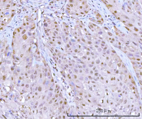
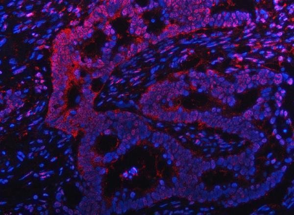
A02867-2 has IHC images from human rectum adenocarcinoma and urothelial carcinoma paraffin sections, plus an IF image from a human intestinal cancer paraffin section (catalog image captions). M02867 lists IF/ICC and human, mouse, and rat reactivity, with no IHC or IF image supplied (catalog application/reactivity/image lists).

Which to pick: Choose A02867-2 for chromogenic tissue IHC: its pictured human paraffin sections used EDTA retrieval at pH 8.0, 2 μg/ml primary antibody, and DAB detection; the fixative is unreported (A02867-2 IHC captions). For IF, A02867-2 has pictured human tissue staining at 5 μg/ml; M02867 is a rabbit monoclonal listed for IF/ICC, with no supplied image (A02867-2 IF caption; M02867 clone/application/image fields). Both SKUs list mouse and rat reactivity, but the supplied tissue images show human samples only (catalog reactivity/image lists).

Each figure is that product's own IHC / IF validation image from its datasheet.

References

  1. UniProt Consortium. UniProt entry P28065 (PSB9_HUMAN, Proteasome subunit beta type-9).
  2. Human Protein Atlas. PSMB9 tissue IHC expression (reliability: Enhanced).
  3. Human Protein Atlas. PSMB9 subcellular location (ICC-IF): Localized to the cytosol..
  4. Human Protein Atlas. PSMB9 antibody validation summary (3 antibodies).
  5. Immunoproteasome subunits are novel signatures for predicting efficacy of immunotherapy in muscle invasive bladder cancer. Journal of translational medicine 2025 — PMC11863778.
  6. PSMB9 exacerbates chondrocyte injury in osteoarthritis via activation of the NF-κB pathway. Journal of orthopaedic surgery and research 2026 — PMC13101164.
  7. PSMB9 Promotes the Malignant Progression of Colorectal Cancer by Regulating the PI3K/Akt Pathway. Oncology research 2026 — PMC13494480.
  8. Parkinson's disease and multiple system atrophy patient iPSC-derived oligodendrocytes exhibit alpha-synuclein-induced changes in maturation and immune reactive properties. Proceedings of the National Academy of Sciences of the United States of America 2022 — PMC8944747.
  9. PubMed PMID:8458375 — UniProt-cited evidence.
  10. PubMed PMID:1453454 — UniProt-cited evidence.
  11. PubMed PMID:1922385 — UniProt-cited evidence.