This website uses cookies to ensure you get the best experience on our website.


- Table of Contents
Plan PSMD11 staining in paraffin sections using the observed cytoplasmic pattern in immune cells and testis basal cells (HPA tissue IHC). Interpret staining with the reported low consistency between antibody staining and RNA expression in mind (HPA tissue IHC).

Expected localisation, validated protocols, controls and antibodies — the at-a-glance facts below, then the full design guide.
| Expected localisation | Cytoplasmic in immune cells and testis basal cells (HPA tissue IHC) | |
| Staining pattern | Cytoplasmic staining in immune and testis basal cells (HPA tissue IHC) | |
| Antigen retrieval | Tris-EDTA pH 9.0 HIER, 95–98 °C, 20 min (rule: nuclear antigen) | |
| Positive control | Testis+4 more · see all | |
| Negative control | Breast+4 more · see all |
| Fixation | Keep fixation consistent across sections. (standard IHC practice; not target-specific) | |
| Caveat | Staining shows low consistency with RNA expression (HPA tissue IHC) | |
| Regulation | Higher in ESCs; falls with differentiation (UniProt) | |
| Isoform / epitope | 2 isoforms; check epitope coverage for each (UniProt) |
The catalog antibody’s IHC-P protocol is followed by three published PSMD11 paraffin-section protocols (PMC11165170; PMC11965110; PMC12541842).
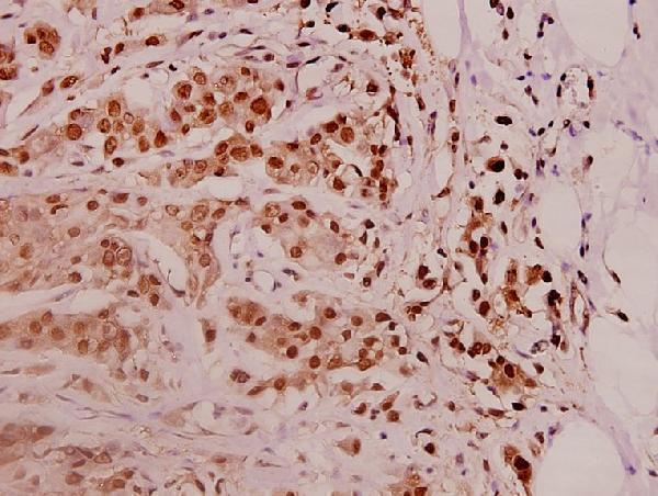
| Sample | Paraffin-embedded human breast carcinoma tissue; fixative not specified (datasheet A08940) |
| Fixation | Image fixative and duration unreported (datasheet A08940); verify before use. |
| Sectioning | 4–5 µm sections on charged slides (standard) |
| Deparaffinisation | Xylene, graded ethanol series to water (standard) |
| Antigen retrieval | Heat-induced epitope retrieval in Tris-EDTA buffer, pH 9.0, 20 min at 95–98 °C (standard rule: nuclear antigen) |
| Peroxidase block | 3% H2O2, 10 min, room temperature (standard) |
| Blocking | 10% normal serum of the secondary host, 30 min, room temperature (standard) |
| Primary antibody | Rabbit anti-PSMD11, 1:50-1:200 (datasheet A08940) |
| Primary incubation | Overnight at 4 °C (standard) |
| Detection | HRP-polymer secondary, DAB chromogen 5–10 min (standard) |
| Counterstain | Hematoxylin, blue, dehydrate and mount (standard) |
| Expected result | PSMD11-positive staining in sertoli cells of testis (HPA tissue IHC: High). HPA tissue profile: Cytoplasmic expression in immune cells and basal cells of testis. No signal in the no-primary control. |
PSMD11 is a proteasome subunit found in the nucleus and cytosol, with no transmembrane segment (UniProt O00231). In tissue IHC, expect chiefly cytoplasmic staining in selected cells, including strong staining in testis Sertoli cells and moderate staining in lung macrophages (HPA tissue IHC). HPA rates the tissue staining “Approved” but reports low consistency with RNA expression; interpret the pattern alongside cell identity and controls (HPA tissue IHC).
| Cytoplasmic staining is strong in testis Sertoli cells; nearby cells show a different intensity. | This matches the reported high Sertoli-cell signal (HPA tissue IHC). Use the named cell population when judging a positive result: the HPA tissue summary also describes cytoplasmic staining in immune cells and basal cells of testis, so confirm the cells on the section rather than assuming all testicular cells should stain equally (HPA tissue IHC). |
| A lung macrophage has moderate cytoplasmic staining, with little staining in surrounding structures. | This is compatible with the reported moderate macrophage signal (HPA tissue IHC). Moderate staining is also reported in bone marrow hematopoietic cells, heart cardiomyocytes, kidney glomerular cells, placenta decidual cells and tonsil non-germinal-center cells (HPA tissue IHC). Differences among these cell types need not indicate assay failure; score each against its reported pattern (HPA tissue IHC). |
| Staining is confined to cell borders or extracellular material, with no convincing intracellular signal. | Treat this as a suspect compartment pattern: PSMD11 has no transmembrane segment and is assigned to the nucleus and cytosol (UniProt O00231). Nuclear staining alone is not automatically wrong, because nuclear localization is reported by UniProt and nucleoplasmic localization by HPA ICC-IF (UniProt O00231; HPA subcellular). Check morphology, detection controls and a known-positive section before calling the border signal specific (general IHC practice). |
| Strong staining appears in a cell population listed as not detected, while expected positive cells remain weak. | Consider cross-reactivity or endogenous detection activity before interpreting the signal as PSMD11 (general IHC practice). HPA lists breast adipocytes and bronchus basal cells among its not-detected examples, but its tissue IHC reliability note reports low staining-to-RNA consistency (HPA tissue IHC). A single unexpected positive cell type therefore warrants controls and review of its identity, not an automatic biological conclusion. |
| The section has diffuse color throughout, or a known-positive Sertoli-cell section has no specific signal. | Uniform haze obscures cell-level localization and suggests background from the detection workflow (general IHC practice). Absent staining in Sertoli cells conflicts with HPA's high testis result, but does not alone identify the failed step (HPA tissue IHC). Review the section, reagent and control results together before scoring the test specimen as negative (general IHC practice). |
| Cell type and tissue | HPA reports high staining in testis Sertoli cells, moderate staining in several named cell populations, low staining in endometrial glandular cells, and not-detected staining in several others (HPA tissue IHC). Choose the comparison cell population explicitly; whole-section color can hide these differences (general IHC practice). |
| Strength of tissue evidence | The tissue IHC profile is “Approved,” with low consistency between antibody staining and RNA expression; HPA advises viewing images for staining in unannotated cells or structures (HPA tissue IHC). Treat the listed levels as observed reference patterns, not guaranteed results for every specimen (HPA tissue IHC). |
| Intracellular location | UniProt places PSMD11 in the nucleus and cytosol and reports no transmembrane segment (UniProt O00231). HPA ICC-IF places it mainly in the nucleoplasm and Golgi apparatus, with additional sperm structures (HPA subcellular). These IF locations inform compartment review but do not specify a separate paraffin IHC pattern. |
| Isoforms and processing | Two isoforms are listed, with no signal peptide or propeptide and a reported chain spanning residues 2–422 (UniProt O00231). The supplied sources do not identify the catalog antibody's epitope or establish whether it recognizes both isoforms; an unexpected IHC pattern cannot be assigned to one isoform from these records. |
| IF/ICC Q&A | Can IF/ICC be read like tissue IHC? HPA reports approved nucleoplasm and Golgi localization in ICC-IF, while its tissue IHC summary emphasizes cytoplasmic staining in selected cells (HPA subcellular; HPA tissue IHC). Use the separate IF/ICC guide for that application; the supplied records do not establish an IF protocol here. |
| Situation | Likely cause | Next action |
|---|---|---|
| No staining in a testis section containing identifiable Sertoli cells. | A failed staining run or an unsuitable section is possible (general IHC practice); HPA reports high Sertoli-cell staining (HPA tissue IHC). | Confirm the Sertoli cells are present and preserved, then review the primary-antibody, retrieval and detection steps against the IHC-validated antibody's instructions; compare with a successfully stained control section (general IHC practice). |
| All cells have pale, even chromogenic haze. | Incomplete blocking, excess detection reagent or insufficient washing can create diffuse background (general IHC practice). | Inspect the no-primary control, check blocking and wash steps, and adjust the detection workflow according to its instructions before judging PSMD11 localization (general IHC practice). |
| Color persists in the no-primary control during enzyme-based detection. | Endogenous enzyme activity or the detection system can contribute color independently of the primary antibody (general IHC practice). | For an HRP system, verify the endogenous-peroxidase block and inspect the detection-only control; correct the background before interpreting cell staining (general IHC practice). |
| Signal appears only at cell borders or in extracellular deposits. | That distribution is difficult to reconcile with the reported nucleus and cytosol localization and absence of a transmembrane segment (UniProt O00231). | Recheck cell boundaries and precipitate-like deposits, then compare the no-primary control and a reported positive cell population on a parallel section (general IHC practice; HPA tissue IHC). |
| A nominally not-detected cell type stains strongly. | Cell misidentification, cross-reactivity or endogenous detection activity are possibilities (general IHC practice); HPA's not-detected calls are observations from its tissue IHC profile (HPA tissue IHC). | Verify the cell type and inspect controls. Compare the result with HPA images and a reported positive population; retain the discrepancy in the record because HPA notes low consistency with RNA expression (HPA tissue IHC). |
| A nuclear signal appears despite an expected cytoplasmic tissue pattern. | Nuclear PSMD11 is supported by UniProt, and HPA ICC-IF reports nucleoplasmic localization (UniProt O00231; HPA subcellular). Tissue IHC emphasizes cytoplasmic staining in selected cells (HPA tissue IHC). | Assess whether the signal is cell-associated and reproducible in controls; record nuclear and cytoplasmic staining separately rather than discarding the section solely for nuclear color (general IHC practice). |


Comprehensive Human Protein Atlas IHC scoring per tissue (reliability: Approved — Low consistency between antibody staining and RNA expression data. Antibody staining in cells/structures not annotated, view images.). Rows are taken directly from the HPA tissue chart — click any row's HPA link to view the source.
| Tissue | Cell type | Level | Evidence | Source |
|---|---|---|---|---|
| Testis | Sertoli cells | High | Protein (IHC) | HPA → |
| Bone marrow | Hematopoietic cells | Medium | Protein (IHC) | HPA → |
| Endometrium | Glandular cells | Low | Protein (IHC) | HPA → |
| Heart muscle | Cardiomyocytes | Medium | Protein (IHC) | HPA → |
| Kidney | Cells in glomeruli | Medium | Protein (IHC) | HPA → |
| Tissue | Cell type | Level | Evidence | Source |
|---|---|---|---|---|
| Breast | Adipocytes | Not detected | Protein (IHC) | HPA → |
| Bronchus | Basal cells | Not detected | Protein (IHC) | HPA → |
| Cerebellum | Cells in granular layer | Not detected | Protein (IHC) | HPA → |
| Cervix | Glandular cells | Not detected | Protein (IHC) | HPA → |
| Duodenum | Glandular cells | Not detected | Protein (IHC) | HPA → |
Troubleshoot PSMD11 staining in paraffin sections by checking retrieval, compartment, cell identity and controls before comparing chromogenic IHC scores.
The catalog includes human- and mouse-reactive PSMD11 antibodies for IHC and IF; only A08940 has a supplied IHC image, from a human paraffin-embedded breast carcinoma section (catalog applications and reactivity; A08940 image caption).
A08940 is the only SKU with a rendered card; its IHC image shows a human paraffin-embedded breast carcinoma section at 1:100 (A08940 image caption). Its listed applications are IHC and WB, and its declared reactivity is human, mouse and rat (catalog: A08940 applications and reactivity).
Which to pick: For tissue IHC, choose A08940: its own image documents staining in a paraffin-embedded human section; the fixative is unreported (A08940 image caption). For IF/ICC, A08940-1 lists IF at 1:50 and human and mouse reactivity, but has no supplied IF image (catalog: A08940-1 applications, dilution, reactivity and image alts). For a rat sample, A08940 is the listed option; both antibodies are rabbit polyclonals, and cross-species staining still needs validation in the intended tissue (catalog: host, clone and reactivity).