This website uses cookies to ensure you get the best experience on our website.


- Table of Contents
Plan chromogenic IHC for PSPC1 in paraffin sections using its predominantly nuclear tissue staining as the expected pattern (HPA tissue IHC). The guide covers fixation consistency, interpretation across cell types, and the limits of the available isoform evidence (UniProt).

Expected localisation, validated protocols, controls and antibodies — the at-a-glance facts below, then the full design guide.
| Expected localisation | Predominantly nuclear in tissue sections (HPA tissue IHC) | |
| Staining pattern | Nuclear staining across a majority of tissues (HPA tissue IHC) | |
| Antigen retrieval | EDTA pH 8.0 HIER, heat-mediated (datasheet A06348-1) | |
| Positive control | Appendix+4 more · see all | |
| Negative control | None in HPA (detected in all 44 tissues); use no-primary + isotype controls |
| Fixation | Keep fixation consistent across sections (standard IHC practice; not target-specific) | |
| Caveat | Ovarian stromal cells show low staining (HPA tissue IHC) | |
| Regulation | No intensity regulator specified (UniProt) | |
| Isoform / epitope | 2 isoforms; epitope coverage is unknown (UniProt) |
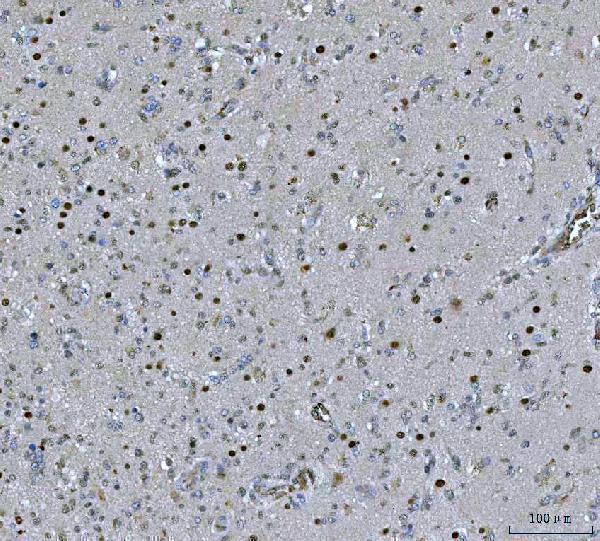
The catalog antibody’s IHC-P protocol (datasheet A06348-1) is accompanied by four published PSPC1 IHC methods (PMC4395133; PMC8053714; PMC9184599; PMC6914800).
| Sample | Paraffin-embedded human glioblastoma tissue; fixative not specified (datasheet A06348-1) |
| Fixation | Image fixative and duration unreported (datasheet A06348-1); verify before use. |
| Sectioning | 4–5 µm sections on charged slides (standard) |
| Deparaffinisation | Xylene, graded ethanol series to water (standard) |
| Antigen retrieval | Heat retrieval: EDTA pH 8.0 (datasheet A06348-1); 20 min, 95–100 °C (standard) |
| Peroxidase block | 3% H2O2, 10 min, room temperature (standard) |
| Blocking | 10% goat serum (datasheet A06348-1) |
| Primary antibody | Rabbit anti-PSPC1, 2-5 μg/ml (datasheet A06348-1) |
| Primary incubation | Overnight at 4 °C (datasheet A06348-1) |
| Detection | HRP-conjugated secondary, DAB chromogen (datasheet A06348-1) |
| Counterstain | Hematoxylin, blue, dehydrate and mount (standard) |
| Expected result | PSPC1-positive staining in glandular cells of appendix (HPA tissue IHC: High). HPA tissue profile: Nuclear expression in a majority of tissues. No signal in the no-primary control. |
PSPC1 is primarily nuclear, including paraspeckles and the nucleolus, with cytoplasmic localization also reported (UniProt Q8WXF1). In paraffin-section IHC, expect nuclear staining across many tissues; HPA reports high staining in selected glandular, endothelial, glial and stromal cells (HPA tissue IHC: Supported; nuclear expression in a majority of tissues). PSPC1 has no transmembrane segment (UniProt Q8WXF1 topology).
| Distinct nuclear chromogen in appendix or duodenum glandular cells. | This fits reported high staining in those cells (HPA tissue IHC: High in appendix and duodenum glandular cells). Judge signal against nearby nuclei and the negative control; a uniformly colored section is less convincing than cell-resolved staining (general IHC practice). |
| Nuclear staining appears punctate, with some nucleolar association. | This is compatible with PSPC1 in paraspeckles and the nucleolus (UniProt Q8WXF1 localization). Routine chromogenic IHC may not resolve individual paraspeckles, so puncta are supportive rather than required (general IHC interpretation). |
| The dominant stain outlines cell membranes or fills extracellular space instead of nuclei. | That distribution conflicts with the mainly nuclear tissue pattern (HPA tissue IHC) and the absence of a transmembrane segment (UniProt Q8WXF1 topology). Consider nonspecific binding, precipitate or detection artefact; compare morphology and control sections (general IHC practice). |
| Strong staining appears in unexpected cells while expected nuclei remain weak. | Cell-specific intensity can vary, but an inverted pattern relative to the selected reference tissue warrants scrutiny (HPA tissue IHC: listed high and low cell groups). Cross-reactivity or endogenous detection activity are possibilities, not diagnoses; assess controls before assigning PSPC1 positivity (general IHC practice). |
| A reference section shows no convincing nuclear signal. | A negative result in appendix or duodenum glandular cells disagrees with their reported high staining (HPA tissue IHC). First check whether the cells are present and the detection run worked; this single section cannot establish target absence (general IHC practice). |
| Reference-cell choice | Appendix and duodenum glandular cells, caudate glia, and cerebral-cortex or colon endothelium are reported high (HPA tissue IHC). Cardiomyocytes and ovarian stromal cells are reported low (HPA tissue IHC); a weak result there is less informative as a run check. |
| Subnuclear resolution | PSPC1 occupies paraspeckles and can associate with the nucleolus (UniProt Q8WXF1 localization). HPA tissue IHC reports nuclear expression broadly, without requiring visible puncta in each chromogenic section; score the nuclear compartment before interpreting fine texture. |
| Antibody evidence | The tissue pattern has Supported reliability with high agreement between staining and RNA data (HPA tissue IHC). HPA038904 is listed as Supported for IHC (HPA antibodies); that status supports use as a reference, while a control section still checks each staining run (general IHC practice). |
| Protein forms and epitope limits | UniProt lists two isoforms and a full-length chain spanning residues 1–523, with no signal peptide or propeptide (UniProt Q8WXF1). The supplied record gives no antibody epitope, so isoform-specific detection or epitope-dependent retrieval cannot be predicted from it. |
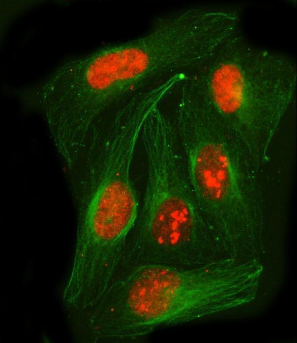
| IF/ICC Q&A: what pattern should IF show? | HPA reports mainly nucleoplasmic signal with additional nucleolar fibrillar-center localization, and lists A-431, U-251MG, U2OS and HEK293 images (HPA subcellular ICC-IF). Use the separate IF/ICC guide for that application; these cell images do not set an IHC scoring threshold. |
| Situation | Likely cause | Next action |
|---|---|---|
| No stain in a section selected as a positive reference. | The sampled field may lack the expected cells, or the IHC run may have failed (general IHC practice). Appendix glandular cells are reported high (HPA tissue IHC). | Locate intact glandular cells, inspect counterstain and tissue preservation, then review the run control and detection steps (general IHC practice). |
| Nuclei are weak, but broad brown haze covers the section. | Background can obscure a nuclear pattern through nonspecific binding or incomplete washing (general IHC practice); HPA describes predominantly nuclear tissue expression (HPA tissue IHC). | Compare the no-primary control, wash and blocking steps, and antibody dilution; retain a setting that reveals cell-resolved nuclei (general IHC practice). |
| Cytoplasm is darker than nuclei throughout the reference tissue. | UniProt also reports cytoplasmic localization, but widespread cytoplasm-dominant IHC conflicts with HPA's nuclear tissue profile (UniProt Q8WXF1; HPA tissue IHC). | Review cell boundaries and the no-primary control; repeat with the IHC-validated antibody conditions before interpreting cytoplasmic signal (general IHC practice). |
| Brown signal persists without primary antibody. | Endogenous detection activity or nonspecific detection reagents can produce color in chromogenic IHC (general IHC practice). Such signal cannot establish PSPC1 localization. | Check the detection chemistry and its blocking controls; compare the affected cells with the primary-antibody section (general IHC practice). |
| A low-reference cell group stains strongly. | Cardiomyocytes and ovarian stromal cells are listed as low, so strong staining merits review, although HPA levels do not define an absolute negative control (HPA tissue IHC). | Compare nuclear location and intensity with an HPA high cell group on the same run; inspect no-primary staining before considering cross-reactivity (general IHC practice). |
| IF images show fine puncta, but the IHC section shows smooth nuclear color. | Chromogenic tissue sections may not resolve the structures visible in ICC-IF (general imaging practice). HPA reports nucleoplasmic and nucleolar fibrillar-center IF signal (HPA subcellular ICC-IF). | Score IHC by convincing nuclear, cell-resolved staining and the chosen tissue reference; do not require individually resolved paraspeckles (HPA tissue IHC; UniProt Q8WXF1 localization). |


Comprehensive Human Protein Atlas IHC scoring per tissue (reliability: Supported — High consistency between antibody staining and RNA expression data.). Rows are taken directly from the HPA tissue chart — click any row's HPA link to view the source.
| Tissue | Cell type | Level | Evidence | Source |
|---|---|---|---|---|
| Appendix | Glandular cells | High | Protein (IHC) | HPA → |
| Breast | Adipocytes | High | Protein (IHC) | HPA → |
| Caudate | Glial cells | High | Protein (IHC) | HPA → |
| Cerebellum | Cells in granular layer | High | Protein (IHC) | HPA → |
| Cerebral cortex | Endothelial cells | High | Protein (IHC) | HPA → |
| Tissue | Cell type | Level | Evidence | Source |
|---|---|---|---|---|
| None in HPA: PSPC1 is detected in all 44 scored tissues. Use a no-primary (secondary-only) and an isotype control instead. | ||||
Use nuclear staining and cell type controls to assess PSPC1 in chromogenic IHC of paraffin sections (HPA tissue IHC; UniProt Q8WXF1).
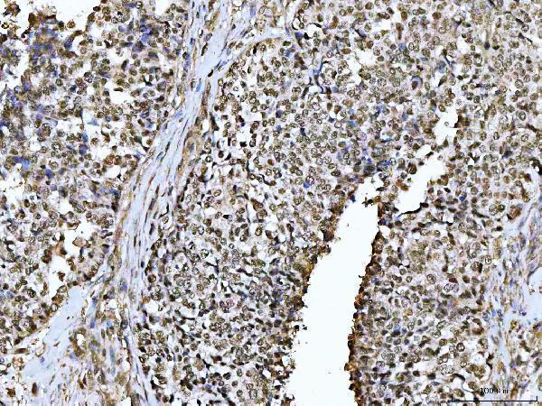
Catalog antibody A06348-1 lists human paraffin-section IHC and human IF/ICC, with image captions for glioblastoma, ovarian serous adenocarcinoma, and U2OS cells (catalog applications; image captions).
A06348-1 lists IHC for human paraffin sections, with captions showing glioblastoma and ovarian serous adenocarcinoma tissue (catalog dilution data; IHC image captions). The same SKU lists human IF/ICC and has an IF caption for U2OS cells (catalog dilution data; IF image caption).
Which to pick: For tissue IHC, A06348-1 lists a starting concentration of 2–5 μg/ml; its paraffin-section captions describe EDTA retrieval at pH 8.0, although they name a different antibody code and do not report the fixative (catalog dilution data; IHC image captions). For IF/ICC, A06348-1 lists 5 μg/ml and has a U2OS IF caption (catalog dilution data; IF image caption). For cross-species work, A06348-1 lists human, mouse, and rat reactivity, while its IHC and IF/ICC dilution entries specify human samples (catalog reactivity; catalog dilution data).