PTGER4 / Prostaglandin E2 receptor EP4 subtype · IHC design guide

Design Immunohistochemistry for PTGER4

Plan paraffin-section PTGER4 IHC with catalog antibody A02153 at 1:25 (datasheet). Compare the observed cytoplasmic tissue pattern (HPA tissue IHC) with the expected membrane location (UniProt), and use specificity controls given the low staining–RNA consistency (HPA tissue IHC).

Evidence assembled Oct 2026 · For research use; verify linked source records and product datasheet before use
Immunohistochemistry protocol sheet for PTGER4 (IHC for PTGER4): expected localisation Cytoplasmic in tissue (HPA tissue IHC); membrane expected (UniProt), antibody A02153, validated IHC image, and IHC protocol steps
Printable PTGER4 IHC protocol sheet — expected localisation Cytoplasmic in tissue (HPA tissue IHC); membrane expected (UniProt), antibody A02153, controls and protocol steps. Open the full PTGER4 IHC guide →

PTGER4 Immunohistochemistry Experimental Design Guide

Expected localisation, validated protocols, controls and antibodies — the at-a-glance facts below, then the full design guide.

Must know before staining
Expected localisation Cytoplasmic in tissue (HPA tissue IHC); membrane expected (UniProt)
Staining pattern Cytoplasmic staining in most tissues (HPA tissue IHC)
Antigen retrieval Citrate pH 6.0 HIER, 95–98 °C, 20 min (rule: cytoplasmic / membrane antigen)
Positive control ⓘ Adrenal gland+4 more · see all
Negative control ⓘ None in HPA (detected in all 45 tissues); use no-primary + isotype controls
Important caveats
Reasons your staining may differ from the expected pattern.
Fixation Keep fixation consistent across paraffin sections. (standard IHC practice; not target-specific)
Caveat Low staining–RNA consistency; verify specificity (HPA tissue IHC)
Regulation Regulation not specified (UniProt)
Isoform / epitope No isoforms annotated; epitope side guides access (UniProt)
Section 1

Recommended PTGER4 IHC & IF Protocols

The catalog antibody’s IHC-P protocol is accompanied by a published PTGER4 IHC protocol for lung cancer tissue (PMC12085179).

Recommended immunohistochemistry (IHC-P) protocol parameters
SampleParaffin-embedded H. brain tissue; fixative not specified (datasheet A02153)
FixationImage fixative and duration unreported (datasheet A02153); verify before use.
Sectioning4–5 µm sections on charged slides (standard)
DeparaffinisationXylene, graded ethanol series to water (standard)
Antigen retrievalHeat-induced epitope retrieval in citrate buffer, pH 6.0, 20 min at 95–98 °C (standard rule: cytoplasmic / membrane antigen)
Peroxidase block3% H2O2, 10 min, room temperature (standard)
Blocking10% normal serum of the secondary host, 30 min, room temperature (standard)
Primary antibodyRabbit anti-PTGER4, 1:25 (datasheet A02153)
Primary incubationOvernight at 4 °C (standard)
DetectionHRP-polymer secondary, DAB chromogen 5–10 min (standard)
CounterstainHematoxylin, blue, dehydrate and mount (standard)
Expected resultPTGER4-positive staining in glandular cells of adrenal gland (HPA tissue IHC: High). HPA tissue profile: Cytoplasmic expression in most tissues. No signal in the no-primary control.
💡Decision noteTry citrate pH 6.0 HIER at 95–98 °C for 20 min first (page antigen retrieval); the published protocol does not specify retrieval (PMC12085179).
Section 2

What Is the Expected PTGER4 Staining Pattern?

PTGER4 is a cell membrane receptor with 7 transmembrane segments (UniProt P35408: topology). In IHC, HPA reports cytoplasmic staining in most tissues and High staining in glandular and bone marrow hematopoietic cells (HPA: tissue IHC). HPA rates its IHC evidence Approved but notes low agreement with RNA and pending external verification (HPA: reliability).

What am I looking at on my slide?
Cytoplasmic stain in glandular or marrow cells (HPA: High).Matches the reported tissue IHC pattern (HPA: tissue IHC).
Nuclear-only stain (UniProt P35408: cell membrane).Suspect nonspecific staining; HPA reports cytoplasm in IHC (HPA: tissue IHC).
Strong adipocyte stain (HPA: Low).Check cross-reactivity or endogenous detection activity (general IHC practice).
Diffuse stain across cells and stroma (general IHC practice).May reflect background from blocking, primary concentration or washes (general IHC practice).
No stain in marrow hematopoietic cells (HPA: High).Check assay performance; HPA notes low agreement with RNA (HPA: reliability).
💡Expected PTGER4 appearanceA positive result shows High cytoplasmic staining in glandular or marrow hematopoietic cells (HPA: tissue IHC); membrane staining is plausible (UniProt P35408: cell membrane), while nuclear-only or diffuse staining is suspect (general IHC practice).
How each factor affects the staining
Topology (UniProt P35408)Cell membrane receptor with 7 transmembrane segments (UniProt P35408: topology).
Tissue IHC (HPA)Cytoplasmic in most tissues; High in listed glandular and marrow cells (HPA: tissue IHC).
Evidence confidence (HPA)IHC Approved; low staining/RNA consistency; external verification pending (HPA: reliability).
IF/ICC: expected location?Membrane summary; no ICC-IF images or main location supplied (HPA: subcellular).
Why is my staining missing, weak or wrong?
SituationLikely causeNext action
No marrow cell signal (HPA: High).Low assay sensitivity or specimen variation (general IHC practice).Check a positive control, primary dilution and detection (general IHC practice).
Nuclear-only signal (UniProt P35408: membrane).Off-target binding or nonspecific detection (general IHC practice).Compare with the other IHC Approved antibody (HPA: antibodies).
Strong adipocyte signal (HPA: Low).Cross-reactivity or endogenous detection activity (general IHC practice).Run a no-primary control and inspect cell identity (general IHC practice).
Diffuse background (general IHC practice).Incomplete blocking or excess primary antibody (general IHC practice).Titrate primary antibody; improve blocking and washes (general IHC practice).
Patchy or edge-only signal (general IHC practice).Uneven reagent coverage or section drying (general IHC practice).Keep sections hydrated and fully covered during staining (general IHC practice).

Sample controls for PTGER4 IHC & IF

🧪Run adrenal gland first and score its glandular cells for staining (HPA: High in adrenal gland glandular cells). HPA detects PTGER4 in all 45 scored tissues, so use no-primary and isotype controls for the negative comparison; assess unstained nonglandular areas on the positive slide as local background, without treating them as a validated negative cell population (HPA: no negative tissue rows).
Positive control tissue: Adrenal gland (Glandular cells, HPA High)
Negative control tissue: None in HPA: PTGER4 is detected in all 45 scored tissues. Use a no-primary (secondary-only) and an isotype control instead.
ICC-IF cell lines (HPA subcellular resource): HPA carries no ICC-IF cell line for PTGER4; derive a cell-line control from the positive tissue's cell type (Glandular cells) and confirm it by RNA or western blot first.
Technical controls: Include a no-primary, secondary-only control, a host- and clonality-matched isotype control, and a PTGER4 knockout specimen or peptide-block control where available (standard IHC practice). For chromogenic detection, block endogenous peroxidase; if using the caption’s biotinylated secondary, control for endogenous biotin (selected A02153 tissue-IHC caption: biotinylated secondary and DAB).
⚠️Feasibility: A target-specific fixation window or fixation effect is unreported, and the selected A02153 paraffin-section caption does not state its fixative (selected A02153 tissue-IHC caption). Antigen-retrieval dependency is unreported in the supplied evidence (selected A02153 tissue-IHC caption). The supplied evidence does not establish that frozen sections or IF are easier for PTGER4; assess adrenal gland background with the specified detection controls (HPA: High in adrenal gland glandular cells; standard IHC practice).

HPA tissue IHC evidence for PTGER4

Comprehensive Human Protein Atlas IHC scoring per tissue (reliability: Approved — Low consistency between antibody staining and RNA expression data. Pending external verification.). Rows are taken directly from the HPA tissue chart — click any row's HPA link to view the source.

Positive expression · recommended positive controls

TissueCell typeLevelEvidenceSource
Adrenal gland Glandular cells High Protein (IHC) HPA →
Appendix Glandular cells High Protein (IHC) HPA →
Bone marrow Hematopoietic cells High Protein (IHC) HPA →
Breast Glandular cells High Protein (IHC) HPA →
Bronchus Respiratory epithelial cells High Protein (IHC) HPA →

Undetected expression · recommended negative controls

TissueCell typeLevelEvidenceSource
None in HPA: PTGER4 is detected in all 45 scored tissues. Use a no-primary (secondary-only) and an isotype control instead.
Section 3

Advanced PTGER4 IHC Tips

Troubleshoot PTGER4 staining in paraffin sections by checking retrieval, compartment, tissue context and controls before assigning biological meaning to chromogenic signal.

What retrieval should I use when PTGER4 staining is weak in paraffin sections?
Start with citrate buffer at pH 6.0, heated to 95–98 °C for 20 min (page retrieval setting). Keep the catalog antibody at the caption’s 1:25 dilution for the first comparison, so a dilution change does not obscure the retrieval result (A02153 tissue-IHC caption). If signal remains weak, compare a cautiously shortened and extended heating time on adjacent sections while holding detection conditions constant (standard IHC practice). Review membrane-associated staining as well as cytoplasmic signal: PTGER4 is a seven-pass cell-membrane receptor, while the HPA tissue profile reports cytoplasmic staining in most tissues (UniProt P35408 topology; HPA tissue-IHC profile).
How should I troubleshoot inconsistent PTGER4 staining across differently processed tissue blocks?
Record each block’s fixative, fixation duration, processing history and section age before comparing PTGER4 staining (standard IHC practice). Target-specific fixation sensitivity is unknown here: the A02153 image caption identifies a paraffin-embedded brain section but does not state its fixative (A02153 tissue-IHC caption). Run blocks in the same staining batch with citrate retrieval at pH 6.0, 95–98 °C for 20 min, and include a control section processed alongside them (page retrieval setting; standard IHC practice). If staining differs, report the processing difference as a possible explanation and test matched material before attributing it to PTGER4 expression (standard IHC practice).
How should I judge cytoplasmic PTGER4 staining when I expect a membrane receptor?
PTGER4 is annotated at the cell membrane and has 7 transmembrane segments, so inspect cell borders when evaluating a chromogenic stain (UniProt P35408 subcellular location and topology). The HPA tissue-IHC profile describes cytoplasmic expression in most tissues, creating a localisation discrepancy that warrants cautious scoring (HPA tissue-IHC profile). Compare staining within intact cells across serial sections and check whether the pattern follows tissue structure rather than section edges or damaged areas (standard IHC practice). Record membrane-associated and cytoplasmic staining separately; neither compartment alone establishes antibody specificity without appropriate controls (UniProt P35408 subcellular location; HPA tissue-IHC profile; standard IHC practice).
What can PTGER4 topology tell me when an antibody’s epitope is unspecified?
Check the catalog antibody’s epitope information before changing retrieval or permeabilisation, because the supplied A02153 tissue-IHC caption does not identify its binding site (A02153 tissue-IHC caption). PTGER4 has extracellular loops, intracellular loops and a cytoplasmic tail spanning residues 333–488; epitope access can therefore depend on which region the antibody recognises (UniProt P35408 topology; standard IHC practice). UniProt lists 0 isoforms and records glycosylation at residue 7 plus phosphoserines at 374, 377, 379 and 382 (UniProt P35408 record). Do not assign weak staining to an isoform or modification without epitope information and a direct validation experiment (standard IHC practice).
How can IF help assess a puzzling PTGER4 IHC pattern?
Use IF on a matched specimen to examine whether PTGER4 signal follows cell boundaries or lies within cells, while treating it as a separate assay requiring its own validation (UniProt P35408 subcellular location; standard IF practice). Multiplex with an independently validated marker of the expected cell type, chosen for the specimen under study, and compare signal within the same cells (standard IF practice). Select a fluorophore channel after checking tissue autofluorescence and include unstained and secondary-only controls (standard IF practice). Permeabilise when probing an intracellular loop or the cytoplasmic tail, but assess surface access when probing an extracellular epitope; the A02153 caption does not specify the epitope (UniProt P35408 topology; A02153 tissue-IHC caption).
What should I check if PTGER4 DAB staining appears diffuse or widespread?
Examine a section lacking primary antibody and check peroxidase blocking, secondary-antibody binding and DAB development time before interpreting diffuse brown signal (standard chromogenic IHC practice). The A02153 paraffin-section caption reports a biotinylated secondary antibody followed by DAB, so include a control that can reveal background from that detection workflow (A02153 tissue-IHC caption; standard IHC practice). Compare a dilution series around the reported 1:25 primary dilution while keeping retrieval and development constant (A02153 tissue-IHC caption; standard IHC practice). Because HPA reports cytoplasmic staining in most tissues with low staining-to-RNA consistency, widespread cytoplasmic color alone is insufficient evidence of specificity (HPA tissue-IHC profile and reliability description).
How should I quantify PTGER4 IHC when membrane and cytoplasmic signals differ? ⚠ ANSWER MARKED FOR VERIFICATION
Define the scored compartment and cell population before measuring PTGER4, and keep those definitions fixed across sections (standard IHC scoring practice). For cell-based scoring, report the percentage of positive cells or an H-score using intensity categories 0–3; score membrane-associated and cytoplasmic signal separately (standard IHC scoring practice; UniProt P35408 subcellular location; HPA tissue-IHC profile). For a cell-rich region, positive-cell density per mm² can complement percentage positive, provided viable tissue area and counting rules are recorded (standard IHC scoring practice). Normalise to the number of eligible cells or viable tissue area, and analyse comparable regions and staining batches together (standard IHC scoring practice).
How do I distinguish plausible PTGER4 positivity from staining artefact?
Look for reproducible staining in intact cells, with compartment and cell identity recorded, across independently stained sections and suitable controls (standard IHC practice). PTGER4 is annotated as a cell-membrane receptor, whereas HPA reports predominantly cytoplasmic tissue staining and low consistency with RNA expression; interpret either pattern with that discrepancy in view (UniProt P35408 subcellular location; HPA tissue-IHC profile and reliability description). HPA reports high staining in bone-marrow hematopoietic cells and cerebral-cortex neuronal cells, which can guide cell-specific review of those tissues without proving specificity (HPA tissue-IHC profile). Discount edge-concentrated color, necrotic areas and signal that persists without primary antibody, and check endogenous peroxidase activity before calling a positive result (standard chromogenic IHC practice).
Boster reagents

Best PTGER4 / Prostaglandin E2 receptor EP4 subtype IHC Antibodies

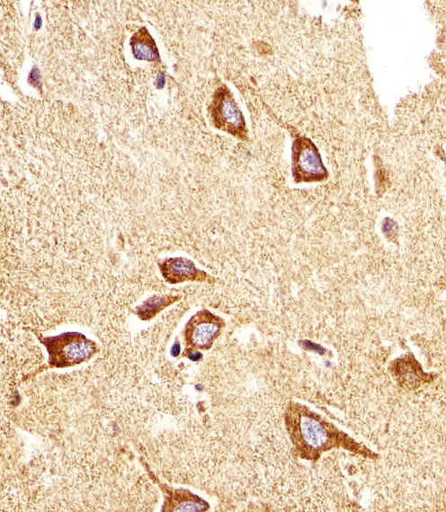
A02153 has real IHC data from a human paraffin-embedded brain section (catalog IHC image caption). Human and mouse reactivity is listed; IF/ICC validation is not listed (catalog: reactivity and applications).

Real IHC data Immunohistochemical analysis of paraffin-embedded H. brain section using PTGER4 Antibody (Center). A02153 was diluted at 1:25 dilution. A undiluted biotinylated goat polyvalent antibody was used as the secondary, followed by DAB staining.
Anti-PTGER4 Antibody (Center)
Cat # A02153

A02153 is listed for IHC-P and human and mouse reactivity (catalog: applications and reactivity). Its IHC figure shows a paraffin-embedded human brain section stained at 1:25 with DAB detection (catalog IHC image caption).

Which to pick: Choose A02153 for paraffin-section tissue IHC: its polyclonal antibody is listed for IHC-P, and its own image documents staining in a human paraffin-embedded brain section (catalog: dilution_raw; catalog IHC image caption). A02153 lists human and mouse reactivity, but the supplied IHC figure documents only a human sample; the fixative is unreported (catalog: reactivity; catalog IHC image caption). No listed SKU has IF/ICC validation, so this payload does not support an IF/ICC pick (catalog: applications).

Each figure is that product's own IHC / IF validation image from its datasheet.

References

  1. UniProt Consortium. UniProt entry P35408 (PE2R4_HUMAN, Prostaglandin E2 receptor EP4 subtype).
  2. Human Protein Atlas. PTGER4 tissue IHC expression (reliability: Approved).
  3. Human Protein Atlas. PTGER4 subcellular location (ICC-IF): Membrane.
  4. Human Protein Atlas. PTGER4 antibody validation summary (2 antibodies).
  5. Expressions of shox2, RASSF1A and PTGER4, and the relationship between their methylation and clinicopathological characteristics in patients with lung cancer. Journal of medical biochemistry 2025 — PMC12085179.
  6. Hypoxia and prostaglandin E receptor 4 signalling pathways synergise to promote endometrial adenocarcinoma cell proliferation and tumour growth. PloS one 2011 — PMC3093383.
  7. TFF3 promotes clonogenic survival of colorectal cancer cells through upregulation of EP4 via activation of STAT3. Translational cancer research 2023 — PMC10331714.
  8. Estradiol-17beta, prostaglandin E2 (PGE2), and the PGE2 receptor are involved in PGE2 positive feedback loop in the porcine endometrium. Endocrinology 2009 — PMC2714846.
  9. PubMed PMID:8163486 — UniProt-cited evidence.
  10. PubMed PMID:8250933 — UniProt-cited evidence.
  11. PubMed PMID:8661119 — UniProt-cited evidence.