PTGES2 / Prostaglandin E synthase 2 · IHC design guide

Design Immunohistochemistry for PTGES2

Plan PTGES2 IHC around granular cytoplasmic tissue staining (HPA tissue IHC) and an antibody starting concentration of 2–5 μg/ml (datasheet A06706-2). Compare high staining in kidney tubules with low staining in hippocampal neurons (HPA tissue IHC), while accounting for cleavage from a Golgi-bound to a cytoplasmic form (UniProt).

Evidence assembled Oct 2026 · For research use; verify linked source records and product datasheet before use
Immunohistochemistry protocol sheet for PTGES2 (IHC for PTGES2): expected localisation Granular cytoplasm (HPA tissue IHC), antibody A06706-2, validated IHC image, and IHC protocol steps
Printable PTGES2 IHC protocol sheet — expected localisation Granular cytoplasm (HPA tissue IHC), antibody A06706-2, controls and protocol steps. Open the full PTGES2 IHC guide →

PTGES2 Immunohistochemistry Experimental Design Guide

Expected localisation, validated protocols, controls and antibodies — the at-a-glance facts below, then the full design guide.

Must know before staining
Expected localisation Granular cytoplasm (HPA tissue IHC)
Staining pattern Granular cytoplasmic staining across tissue cell types (HPA tissue IHC)
Antigen retrieval EDTA pH 8.0 HIER, heat-mediated (datasheet A06706-2)
Positive control ⓘ Adrenal gland+4 more · see all
Negative control ⓘ Soft tissue
Important caveats
Reasons your staining may differ from the expected pattern.
Fixation Keep fixation consistent across sections. (standard IHC practice; not target-specific)
Caveat Cleavage can shift signal from Golgi to cytoplasm (UniProt)
Regulation Tissue-dependent expression (UniProt)
Isoform / epitope No isoforms; cleavage removes aa 1–87, retaining aa 88–377 (UniProt)
Section 1

Recommended PTGES2 IHC & IF Protocols

The catalog antibody protocol uses EDTA pH 8.0 retrieval (datasheet A06706-2). Two published PTGES2 IHC protocols provide additional conditions for renal carcinoma and endometrial specimens (PMC12484182; PMC5080328).

Recommended immunohistochemistry (IHC-P) protocol parameters
SampleParaffin-embedded human breast cancer tissue; fixative not specified (datasheet A06706-2)
FixationImage fixative and duration unreported (datasheet A06706-2); verify before use.
Sectioning4–5 µm sections on charged slides (standard)
DeparaffinisationXylene, graded ethanol series to water (standard)
Antigen retrievalHeat retrieval: EDTA pH 8.0 (datasheet A06706-2); 20 min, 95–100 °C (standard)
Peroxidase block3% H2O2, 10 min, room temperature (standard)
Blocking10% goat serum (datasheet A06706-2)
Primary antibodyRabbit anti-PTGES2, 2-5 μg/ml (datasheet A06706-2)
Primary incubationOvernight at 4 °C (datasheet A06706-2)
DetectionStreptavidin-biotin complex (SABC), DAB chromogen (datasheet A06706-2)
CounterstainHematoxylin, blue, dehydrate and mount (standard)
Expected resultPTGES2-positive staining in glandular cells of adrenal gland (HPA tissue IHC: High). HPA tissue profile: General cytoplasmic expression with a granular pattern. No signal in the no-primary control.
💡Decision noteStart with EDTA pH 8.0 heat retrieval (datasheet A06706-2). If staining needs optimization, compare the published Tris-EDTA pH 9.0 condition (PMC12484182).
Section 2

What Is the Expected PTGES2 Staining Pattern?

PTGES2 IHC should show mainly granular cytoplasmic staining in appropriate cells (HPA tissue IHC: Approved; medium consistency with RNA). A perinuclear emphasis is plausible because the predominant cleaved form is cytoplasmic and enriched there; the precursor is Golgi membrane bound, with one transmembrane segment at residues 58–74 (UniProt Q9H7Z7: processing and topology). Judge the pattern within each tissue’s documented cell type, rather than treating every cell as equally positive (HPA tissue IHC).

What am I looking at on my slide?
Granular cytoplasmic signal, sometimes concentrated around the nucleus, in kidney tubule cells or heart cardiomyocytes.This fits the reported tissue pattern and High staining in those cell types (HPA tissue IHC). Perinuclear enrichment also fits the predominant soluble, cleaved form (UniProt Q9H7Z7: processing). Compare cell identity, compartment, and intensity together; a brown deposit alone is insufficient to establish a PTGES2-positive cell (general IHC interpretation).
Predominantly nuclear, sharply membranous, or extracellular deposit, with little granular cytoplasmic signal.The dominant compartment conflicts with the reported granular cytoplasmic IHC profile (HPA tissue IHC) and the cytoplasmic, perinuclear cleaved form (UniProt Q9H7Z7: subcellular location). Review morphology and controls before calling it specific; the Golgi-bound precursor permits intracellular membrane-associated signal, but does not by itself explain strong surface or extracellular staining (UniProt Q9H7Z7: processing and topology).
Strong staining in peripheral nerve within soft tissue, or in cells other than those expected in a positive tissue.Peripheral nerve was Not detected in the HPA soft-tissue example (HPA tissue IHC). Unexpected staining can reflect cross-reactivity or endogenous chromogen activity (general IHC practice); it is a reason to check controls and cell identity, not proof of either cause. Tissue context matters because PTGES2 expression is broad and staining differs among cell types (UniProt Q9H7Z7: tissue specificity; HPA tissue IHC).
Uniform haze, precipitate, or colour across multiple compartments and cell types.This obscures the granular cytoplasmic pattern reported for PTGES2 (HPA tissue IHC). Assess reagent-only and negative-control sections for detection background, incomplete blocking, or chromogen deposit (general IHC practice). Do not score diffuse colour as target expression solely because the specimen contains an HPA-listed positive cell type (HPA tissue IHC).
No staining in kidney tubule cells or heart cardiomyocytes on an otherwise interpretable section.Both are reported as High IHC cell populations, so an absent signal raises concern about assay performance (HPA tissue IHC). Check the positive control, antibody use, retrieval, and detection steps (general IHC practice). HPA’s Approved rating has medium consistency with RNA, so one negative section should not be treated as definitive evidence that the target is absent (HPA tissue IHC).
💡Expected PTGES2 appearanceCall positive a predominantly granular cytoplasmic, possibly perinuclear signal in documented cells such as High kidney tubule cells; dominant nuclear or diffuse extracellular colour is suspicious (HPA tissue IHC; UniProt Q9H7Z7: subcellular location).
How each factor affects the staining
Processing and compartmentPTGES2 is synthesized as a Golgi membrane-bound 1–377 protein and cleaved to a predominant soluble 88–377 form enriched in perinuclear cytoplasm (UniProt Q9H7Z7: chains and subcellular location). Interpret mixed intracellular staining in that context; these records do not give the catalog antibody’s epitope or prove which form its IHC signal detects.
Cell-type selectionHPA reports High staining in adrenal and endometrial glandular cells, kidney tubule cells, cardiomyocytes, pancreatic exocrine cells, parathyroid and rectal glandular cells, and Leydig cells (HPA tissue IHC). It reports Low staining in several other cell types, so match intensity to the cell population when choosing a comparison section (HPA tissue IHC).
Strength of validationThe tissue IHC profile is Approved, with medium consistency between antibody staining and RNA expression; HPA020733 is listed as IHC Approved (HPA tissue IHC; HPA antibodies). These labels support pattern comparison but do not establish that every brown cell is specific. No IHC Enhanced designation is supplied for this antibody (HPA antibodies).
Antigen retrieval and epitopeNo retrieval condition or antibody epitope is supplied here. The transmembrane span is 58–74, while the cleaved chain begins at 88 (UniProt Q9H7Z7: topology and chains). Whether processing changes recognition depends on the unknown epitope; choose and assess retrieval with suitable controls as general IHC optimization, without assuming a PTGES2-specific fixation effect.
Chromogenic detectionEndogenous enzyme activity and nonspecific reagent binding can add colour in chromogenic IHC (general IHC practice). Use detection and negative controls to separate those signals from the reported granular cytoplasmic profile (HPA tissue IHC). The supplied HPA and UniProt records provide no target-specific claim about endogenous activity or fixation sensitivity.
Why is my staining missing, weak or wrong?
SituationLikely causeNext action
Positive-control tubules or cardiomyocytes show no signal.A failed staining step is possible; these populations are High in HPA IHC (HPA tissue IHC).Check control section quality, primary-antibody application, retrieval, and detection reagents (general IHC practice). Record the actual conditions tested; no PTGES2-specific retrieval setting is supplied.
Only nuclei stain strongly.Nuclear dominance conflicts with the reported granular cytoplasmic pattern (HPA tissue IHC).Check counterstain and negative-control sections, then reassess whether true cytoplasmic granules are present (general IHC practice). Do not score nuclear colour alone as PTGES2-positive.
Brown colour appears in peripheral nerve or many unexpected cells.Peripheral nerve was Not detected in the HPA soft-tissue example; cross-reactivity or detection background is possible (HPA tissue IHC; general IHC practice).Compare matched negative and detection controls, inspect morphology, and require the expected intracellular pattern before assigning positivity (general IHC practice).
Granular signal is buried under diffuse background.Background can conceal the reported cytoplasmic granules (HPA tissue IHC; general IHC practice).Review blocking, washes, chromogen development, and detection-only controls (general IHC practice). Adjust the step implicated by the control, then compare the same documented cell type.
A faint sample is being called negative.Intensity varies by cell type; HPA reports Low staining in neuronal, squamous epithelial, salivary glandular, and smooth muscle cells (HPA tissue IHC).Compare with a documented High cell population and the negative control on the same run (HPA tissue IHC; general IHC practice). Report faint, localized signal separately from complete absence.
Can IF/ICC confirm the IHC compartment?HPA supplies no main ICC-IF location, cell-line images, or ICC validation entry for HPA020733 (HPA subcellular; HPA antibodies).Treat IF/ICC as a separate assay requiring its own controls and validation (general IF practice). Use the IHC tissue pattern for IHC interpretation; no IF/ICC protocol is established by these records.

Sample controls for PTGES2 IHC & IF

🧪Run kidney first: tubular cells should stain (HPA: High in kidney cells in tubules). Use peripheral nerve in soft tissue as the negative tissue (HPA: Not detected in peripheral nerve); on the kidney slide, assess nontubular cells as an internal background reference without assuming they are PTGES2-negative.
Positive control tissue: Adrenal gland (Glandular cells, HPA High)
Negative control tissue: Soft tissue (HPA Not detected)
ICC-IF cell lines (HPA subcellular resource): HPA carries no ICC-IF cell line for PTGES2; derive a cell-line control from the positive tissue's cell type (Glandular cells) and confirm it by RNA or western blot first.
Technical controls: Include a no-primary, secondary-only control, a host-matched rabbit IgG isotype control, and PTGES2 knockout tissue if available (selected-SKU caption: rabbit primary antibody). With the caption’s biotin-based, DAB detection, control endogenous biotin and peroxidase activity in kidney sections (selected-SKU caption: biotinylated secondary, SABC and DAB; standard IHC practice).
⚠️Feasibility: No target-specific fixation window or fixation effect is reported, and the selected-SKU paraffin-section caption does not state the fixative (selected-SKU caption: fixative unreported). The caption uses heat retrieval in EDTA at pH 8.0, but does not establish that retrieval is required (selected-SKU caption: EDTA retrieval). Whether frozen sections or IF are easier is unestablished; no HPA ICC-IF image-bearing cell lines are supplied (HPA subcellular: none), and endogenous biotin may complicate the caption’s detection method in kidney (selected-SKU caption: SABC; standard IHC practice).

HPA tissue IHC evidence for PTGES2

Comprehensive Human Protein Atlas IHC scoring per tissue (reliability: Approved — Medium consistency between antibody staining and RNA expression data.). Rows are taken directly from the HPA tissue chart — click any row's HPA link to view the source.

Positive expression · recommended positive controls

TissueCell typeLevelEvidenceSource
Adrenal gland Glandular cells High Protein (IHC) HPA →
Endometrium Glandular cells High Protein (IHC) HPA →
Heart muscle Cardiomyocytes High Protein (IHC) HPA →
Kidney Cells in tubules High Protein (IHC) HPA →
Pancreas Exocrine glandular cells High Protein (IHC) HPA →

Undetected expression · recommended negative controls

TissueCell typeLevelEvidenceSource
Soft tissue Peripheral nerve Not detected Protein (IHC) HPA →
Section 3

Advanced PTGES2 IHC Tips

Troubleshoot PTGES2 staining in paraffin sections by checking retrieval, controls, compartment, and cell type before interpreting chromogenic signal.

Which retrieval condition should I try first when PTGES2 staining is weak?
Start with heat-mediated retrieval in EDTA at pH 8.0 for paraffin sections (datasheet A06706-2). The selected tissue image used this condition before incubation with 2 μg/ml primary antibody overnight at 4°C, so keep those steps consistent when assessing retrieval (datasheet A06706-2). Compare a known high-staining cell population, such as kidney tubule cells, across runs while holding detection and exposure to DAB constant (HPA: High in kidney tubule cells; standard IHC practice). If staining remains weak, test an alternative retrieval buffer as a controlled fallback, changing only one variable at a time (standard IHC practice). Excess retrieval can damage tissue morphology or increase background, so assess both signal and section integrity (standard IHC practice).
Can fixation explain weak PTGES2 staining in paraffin sections?
Target-specific fixation sensitivity is unknown because the selected paraffin-section caption does not report a fixative (datasheet A06706-2). Record the fixative and processing history for each block, then compare sections processed alike before attributing intensity differences to PTGES2 biology (standard IHC practice). Use the documented EDTA retrieval at pH 8.0 and the same antibody concentration, 2 μg/ml, when comparing blocks (datasheet A06706-2). Inspect nuclear detail and tissue preservation alongside the cytoplasmic signal; poorly preserved sections make staining intensity difficult to interpret (standard IHC practice). The reported HPA staining pattern establishes neither an optimal fixation condition nor a PTGES2-specific response to fixation (HPA: general granular cytoplasmic expression; datasheet A06706-2: fixative unreported).
Should PTGES2 appear in the nucleus or around it?
Evaluate chromogenic staining primarily in the cytoplasm, including a granular or perinuclear distribution (HPA: general granular cytoplasmic expression; UniProt Q9H7Z7: perinuclear cytoplasm). Full-length PTGES2 is synthesized at the Golgi membrane, while cleavage yields a predominant soluble form spanning residues 88–377 that is enriched near the nucleus (UniProt Q9H7Z7: processing and localisation). A perinuclear accent can therefore fit the record, but DAB alone cannot establish that individual granules are Golgi structures (UniProt Q9H7Z7: localisation; standard IHC practice). Treat isolated nuclear staining as questionable and compare it with a no-primary control, adjacent morphology, and a known positive cell population (HPA: cytoplasmic profile; standard IHC practice). Keep staining and counterstain settings comparable across sections before assigning a compartment pattern (standard IHC practice).
Could antibody epitope placement change which PTGES2 form I detect?
Check the catalog antibody’s stated immunogen or epitope before interpreting differences between the membrane-associated and soluble forms (UniProt Q9H7Z7: processing; standard IHC practice). PTGES2 has one annotated full-length chain, residues 1–377, and a processed chain, residues 88–377; the record lists 0 alternative isoforms (UniProt Q9H7Z7: processing and isoforms). An epitope within residues 1–87 would not occur in the processed chain, whereas an epitope within 88–377 could occur in both (UniProt Q9H7Z7: processing). The transmembrane segment spans residues 58–74, with residues 75–377 on the cytoplasmic side (UniProt Q9H7Z7: topology). If the antibody epitope is undisclosed, report the observed IHC pattern without assigning it to either chain (standard IHC practice).
How can I investigate PTGES2 localisation by IF after chromogenic IHC?
Use IF/ICC as a separate optimisation experiment; the selected antibody caption documents paraffin-section chromogenic IHC, with no IF/ICC conditions supplied here (datasheet A06706-2). Multiplex PTGES2 with a marker of the expected cell type, such as a cardiomyocyte marker in heart muscle, and include single-label controls to check channel overlap (HPA: High in cardiomyocytes; standard IF practice). Choose fluorophores after inspecting unstained tissue autofluorescence; a longer-wavelength channel may improve separation where shorter-wavelength background is strong (standard IF practice). Set permeabilisation according to the verified epitope: residues 75–377 face the cytoplasm, whereas residues 1–57 are lumenal in the membrane-bound form (UniProt Q9H7Z7: topology). Confirm any apparent perinuclear signal with morphology and controls before comparing it with DAB staining (UniProt Q9H7Z7: localisation; standard IF/IHC practice).
How do I distinguish diffuse background from PTGES2 staining?
Compare a no-primary control with the complete staining run to identify signal from the secondary and detection reagents (standard IHC practice). The selected paraffin-section protocol used 10% goat serum block, biotinylated goat anti-rabbit secondary, streptavidin-biotin detection, and DAB (datasheet A06706-2). Check endogenous peroxidase blocking and consider endogenous biotin when background follows tissue structures independently of the primary antibody (standard chromogenic IHC practice). Titrate the primary around the documented 2 μg/ml condition while keeping retrieval at EDTA pH 8.0 and development conditions consistent (datasheet A06706-2; standard IHC practice). Prefer reproducible granular cytoplasmic signal in appropriate cells over uniform haze or staining confined to section edges (HPA: granular cytoplasmic profile; standard IHC practice).
What should I score when comparing PTGES2 across paraffin sections? ⚠ ANSWER MARKED FOR VERIFICATION
Define the cell population and tissue region before scoring PTGES2, then apply the same rules to every paraffin section (standard IHC practice). For cytoplasmic DAB, record the percentage of positive cells and staining intensity, combining them as an H-score if that scale suits the study (HPA: general cytoplasmic expression; standard IHC practice). Normalise counts to the number of eligible cells, or report positive-cell density per mm² of viable tissue when sampling areas differ (standard IHC practice). Exclude necrosis, folds, and edges using prespecified criteria, and keep retrieval, antibody incubation, and DAB development consistent across batches (standard IHC practice). Report cell types separately because HPA describes High staining in kidney tubule cells and Low staining in hippocampal neuronal cells (HPA: tissue IHC).
When is an apparent PTGES2-positive result likely to be artefactual?
Treat reproducible granular cytoplasmic staining in an appropriate cell population as more credible than isolated nuclear or edge-confined DAB (HPA: general granular cytoplasmic expression; standard IHC practice). Kidney tubule cells and cardiomyocytes are reported as High, while peripheral nerve in soft tissue is reported as Not detected; use such contrasts to assess assay behaviour without treating them as absolute biological rules (HPA: tissue IHC). Check whether signal tracks necrosis, damaged section edges, or endogenous enzyme activity in the no-primary control (standard chromogenic IHC practice). A perinuclear accent is plausible for the processed PTGES2 form, but compartment appearance alone does not prove which chain is present (UniProt Q9H7Z7: processing and localisation). Interpret intensity cautiously because HPA assigns Approved reliability with medium consistency between antibody staining and RNA expression (HPA: tissue IHC reliability).
Boster reagents

Best PTGES2 / Prostaglandin E synthase 2 IHC Antibodies

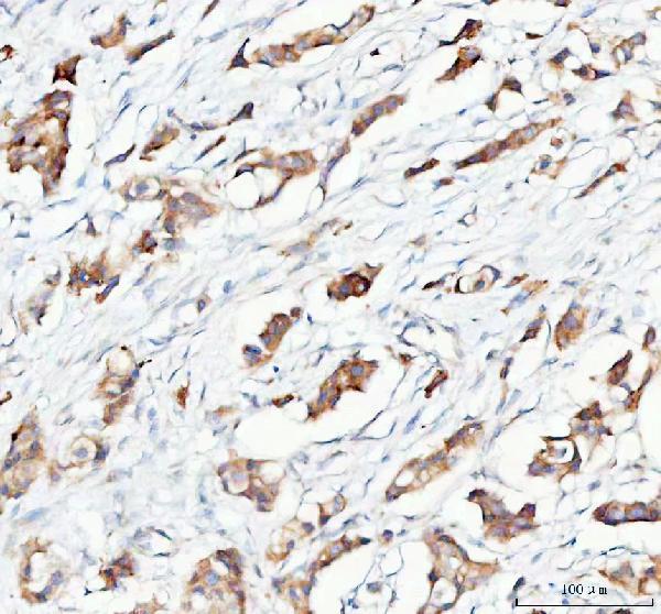
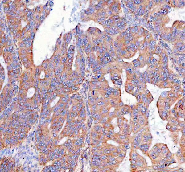
A06706-2 has real IHC data from paraffin-embedded human breast and ovarian cancer sections and IF data from Hela cells (catalog image captions). Listed reactivity includes human, mouse and rat (catalog reactivity).

Real IHC data IHC analysis of PTGES2/Gbf1 using anti-PTGES2/Gbf1 antibody (A06706-2). PTGES2/Gbf1 was detected in a paraffin-embedded section of human breast cancer tissue. Heat mediated antigen retrieval was performed in EDTA buffer (pH 8.0, epitope retrieval solution). The tissue section was blocked with 10% goat serum. The tissue section was then incubated with 2 μg/ml rabbit anti-PTGES2/Gbf1 Antibody (A06706-2) overnight at 4°C. Biotinylated goat anti-rabbit IgG was used as secondary antibody and incubated for 30 minutes at 37°C. The tissue section was developed using Strepavidin-Biotin-Complex (SABC) (Catalog # SA1022) with DAB as the chromogen.
Anti-PTGES2/Gbf1 Antibody ®
Cat # A06706-2

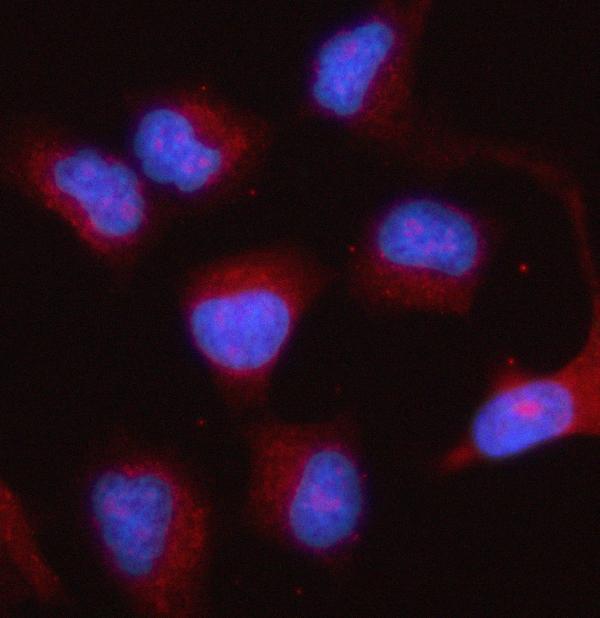
A06706-2 shows IHC staining in paraffin-embedded human breast and ovarian cancer sections (catalog IHC captions). The same SKU shows IF staining in Hela cells and lists both IF and ICC applications (catalog IF caption; catalog applications).

Which to pick: For tissue IHC, choose A06706-2: its own images document paraffin-embedded human sections stained at 2 μg/ml; the fixative is unreported (catalog IHC captions). For IF/ICC, the same rabbit-host SKU lists both applications and shows IF staining in Hela cells at 5 μg/ml; clonality is unspecified (catalog host/clone; catalog applications; catalog IF caption). For mouse or rat samples, A06706-2 lists reactivity with both species, while its supplied IHC and IF images document human samples (catalog reactivity; catalog image captions).

Each figure is that product's own IHC / IF validation image from its datasheet.

References

  1. UniProt Consortium. UniProt entry Q9H7Z7 (PGES2_HUMAN, Prostaglandin E synthase 2).
  2. Human Protein Atlas. PTGES2 tissue IHC expression (reliability: Approved).
  3. Human Protein Atlas. PTGES2 subcellular location (ICC-IF): Variation in transcript expression is not correlated to the cell cycle..
  4. Human Protein Atlas. PTGES2 antibody validation summary (1 antibodies).
  5. PTGES Expression Is Associated with Metabolic and Immune Reprogramming in Pancreatic Ductal Adenocarcinoma. International journal of molecular sciences 2023 — PMC10138618.
  6. Comprehensive analysis of metabolic patterns in renal cell carcinoma: implications for prognosis and treatment. Frontiers in immunology 2025 — PMC12484182.
  7. Prostaglandin E2 (PGE2) promotes proliferation and invasion by enhancing SUMO-1 activity via EP4 receptor in endometrial cancer. Tumour biology : the journal of the International Society for Oncodevelopmental Biology and Medicine 2016 — PMC5080328.
  8. Inhibiting cholesterol de novo synthesis promotes hepatocellular carcinoma progression by upregulating prostaglandin E synthase 2-mediated arachidonic acid metabolism under high fatty acid conditions. Cancer science 2024 — PMC10859596.
  9. PubMed PMID:14702039 — UniProt-cited evidence.
  10. PubMed PMID:15164053 — UniProt-cited evidence.
  11. PubMed PMID:15489334 — UniProt-cited evidence.