This website uses cookies to ensure you get the best experience on our website.


- Table of Contents
Plan PTP4A2 paraffin-section IHC using the expected cytoplasmic tissue pattern and high staining in colon glandular cells (HPA tissue IHC). Interpret staining with the reported multi-gene targeting caution (HPA tissue IHC) and four isoforms in mind (UniProt).

Expected localisation, validated protocols, controls and antibodies — the at-a-glance facts below, then the full design guide.
| Expected localisation | Cytoplasmic staining in most tissues (HPA tissue IHC) | |
| Staining pattern | Cytoplasmic staining in colon glandular cells (HPA tissue IHC) | |
| Antigen retrieval | Citrate pH 6 HIER, heat-mediated (datasheet PB9740) | |
| Positive control | Appendix+4 more · see all | |
| Negative control | Adipose tissue+4 more · see all |
| Fixation | Keep fixation consistent across sections. (standard IHC practice; not target-specific) | |
| Caveat | Staining may reflect protein from more than one gene (HPA tissue IHC) | |
| Regulation | Overexpressed in prostate tumor tissue (UniProt) | |
| Isoform / epitope | Four isoforms; check epitope coverage across variants (UniProt) |
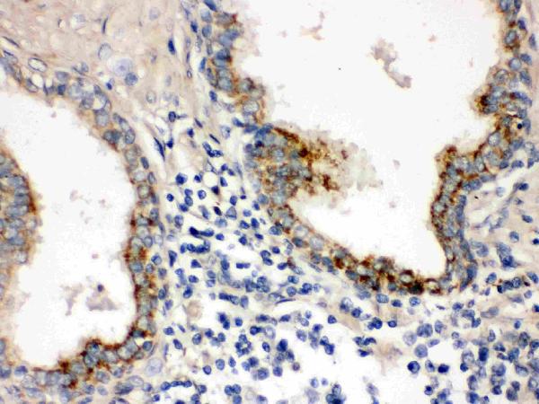
The catalog antibody's IHC-P protocol (datasheet: PB9740) is paired with a published nasopharyngeal carcinoma tissue protocol (PMC5649923).
| Sample | Paraffin-embedded Human Prostatic Cancer tissue; fixative not specified (datasheet PB9740) |
| Fixation | Image fixative and duration unreported (datasheet PB9740); verify before use. |
| Sectioning | 4–5 µm sections on charged slides (standard) |
| Deparaffinisation | Xylene, graded ethanol series to water (standard) |
| Antigen retrieval | Heat retrieval: Citrate pH 6, 20 min (datasheet PB9740) |
| Peroxidase block | 3% H2O2, 10 min, room temperature (standard) |
| Blocking | 10% goat serum (datasheet PB9740) |
| Primary antibody | Rabbit anti-PTP4A2, 0.5-1μg/ml (datasheet PB9740) |
| Primary incubation | Overnight at 4 °C (datasheet PB9740) |
| Detection | Streptavidin-biotin complex (SABC), DAB chromogen (datasheet PB9740) |
| Counterstain | Hematoxylin, blue, dehydrate and mount (standard) |
| Expected result | PTP4A2-positive staining in glandular cells of appendix (HPA tissue IHC: High). HPA tissue profile: Cytoplasmic expression in most tissues. No signal in the no-primary control. |
PTP4A2 is reported in the cytoplasm, early endosome and cell membrane, with no transmembrane segment (UniProt Q12974: subcellular location and topology). In paraffin-section IHC, expect mainly cytoplasmic staining in glandular epithelium and kidney tubules (HPA: cytoplasmic expression in most tissues; High in glandular cells and kidney tubule cells). Treat this as a provisional pattern: HPA rates tissue IHC reliability Uncertain and cautions that the antibodies target protein from more than one gene (HPA: reliability).
| Cytoplasmic signal in appendix, colon or rectum glandular cells, or kidney tubule cells, with preserved tissue detail (HPA: High in these cells). | This matches the reported tissue and compartment pattern (HPA: cytoplasmic expression; High in these cells). It supports a plausible positive result, but staining alone cannot establish PTP4A2 specificity because HPA rates the IHC evidence Uncertain and flags detection of protein from more than one gene (HPA: reliability). |
| Predominantly nuclear staining, especially where cytoplasm remains clear. | A nuclear-only pattern conflicts with the supplied locations: cytoplasm, early endosome and cell membrane (UniProt Q12974: subcellular location; HPA: cytoplasmic tissue expression). Investigate artefact or nonspecific signal before scoring those cells as positive; weak membrane-associated or punctate staining cannot be dismissed solely from compartment because UniProt also reports membrane and endosomal locations (UniProt Q12974: subcellular location). |
| Strong staining in adipocytes, soft-tissue fibroblasts or skeletal-muscle myocytes. | These cell types were Not detected in the supplied HPA tissue IHC observations (HPA: adipose tissue, soft tissue and skeletal muscle). Check cross-reactivity and endogenous chromogen-producing activity as general IHC possibilities. Skeletal muscle needs special caution: UniProt reports high tissue expression there, while HPA reports myocytes Not detected; the sources do not resolve that difference (UniProt Q12974: tissue specificity; HPA: skeletal muscle). |
| Diffuse colour over epithelium, stroma and empty spaces, without cell boundaries or a clear cytoplasmic distribution. | That distribution does not resemble HPA's cellular cytoplasmic pattern (HPA: tissue IHC profile). Treat it as background until controls separate staining from chromogen deposition or endogenous detection activity; assessing tissue-free regions and a primary-antibody omission control is general IHC practice. |
| No signal in well-preserved appendix or colon glandular cells, or kidney tubules. | A negative result conflicts with the supplied High observations in those cells (HPA: appendix, colon and kidney). First assess the detection run and tissue identity using controls, then review the antibody's documented IHC-P conditions if available (general IHC practice). HPA's Uncertain reliability means a single negative section does not settle true absence or antibody performance (HPA: reliability). |
| Tissue and cell selection (HPA: tissue IHC profile). | Appendix, colon and rectum glandular cells and kidney tubule cells were High; adipocytes and soft-tissue fibroblasts were Not detected (HPA: named tissues and cells). Compare the named cell types within intact tissue rather than assigning one expected intensity to every cell (general IHC interpretation practice). |
| Compartment interpretation (UniProt Q12974: subcellular location and topology). | HPA describes mainly cytoplasmic tissue expression (HPA: tissue IHC profile). UniProt also places PTP4A2 at the cell membrane and early endosome and lists no transmembrane segment (UniProt Q12974: subcellular location and topology). The supplied HPA IHC data do not verify a separate membranous or punctate pattern. |
| Antibody evidence (HPA: antibody validation and tissue reliability). | Both listed antibodies, HPA003281 and CAB011204, have IHC status Uncertain; neither has an Enhanced IHC designation in the supplied record (HPA: antibodies). HPA also cautions that the tissue-staining antibodies target protein from more than one gene, so matching morphology is supportive but not proof of PTP4A2 identity (HPA: reliability). |
| Isoforms and epitope coverage (UniProt Q12974: isoforms and processing). | UniProt lists four isoforms and a chain spanning residues 1–164, with a modified cysteine at 164 (UniProt Q12974: isoforms, processing and modified residues). No antibody epitope is supplied, so the fraction of isoforms detected and any effect of that modification on staining are unknown; do not infer either from signal intensity. |
| IF/ICC question: Is a cellular fluorescence pattern established here? | No: HPA supplies no ICC-IF images or main subcellular location for this record (HPA: subcellular record). UniProt lists cytoplasm, early endosome and cell membrane as locations, but these do not validate an IF pattern for a particular antibody (UniProt Q12974: subcellular location; HPA: subcellular record). |
| Situation | Likely cause | Next action |
|---|---|---|
| High HPA reference cells appear blank (HPA: appendix, colon or kidney). | The detection run, tissue selection or antibody conditions may have failed; absence is difficult to interpret with Uncertain IHC evidence (HPA: tissue profile and reliability; general IHC practice). | Confirm the named cells on the section, check a working positive control and review the antibody's documented IHC-P detection and retrieval conditions if available (general IHC practice). Avoid declaring biological absence from one section. |
| Most signal is nuclear despite clear cytoplasm. | The pattern conflicts with the supplied cytoplasmic tissue profile and UniProt locations (HPA: tissue IHC profile; UniProt Q12974: subcellular location). Nonspecific staining is a possible general IHC explanation. | Compare the primary-antibody omission control and cell morphology; repeat evaluation on a named HPA High tissue before assigning a positive score (HPA: tissue IHC profile; general IHC practice). |
| Adipocytes or soft-tissue fibroblasts stain as strongly as nearby epithelium. | HPA records those specific cell types as Not detected (HPA: adipose tissue and soft tissue). Cross-reactivity or endogenous detection activity is possible in chromogenic IHC (general IHC practice). | Inspect control sections for endogenous colour, use the appropriate enzyme-blocking step for the detection chemistry, and score named cell types separately (general IHC practice). |
| Colour is diffuse across tissue and blank spaces. | The distribution lacks the cellular cytoplasmic pattern reported by HPA (HPA: tissue IHC profile). General IHC causes include excess background from detection reagents or inadequate washing. | Check the primary-antibody omission control, reagent-only areas and washes; interpret only signal that resolves to intact cells (general IHC practice). |
| Skeletal-muscle myocytes are negative, but another source suggests high expression. | The sources differ: UniProt reports high skeletal-muscle expression, whereas HPA reports myocytes Not detected by tissue IHC (UniProt Q12974: tissue specificity; HPA: skeletal muscle). The supplied record cannot explain the discrepancy. | Document the discrepancy and use HPA High glandular cells or kidney tubules to assess this IHC run; do not force skeletal muscle into a positive-control role (HPA: tissue IHC profile; general IHC practice). |
| A positive section shows membrane-edge or punctate signal alongside cytoplasm. | Membrane and early-endosome localization are compatible with UniProt, but HPA's supplied tissue profile reports cytoplasmic expression without validating those finer patterns (UniProt Q12974: subcellular location; HPA: tissue IHC profile). | Record the compartment separately from the main cytoplasmic result, compare controls, and avoid calling the fine pattern PTP4A2-specific without further antibody validation (HPA: Uncertain IHC reliability; general IHC practice). |


Comprehensive Human Protein Atlas IHC scoring per tissue (reliability: Uncertain — Medium consistency between antibody staining and RNA expression data. Caution, targets protein from more than one gene.). Rows are taken directly from the HPA tissue chart — click any row's HPA link to view the source.
| Tissue | Cell type | Level | Evidence | Source |
|---|---|---|---|---|
| Appendix | Glandular cells | High | Protein (IHC) | HPA → |
| Colon | Glandular cells | High | Protein (IHC) | HPA → |
| Epididymis | Glandular cells | High | Protein (IHC) | HPA → |
| Kidney | Cells in tubules | High | Protein (IHC) | HPA → |
| Rectum | Glandular cells | High | Protein (IHC) | HPA → |
| Tissue | Cell type | Level | Evidence | Source |
|---|---|---|---|---|
| Adipose tissue | Adipocytes | Not detected | Protein (IHC) | HPA → |
| Cerebral cortex | Endothelial cells | Not detected | Protein (IHC) | HPA → |
| Lymph node | Germinal center cells | Not detected | Protein (IHC) | HPA → |
| Skeletal muscle | Myocytes | Not detected | Protein (IHC) | HPA → |
| Smooth muscle | Smooth muscle cells | Not detected | Protein (IHC) | HPA → |
Troubleshoot PTP4A2 chromogenic IHC by checking retrieval, tissue processing, cellular localisation, controls, and scoring against the documented paraffin-section workflow.
PB9740 has real IHC data from a human prostatic cancer paraffin section (PB9740 image caption). Human and rat are listed as reactive species (catalog: PB9740 reactivity).
PB9740 is the only SKU that will render; its IHC figure shows staining in a human prostatic cancer paraffin section (PB9740 image caption). Its listed applications include IHC, while no IF figure or IF/ICC application is provided (catalog: PB9740 applications and IF images).
Which to pick: Choose PB9740 for paraffin-section tissue IHC based on its own human tissue figure; the fixative is unreported (PB9740 image caption). For IF/ICC, PB9740 has no listed application or figure, and its clonality is unspecified (catalog: PB9740 applications, IF images, clone). For cross-species planning, PB9740 lists human and rat reactivity, but its shown IHC evidence covers human tissue only (catalog: PB9740 reactivity; PB9740 image caption).