PTPN1 / Tyrosine-protein phosphatase non-receptor type 1 · IHC design guide

Design Immunohistochemistry for PTPN1

Plan paraffin-section PTPN1 IHC around cytoplasmic staining, mainly in immune cells (HPA tissue IHC). Lung macrophages and lymph-node germinal-center cells show high staining, while heart cardiomyocytes are not detected (HPA tissue IHC).

Evidence assembled Oct 2026 · For research use; verify linked source records and product datasheet before use
Immunohistochemistry protocol sheet for PTPN1 (IHC for PTPN1): expected localisation Cytoplasm (HPA tissue IHC); ER membrane (UniProt), antibody A00613-1, validated IHC image, and IHC protocol steps
Printable PTPN1 IHC protocol sheet — expected localisation Cytoplasm (HPA tissue IHC); ER membrane (UniProt), antibody A00613-1, controls and protocol steps. Open the full PTPN1 IHC guide →

PTPN1 Immunohistochemistry Experimental Design Guide

Expected localisation, validated protocols, controls and antibodies — the at-a-glance facts below, then the full design guide.

Must know before staining
Expected localisation Cytoplasm (HPA tissue IHC); ER membrane (UniProt)
Staining pattern Cytoplasmic staining, mainly in immune cells (HPA tissue IHC)
Antigen retrieval EDTA pH 8.0 HIER, heat-mediated (datasheet A00613-1)
Positive control ⓘ Appendix+4 more · see all
Negative control ⓘ Adipose tissue+4 more · see all
Important caveats
Reasons your staining may differ from the expected pattern.
Fixation Keep fixation consistent across sections. (standard IHC practice; not target-specific)
Caveat Immune-cell staining may obscure other cell populations (HPA tissue IHC)
Regulation Cell-type-dependent intensity (HPA tissue IHC)
Isoform / epitope No isoforms or cleavage reported; map epitopes to 1–435 (UniProt)
Section 1

Recommended PTPN1 IHC & IF Protocols

Compare the catalog antibody’s IHC-P protocol (datasheet: A00613-1) with three published PTPN1 paraffin-section protocols (PMC10272960; PMC6911276; PMC10626546).

Recommended immunohistochemistry (IHC-P) protocol parameters
SampleParaffin-embedded rat spleen tissue; fixative not specified (datasheet A00613-1)
FixationImage fixative and duration unreported (datasheet A00613-1); verify before use.
Sectioning4–5 µm sections on charged slides (standard)
DeparaffinisationXylene, graded ethanol series to water (standard)
Antigen retrievalHeat retrieval: EDTA pH 8.0 (datasheet A00613-1); 20 min, 95–100 °C (standard)
Peroxidase block3% H2O2, 10 min, room temperature (standard)
Blocking10% goat serum (datasheet A00613-1)
Primary antibodyRabbit anti-PTPN1, 2-5μg/ml (datasheet A00613-1)
Primary incubationOvernight at 4 °C (datasheet A00613-1)
DetectionHRP-conjugated secondary, DAB chromogen (datasheet A00613-1)
CounterstainHematoxylin, blue, dehydrate and mount (standard)
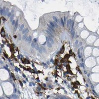
Expected resultPTPN1-positive staining in lymphoid tissue of appendix (HPA tissue IHC: High). HPA tissue profile: Cytoplasmic expression mainly in cells of the immune system. No signal in the no-primary control.
💡Decision noteStart with heat-mediated EDTA, pH 8.0 (datasheet: A00613-1); citrate, pH 6.0, is a published alternative (PMC10272960).
Section 2

What Is the Expected PTPN1 Staining Pattern?

PTPN1 is associated with the endoplasmic reticulum membrane despite having no predicted transmembrane segment (UniProt P18031 topology). In paraffin-section IHC, expect predominantly cytoplasmic staining, especially in immune cells, while assessing each tissue by cell type (HPA: tissue IHC profile). HPA rates its tissue staining Approved, with medium consistency between antibody staining and RNA expression; treat the pattern as a guide for interpretation, not a guarantee for every specimen (HPA: tissue IHC reliability).

What am I looking at on my slide?
Cytoplasmic staining in lung macrophages or lymph node germinal center cells, with identifiable tissue structure.This matches cell populations scored High by HPA and its predominantly cytoplasmic IHC profile (HPA: High in lung macrophages; High in lymph node germinal center cells). Judge the stained cells individually: a positive immune cell does not imply that every neighboring cell should stain.
Predominantly nuclear or sharply surface-restricted staining replaces the expected cytoplasmic pattern.Treat this as a compartment mismatch requiring verification, rather than a convincing positive result: UniProt places PTPN1 at the ER membrane, and HPA describes cytoplasmic tissue staining (UniProt P18031 subcellular location; HPA: tissue IHC profile). UniProt also reports interaction with EPHA3 at the cell membrane, so an isolated surface signal is not sufficient evidence of PTPN1.
Strong staining appears in an HPA-listed negative cell population, such as adipocytes, while expected positive cells are weak.Consider antibody cross-reactivity or endogenous chromogenic detection activity before assigning biological expression (HPA: Not detected in adipocytes; general IHC practice). HPA scores specified cells, not an entire tissue, so check morphology and cell identity before calling a specimen discordant (HPA: tissue IHC cell-level entries).
Uniform color covers stroma, empty spaces, and many unrelated cell types, obscuring boundaries.This is diffuse background rather than an interpretable cell-specific pattern (general IHC practice). Compare a no-primary control and inspect detection and blocking conditions; the HPA cytoplasmic profile cannot validate indiscriminate staining (general IHC practice; HPA: tissue IHC profile).
A section containing identifiable lung macrophages or lymph node germinal center cells shows no signal.HPA scores these cells High, making them useful reference populations, but a negative run alone cannot establish biological absence (HPA: High in lung macrophages; High in lymph node germinal center cells). Check tissue preservation, retrieval, antibody application, and detection with appropriate controls (general IHC practice).
💡Expected PTPN1 appearanceA convincing positive is cell-associated cytoplasmic staining in an expected population, potentially strong in HPA High cells; widespread staining of unrelated cells or a dominant nuclear signal warrants investigation (HPA: tissue IHC profile and cell-level scores; general IHC practice).
How each factor affects the staining
Cell-type distributionHPA scores High staining in lung macrophages, appendix lymphoid tissue, colon endothelial cells, duodenal and fallopian-tube glandular cells, and kidney glomerular cells (HPA: tissue IHC). Its RNA assessment is low tissue specificity, so use cell-level staining observations rather than a tissue-wide positive/negative rule (HPA: RNA specificity).
Subcellular interpretationUniProt places PTPN1 at the ER membrane and records no transmembrane segment; HPA reports predominantly cytoplasmic tissue staining (UniProt P18031 topology and location; HPA: tissue IHC profile). These records support a compartment expectation but do not establish which retrieval condition best exposes an antibody epitope.
Strength of IHC evidenceHPA labels its tissue IHC assessment Approved and reports medium agreement between antibody staining and RNA data (HPA: tissue IHC reliability). The listed antibodies have Approved IHC status; that status does not turn every visible signal into a validated cell-specific result (HPA: antibody validation; general IHC practice).
IF/ICC: What pattern is expected?ER localization is supported, while additional cytosolic localization is uncertain in HPA ICC-IF images (HPA: subcellular location). Use that distinction when interpreting an IF image; the IF/ICC experiment has its own guide, and these localization observations supply no IF protocol conditions.
Why is my staining missing, weak or wrong?
SituationLikely causeNext action
Known-positive reference cells show little or no chromogen.A run or specimen issue may have reduced detectable signal; HPA High is an observed cell-level score, not a promise for each section (HPA: tissue IHC; general IHC practice).Confirm the cells are present, then review the run control, retrieval, antibody application, and detection steps (general IHC practice). Avoid declaring PTPN1 absent from one failed-looking section.
Signal is diffuse across tissue and no-primary control.Endogenous detection activity or nonspecific detection can produce color without a PTPN1-specific primary-antibody signal (general IHC practice).Inspect the no-primary result and the detection system; review the relevant endogenous-activity blocking step before interpreting cell staining (general IHC practice).
Negative-reference adipocytes stain strongly.Cross-reactivity or background is plausible because HPA reports PTPN1 as Not detected in adipocytes (HPA: adipocytes; general IHC practice).Verify adipocyte identity and compare positive and no-primary controls; assess whether staining follows cell boundaries or a broader artifact pattern (general IHC practice).
Only nuclei are prominent in the positive reference.The dominant compartment conflicts with the ER-associated and cytoplasmic expectations (UniProt P18031 location; HPA: tissue IHC profile).Check counterstain versus chromogen, review controls, and reassess antibody and detection specificity before scoring nuclear staining as PTPN1 (general IHC practice).
Expected cell groups appear inconsistent across tissues.HPA reports medium staining/RNA consistency and cell-specific High, Low, and Not detected entries (HPA: tissue IHC reliability and cell-level scores).Record tissue, cell identity, compartment, intensity, and control performance separately; compare each population with its HPA entry rather than averaging whole sections (HPA: tissue IHC; general IHC practice).
A different retrieval condition seems to change intensity.Retrieval can affect IHC staining generally, but target-specific fixation or retrieval sensitivity is unreported in the supplied UniProt and HPA records (general IHC practice; UniProt P18031; HPA: tissue IHC).Evaluate conditions with matched positive and negative reference cells and run controls; report the observed change without attributing it to a proven PTPN1-specific epitope effect (general IHC practice).

Sample controls for PTPN1 IHC & IF

🧪Run appendix first and expect staining in its lymphoid tissue (HPA: Appendix, lymphoid tissue, High). Use adipose tissue adipocytes as the negative comparison (HPA: Adipose tissue, adipocytes, Not detected); candidate internal negative cells outside the appendix lymphoid regions should be unstained or appreciably lighter, but their negativity requires verification because the HPA row specifies only lymphoid tissue (HPA: Appendix, lymphoid tissue, High).
Positive control tissue: Appendix (Lymphoid tissue, HPA High)
Negative control tissue: Adipose tissue (HPA Not detected)
ICC-IF cell lines (HPA subcellular resource): HPA ICC-IF images show PTPN1 in A-431, U-251MG, U2OS, ASC52telo, RPTEC/TERT1, hTERT-RPE1 (serum starved), KOLF2.1J, with annotated localisation: Endoplasmic reticulum (supported) (HPA subcellular).
Technical controls: Include a no-primary, secondary-only control, matched nonimmune rabbit IgG control, and PTPN1 knockout material or a validated immunizing-peptide block as a biological specificity control (caption: rabbit primary and peroxidase-conjugated anti-rabbit secondary; standard IHC practice). Quench endogenous peroxidase and inspect the no-primary appendix section for residual DAB signal in lymphoid regions (HPA: Appendix, lymphoid tissue, High; standard chromogenic IHC practice).
⚠️Feasibility: A target-specific fixation window or fixation effect is unreported, and the selected A00613-1 paraffin-section caption does not state a fixative (selected IHC caption: fixative not stated). The documented IHC workflow uses heat retrieval in EDTA at pH 8.0; retrieval dependence beyond that workflow is unreported (selected IHC caption). Whether frozen sections or tissue IF are easier is unreported; for appendix, endogenous peroxidase can complicate DAB interpretation (selected IHC caption: paraffin section with HRP/DAB; standard IHC practice).

HPA tissue IHC evidence for PTPN1

Comprehensive Human Protein Atlas IHC scoring per tissue (reliability: Approved — Medium consistency between antibody staining and RNA expression data. Antibody staining in cells/structures not annotated, view images.). Rows are taken directly from the HPA tissue chart — click any row's HPA link to view the source.

Positive expression · recommended positive controls

TissueCell typeLevelEvidenceSource
Appendix Lymphoid tissue High Protein (IHC) HPA →
Caudate Glial cells High Protein (IHC) HPA →
Colon Endothelial cells High Protein (IHC) HPA →
Duodenum Glandular cells High Protein (IHC) HPA →
Fallopian tube Glandular cells High Protein (IHC) HPA →

Undetected expression · recommended negative controls

TissueCell typeLevelEvidenceSource
Adipose tissue Adipocytes Not detected Protein (IHC) HPA →
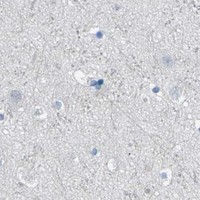
Cerebellum Cells in granular layer Not detected Protein (IHC) HPA →
Endometrium Cells in endometrial stroma Not detected Protein (IHC) HPA →
Gallbladder Glandular cells Not detected Protein (IHC) HPA →
Heart muscle Cardiomyocytes Not detected Protein (IHC) HPA →
Section 3

Advanced PTPN1 IHC Tips

Troubleshoot chromogenic PTPN1 staining in paraffin sections using the selected antibody’s tissue protocol and the expected cellular distribution.

What should I change when PTPN1 staining is weak after retrieval?
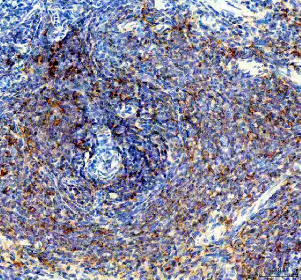
Start with heat-mediated retrieval in EDTA pH 8.0 for this catalog antibody (datasheet A00613-1). The demonstrated rat spleen section used that retrieval before 2 μg/ml primary antibody overnight at 4°C, so check those conditions and the positive control before changing the buffer (datasheet A00613-1). If staining remains weak, vary heating time on adjacent sections while keeping the buffer at pH 8.0, and inspect tissue integrity and background together (standard IHC practice). Score signal in identifiable immune cells rather than treating uniform tissue darkening as improved retrieval (HPA: cytoplasmic expression mainly in immune-system cells; standard IHC practice).
How can I troubleshoot weak PTPN1 staining when fixation history is uncertain?
The selected rat spleen caption identifies a paraffin section but does not state its fixative, so target-specific fixation sensitivity is unknown (datasheet A00613-1). Record the available fixation history for each specimen and compare adjacent sections processed with the same EDTA pH 8.0 retrieval and antibody incubation (datasheet A00613-1; standard IHC practice). Include a section expected to contain PTPN1-positive cells in the same staining run, then assess morphology and background alongside signal (HPA: high in lung macrophages and lymph-node germinal center cells; standard IHC practice). Do not assign weak staining to a particular fixative without a controlled comparison (standard IHC practice).
Where should convincing PTPN1 signal appear in a paraffin section?
Look for cytoplasmic staining in identifiable cells, with a distribution compatible with PTPN1’s endoplasmic reticulum association (HPA: cytoplasmic tissue expression; HPA: supported endoplasmic reticulum location; UniProt P18031: endoplasmic reticulum membrane). Prioritise cell identity over apparent organelle resolution in a chromogenic section, since DAB staining may not resolve the endoplasmic reticulum directly (standard IHC practice). The protein has no annotated transmembrane segment, and its interaction with EPHA3 at the cell membrane does not establish diffuse cell-surface staining as the expected IHC pattern (UniProt P18031 topology and subunit). Compare suspect membrane-only or nuclear-only signal with tissue morphology, controls and the cytoplasmic pattern in the same run (standard IHC practice; HPA: tissue IHC profile).
Could epitope placement or modification explain inconsistent PTPN1 IHC staining?
The supplied record lists one 435-aa chain and 0 isoforms, so it provides no basis for assigning different tissue patterns to annotated isoforms (UniProt P18031). Its protein tyrosine phosphatase domain spans residues 3–277, while several modified residues are annotated, including phosphotyrosine at 20 and 66 (UniProt P18031 domains and modified residues). The antibody epitope is not specified here, so neither a particular modification nor epitope masking can be identified as the cause of weak staining (supplied datasheet A00613-1; UniProt P18031). Compare matched sections under the same retrieval and detection conditions before attributing differences to molecular processing (standard IHC practice).
How should I check a PTPN1 pattern by IF alongside chromogenic IHC?
For a separate IF experiment, multiplex PTPN1 with a marker that identifies the expected cell population, such as a validated macrophage marker when examining lung macrophages (HPA: high PTPN1 in lung macrophages; standard IF practice). Choose fluorophores after inspecting an unstained section for tissue autofluorescence, and place the weaker signal in a cleaner spectral channel (standard IF practice). Permeabilise sufficiently to expose an intracellular epitope associated with the endoplasmic reticulum, while optimising detergent conditions for the antibody’s unknown epitope (HPA: supported endoplasmic reticulum location; UniProt P18031 topology; standard IF practice). Compare cell identity and intracellular distribution with IHC, rather than equating fluorescence brightness with DAB intensity (standard IHC/IF practice).
How do I reduce diffuse brown background without losing PTPN1 signal?
Start by comparing a no-primary control with the stained section, and check whether brown deposits follow tissue structure or appear throughout the slide (standard IHC practice). The demonstrated protocol blocked with 10% goat serum, used 2 μg/ml primary antibody overnight at 4°C, and developed signal with HRP and DAB (datasheet A00613-1). Apply an endogenous peroxidase block and optimise washes and DAB development as general chromogenic IHC steps, checking morphology after each adjustment (standard IHC practice). Preserve interpretable cytoplasmic staining in expected cells while reducing diffuse extracellular or tissue-edge colour (HPA: cytoplasmic expression mainly in immune-system cells; standard IHC practice).
What should I measure when comparing PTPN1 staining between sections? ⚠ ANSWER MARKED FOR VERIFICATION
Define the cell population and tissue region before scoring, because PTPN1 tissue staining varies across cell types (HPA: tissue IHC profile; standard IHC practice). For chromogenic IHC, report the percentage of positive cells and an H-score based on staining intensity within that population; use positive-cell density per mm² when cell abundance itself is the question (standard IHC practice). Normalise counts to the number of eligible cells or the analysed tissue area, and keep retrieval, antibody incubation, DAB development and imaging settings consistent across samples (standard IHC practice; datasheet A00613-1). Exclude folds, necrosis and tissue edges using prespecified criteria before comparing groups (standard IHC practice).
When is a strong PTPN1 stain more likely to be artefact?
A convincing result places signal in the cytoplasm of morphologically identifiable cells, consistent with the tissue and subcellular evidence for PTPN1 (HPA: cytoplasmic expression mainly in immune-system cells; HPA: supported endoplasmic reticulum location). Strong signal limited to nuclei or extracellular spaces warrants scrutiny against a no-primary control and an expected-positive region (HPA: tissue IHC profile; standard IHC practice). Also check whether colour concentrates at section edges, damaged areas or necrosis, or persists without primary antibody, which can indicate processing artefact or endogenous peroxidase activity (standard IHC practice). Interpret intensity in the named cell population rather than treating all brown staining in a mixed tissue as PTPN1-positive cells (standard IHC practice).
Boster reagents

Best PTPN1 / Tyrosine-protein phosphatase non-receptor type 1 IHC Antibodies

The catalog includes an IHC image from rat paraffin-embedded spleen for A00613-1 (catalog image caption); M00613 lists human IHC and IF/ICC applications without an accompanying figure (catalog applications and image records).

Real IHC data IHC analysis of PTP1B/PTPN1 using anti-PTP1B/PTPN1 antibody (A00613-1). PTP1B/PTPN1 was detected in a paraffin-embedded section of rat spleen tissue. Heat mediated antigen retrieval was performed in EDTA buffer (pH 8.0, epitope retrieval solution). The tissue section was blocked with 10% goat serum. The tissue section was then incubated with 2 μg/ml rabbit anti-PTP1B/PTPN1 Antibody (A00613-1) overnight at 4°C. Peroxidase Conjugated Goat Anti-rabbit IgG was used as secondary antibody and incubated for 30 minutes at 37°C. The tissue section was developed using HRP Conjugated Rabbit IgG Super Vision Assay Kit (Catalog # SV0002) with DAB as the chromogen.
Anti-PTP1B/PTPN1 Antibody ®
Cat # A00613-1

A00613-1 has an IHC image from rat paraffin-embedded spleen and lists human, mouse and rat reactivity (catalog image caption; catalog reactivity). M00613 lists human reactivity and IHC and IF/ICC applications, but has no IHC or IF image in the payload (catalog applications, reactivity and image records).

Which to pick: For tissue IHC with an illustrated starting point, choose A00613-1: its rat spleen paraffin-section caption reports EDTA retrieval at pH 8.0 and 2 μg/ml primary antibody; the fixative is unreported (catalog image caption). For IF/ICC, M00613 is the listed option because it is a rabbit monoclonal with human reactivity and IF/ICC on its application list (catalog host, clone, reactivity and applications). For work across human, mouse and rat, A00613-1 lists all three species, though its supplied IHC image documents rat tissue only (catalog reactivity; catalog image caption).

Each figure is that product's own IHC / IF validation image from its datasheet.

References

  1. UniProt Consortium. UniProt entry P18031 (PTN1_HUMAN, Tyrosine-protein phosphatase non-receptor type 1).
  2. Human Protein Atlas. PTPN1 tissue IHC expression (reliability: Approved).
  3. Human Protein Atlas. PTPN1 subcellular location (ICC-IF): Mainly localized to the endoplasmic reticulum. In addition localized to the cytosol..
  4. Human Protein Atlas. PTPN1 antibody validation summary (3 antibodies).
  5. Curcumin inhibits proliferation of hepatocellular carcinoma cells by blocking PTPN1 and PTPN11 expression. Oncology letters 2023 — PMC10272960.
  6. Protein tyrosine phosphatase PTPN1 modulates cell growth and associates with poor outcome in human neuroblastoma. Diagnostic pathology 2019 — PMC6911276.
  7. CAPN1 promotes malignant behavior and erlotinib resistance mediated by phosphorylation of c-Met and PIK3R2 via degrading PTPN1 in lung adenocarcinoma. Thoracic cancer 2020 — PMC7327690.
  8. PTPN1 is a prognostic biomarker related to cancer immunity and drug sensitivity: from pan-cancer analysis to validation in breast cancer. Frontiers in immunology 2023 — PMC10626546.
  9. PubMed PMID:2157211 — UniProt-cited evidence.
  10. PubMed PMID:2164224 — UniProt-cited evidence.
  11. PubMed PMID:11780052 — UniProt-cited evidence.