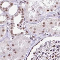
PTPN2 / Tyrosine-protein phosphatase non-receptor type 2 · IHC design guide

Design Immunohistochemistry for PTPN2

Plan chromogenic IHC on paraffin sections using the catalog antibody at 0.5–1 μg/mL (datasheet: PB9501). Assess nuclear staining by cell type, using the reported high staining in cervical glandular cells and undetected staining in adipocytes as references (HPA tissue IHC).

Evidence assembled Oct 2026 · For research use; verify linked source records and product datasheet before use
Immunohistochemistry protocol sheet for PTPN2 (IHC for PTPN2): expected localisation General nuclear staining (HPA tissue IHC), antibody PB9501, validated IHC image, and IHC protocol steps
Printable PTPN2 IHC protocol sheet — expected localisation General nuclear staining (HPA tissue IHC), antibody PB9501, controls and protocol steps. Open the full PTPN2 IHC guide →

PTPN2 Immunohistochemistry Experimental Design Guide

Expected localisation, validated protocols, controls and antibodies — the at-a-glance facts below, then the full design guide.

Must know before staining
Expected localisation General nuclear staining (HPA tissue IHC)
Staining pattern Nuclear staining across tissue cell types (HPA tissue IHC)
Antigen retrieval EDTA pH 8.0 HIER, heat-mediated (datasheet PB9501)
Positive control ⓘ Cerebellum+4 more · see all
Negative control ⓘ Adipose tissue
Important caveats
Reasons your staining may differ from the expected pattern.
Fixation Keep fixation consistent across sections (standard IHC practice; not target-specific)
Caveat Adipocytes have no detected staining (HPA tissue IHC)
Regulation Lymphoid tissue enhanced at RNA level (HPA tissue RNA)
Isoform / epitope 4 isoforms; antibody epitope coverage is unspecified (UniProt)
Section 1

Recommended PTPN2 IHC & IF Protocols

The catalog antibody’s IHC-P protocol is paired with three published PTPN2 IHC protocols that report usable staining parameters (PMC8865802; PMC6394658; PMC10599764).

Recommended immunohistochemistry (IHC-P) protocol parameters
SampleParaffin-embedded rat kidney tissue; fixative not specified (datasheet PB9501)
FixationImage fixative and duration unreported (datasheet PB9501); verify before use.
Sectioning4–5 µm sections on charged slides (standard)
DeparaffinisationXylene, graded ethanol series to water (standard)
Antigen retrievalHeat retrieval: EDTA pH 8.0 (datasheet PB9501); 20 min, 95–100 °C (standard)
Peroxidase block3% H2O2, 10 min, room temperature (standard)
Blocking10% goat serum (datasheet PB9501)
Primary antibodyRabbit anti-PTPN2, 0.5-1μg/ml (datasheet PB9501)
Primary incubationOvernight at 4 °C (datasheet PB9501)
DetectionStreptavidin-biotin complex (SABC), DAB chromogen (datasheet PB9501)
CounterstainHematoxylin, blue, dehydrate and mount (standard)
Expected resultPTPN2-positive staining in purkinje cells - dendrites of cerebellum (HPA tissue IHC: High). HPA tissue profile: General nuclear expression. No signal in the no-primary control.
💡Decision noteStart with heat-mediated EDTA pH 8.0 retrieval for the catalog antibody (datasheet: PB9501); the breast tumor study used low-pH retrieval with a different antibody (PMC6394658).
Section 2

What Is the Expected PTPN2 Staining Pattern?

PTPN2 should show predominantly nuclear staining in tissue IHC, with cell type and intensity varying by tissue (HPA tissue IHC: general nuclear expression; Enhanced reliability with medium RNA concordance). Nucleoplasmic and additional cytosolic signal are supported in ICC-IF (HPA subcellular: supported). UniProt also places PTPN2 in the ER, cytoplasm and cell membrane, and reports no transmembrane segment (UniProt P17706: subcellular location and topology).

What am I looking at on my slide?
Nuclear staining in cervical glandular cells or pancreatic exocrine glandular cells.This fits the general nuclear IHC profile and high staining reported for both cell populations (HPA tissue IHC: general nuclear expression; High in both). Compare intensity within the same tissue and cell type; high staining in these examples does not set a universal intensity threshold (HPA tissue IHC: tissue-specific levels).
Cytoplasmic signal accompanies a clear nuclear pattern.Additional cytosolic staining can be plausible: ICC-IF supports nucleoplasm as the main location and cytosol as an additional location (HPA subcellular: supported). UniProt also lists cytoplasm and ER (UniProt P17706: subcellular location). A cytoplasmic-only IHC result warrants review against the general nuclear tissue profile (HPA tissue IHC).
Strong membrane-only staining with little or no nuclear signal.Treat this as discordant with the general nuclear IHC profile and check assay specificity (HPA tissue IHC: general nuclear expression; standard IHC practice). Membrane signal alone cannot prove an artefact, because cell membrane is among UniProt's reported locations (UniProt P17706: subcellular location).
Prominent adipocyte staining or colour spread across unrelated cells.Adipocytes were not detected in the supplied tissue IHC record, so prominent staining merits a control review (HPA tissue IHC: adipocytes Not detected). Widespread chromogen can reflect background or endogenous detection activity; localisation and controls determine whether it represents specific staining (standard IHC practice).
No signal in cervical glandular cells or pancreatic exocrine glandular cells.An absent result conflicts with the high staining reported in these cell populations (HPA tissue IHC: High). Check a known-positive section, antibody conditions and detection controls before interpreting absence as biological loss (standard IHC practice). HPA's Enhanced reliability still has medium consistency with RNA expression (HPA tissue IHC).
💡Expected PTPN2 appearanceA convincing positive shows predominantly nuclear chromogen in an appropriate cell population, potentially strong in cervical glandular or pancreatic exocrine cells (HPA tissue IHC: general nuclear expression; High), whereas diffuse, compartment-free colour suggests background (standard IHC practice).
How each factor affects the staining
Cell population and reference intensity (HPA tissue IHC)High staining is reported in Purkinje dendrites, cervical glandular cells, pancreatic exocrine cells, Leydig cells, urothelial cells and vaginal squamous cells; skin fibroblasts and adrenal glandular cells are Medium, skeletal myocytes and marrow hematopoietic cells Low, and adipocytes Not detected (HPA tissue IHC). Score against the selected cell population, including the dendritic exception to the general nuclear profile (HPA tissue IHC).
Compartment and topology (UniProt P17706; HPA subcellular)UniProt lists ER, ER-Golgi intermediate compartment, nucleus, cytoplasm and cell membrane, with ER targeting by a C-terminal hydrophobic region and no transmembrane segment (UniProt P17706). ICC-IF supports mainly nucleoplasmic plus cytosolic localisation (HPA subcellular). These records allow secondary signal; they do not make an isolated membrane-only IHC pattern the expected result (HPA tissue IHC: general nuclear expression).
Isoforms and antibody interpretation (UniProt P17706)Four isoforms are listed; isoform 2 is probably major, while isoform 1 is reported in T-cells and placenta (UniProt P17706). Interpret unexpected differences cautiously because the supplied record gives no antibody epitope or isoform coverage (UniProt P17706: isoforms; supplied evidence). Do not assign an IHC compartment or intensity to a particular isoform from these data alone.
IF/ICC: what localisation should I expect? (HPA subcellular)Expect mainly nucleoplasmic signal with additional cytosolic signal; both locations are supported in ICC-IF (HPA subcellular). This is localisation evidence, not an IHC-P protocol or a guarantee that every tissue cell has the same intensity (HPA subcellular; HPA tissue IHC: variable cell-specific levels).
Why is my staining missing, weak or wrong?
SituationLikely causeNext action
Expected-positive tissue is blank.Assay performance or detection may be inadequate; cervical glandular and pancreatic exocrine cells are reported High (HPA tissue IHC; standard IHC practice).Run a known-positive section with the same staining batch and check antibody dilution, retrieval, chromogen and detection reagents using the chosen assay's instructions (standard IHC practice). No PTPN2-specific retrieval setting is supplied.
Nuclei are pale but the slide has broad background.Weak specific staining can be obscured by nonspecific chromogen deposition (standard IHC practice); the tissue reference is generally nuclear (HPA tissue IHC).Compare with a no-primary control, review blocking and washing, and adjust detection conditions one variable at a time (standard IHC practice). Judge improvement by clearer localisation in the expected cells (HPA tissue IHC: general nuclear expression and cell-specific levels).
Adipocytes stain strongly.That conflicts with the supplied Not detected adipocyte observation; background or endogenous activity is possible (HPA tissue IHC: adipocytes Not detected; standard IHC practice).Check no-primary and detection controls, and assess cell borders and chromogen distribution before calling adipocytes positive (standard IHC practice). A single discordant cell type does not establish antibody cross-reactivity.
Signal appears only at cell membranes.The pattern differs from general nuclear tissue IHC, although UniProt also lists cell membrane localisation (HPA tissue IHC; UniProt P17706: subcellular location).Inspect nuclei in a known-positive population and compare control sections and, if available, an independently validated antibody (standard IHC practice). HPA lists HPA015004 and HPA046176 as IHC Enhanced (HPA antibodies).
A cytoplasmic pattern is scored as a failed stain.Additional cytosolic localisation is supported in ICC-IF, and UniProt lists cytoplasm and ER (HPA subcellular; UniProt P17706: subcellular location).Reassess whether nuclei also stain and whether signal follows a plausible cell compartment; treat cytoplasmic-only tissue IHC as uncertain against HPA's general nuclear profile (HPA tissue IHC; standard IHC practice).
All tissues are scored against one intensity cutoff.The supplied IHC levels range from High to Not detected across named cell populations (HPA tissue IHC).Record compartment, cell identity, extent and intensity separately, using the relevant HPA tissue and cell reference for comparison (HPA tissue IHC; standard IHC practice).

Sample controls for PTPN2 IHC & IF

🧪Start with cervix and require staining in glandular cells (HPA: High in cervical glandular cells). Use adipose tissue as the negative comparator, focusing on adipocytes (HPA: Not detected in adipocytes); neighboring cells without visible signal on the cervical slide should show only background staining, but are not validated biological negatives (UniProt P17706: ubiquitously expressed).
Positive control tissue: Cerebellum (Purkinje cells - dendrites, HPA High)
Negative control tissue: Adipose tissue (HPA Not detected)
ICC-IF cell lines (HPA subcellular resource): HPA ICC-IF images show PTPN2 in SiHa, U-251MG, U2OS, A-431, CACO-2, with annotated localisation: Nucleoplasm (supported) (HPA subcellular).
Technical controls: Run no-primary and secondary-only controls, a rabbit IgG control matched to the primary antibody’s clonality, and a PTPN2-knockout biological negative if available (PB9501 caption: rabbit primary and biotinylated goat anti-rabbit secondary; standard IHC practice). Block endogenous peroxidase and check for endogenous biotin background in the cervical section (PB9501 caption: biotinylated secondary, SABC and DAB; standard IHC practice).
⚠️Feasibility: A target-specific fixation window or fixation effect is unreported, and the selected PB9501 tissue-IHC caption does not state a fixative (PB9501 caption: fixative not stated). Heat retrieval with EDTA at pH 8.0 was used for paraffin-embedded rat kidney, but the caption does not establish that retrieval is required for cervical tissue (PB9501 caption: EDTA retrieval in rat kidney). The supplied evidence does not establish whether frozen sections or IF would be easier; inspect cervical glandular luminal material for trapped chromogen when scoring (HPA: High in cervical glandular cells; standard IHC practice).

HPA tissue IHC evidence for PTPN2

Comprehensive Human Protein Atlas IHC scoring per tissue (reliability: Enhanced — Medium consistency between antibody staining and RNA expression data.). Rows are taken directly from the HPA tissue chart — click any row's HPA link to view the source.

Positive expression · recommended positive controls

TissueCell typeLevelEvidenceSource
Cerebellum Purkinje cells - dendrites High Protein (IHC) HPA →
Cervix Glandular cells High Protein (IHC) HPA →
Pancreas Exocrine glandular cells High Protein (IHC) HPA →
Skin Fibroblasts Medium Protein (IHC) HPA →
Testis Leydig cells High Protein (IHC) HPA →

Undetected expression · recommended negative controls

TissueCell typeLevelEvidenceSource
Adipose tissue Adipocytes Not detected Protein (IHC) HPA →
Section 3

Advanced PTPN2 IHC Tips

Troubleshoot PTPN2 staining in paraffin sections by checking retrieval, compartment patterns, background, and cell-specific scoring against the supplied evidence.

Which retrieval condition should I try first for weak PTPN2 staining?
Start with heat-mediated retrieval in EDTA at pH 8.0 for PB9501 paraffin sections (datasheet PB9501). The reported rat kidney example used this condition, followed by 1 μg/ml primary antibody overnight at 4°C (datasheet PB9501). If staining is weak, compare retrieval duration or a different buffer on adjacent sections while keeping antibody concentration and detection constant (standard IHC practice). Record tissue preservation and nuclear versus cytoplasmic signal separately, since both compartments are plausible for PTPN2 (HPA subcellular; UniProt P17706 localization). Include a no-primary section to distinguish retrieval-related background from specific staining (standard IHC practice).
How should I assess whether fixation is causing weak staining?
The PB9501 rat kidney caption identifies a paraffin section but does not report the fixative (datasheet PB9501). Target-specific PTPN2 sensitivity to fixative type or fixation duration is therefore unknown from this evidence (datasheet PB9501). Compare sections with documented processing histories, using the same EDTA retrieval at pH 8.0 and the same detection run (datasheet PB9501; standard IHC practice). Evaluate morphology alongside staining, because damaged or poorly preserved areas can confound an intensity comparison (standard IHC practice). Do not attribute a difference to fixation unless tissue handling, retrieval, and detection have been accounted for (standard IHC practice).
Should PTPN2 stain nuclei, cytoplasm, or membranes in tissue?
Expect a prominent nuclear pattern as one useful reference: tissue IHC reports general nuclear expression, and cell imaging supports nucleoplasmic localization (HPA tissue IHC; HPA subcellular). Cytosolic staining can also be plausible, while the protein record additionally lists endoplasmic reticulum, intermediate compartment, and cell membrane locations (HPA subcellular; UniProt P17706 localization). Its C-terminal hydrophobic region targets the endoplasmic reticulum, despite no annotated transmembrane segment (UniProt P17706 topology and localization). Score nuclear and cytoplasmic staining separately rather than combining them into one intensity value (standard IHC practice). Treat an isolated membrane outline cautiously until appropriate controls support it (UniProt P17706 localization; standard IHC practice).
Could isoform choice or epitope accessibility explain discordant staining?
PTPN2 has 4 annotated isoforms, and isoform 2 is probably the major form (UniProt P17706 isoforms and tissue specificity). Isoform 1 is reported in T-cells and placenta, so cell composition may affect comparisons between sections (UniProt P17706 tissue specificity). The phosphatase domain spans residues 5–275, but the supplied PB9501 caption does not identify its immunogen or epitope (UniProt P17706 domains; datasheet PB9501). Check the catalog antibody’s epitope documentation before claiming that it detects every isoform (standard IHC practice). Compare matched sections under identical retrieval and detection conditions before assigning a staining difference to isoform expression (standard IHC practice).
How can I adapt this tissue finding to a multiplex IF experiment?
For an IF experiment, pair PTPN2 with a marker identifying the intended cell population; urothelial cells are one tissue IHC population reported as high (HPA tissue IHC). Choose spectrally separated fluorophores, favoring a longer-wavelength channel for a dim target when the specimen shows autofluorescence (standard IF practice). Use single-stain and no-primary controls to assess bleed-through and background before interpreting colocalization (standard IF practice). Permeabilize sufficiently for a nuclear or cytosolic epitope, while checking epitope documentation before assuming access to an endoplasmic reticulum-associated region (HPA subcellular; UniProt P17706 localization; standard IF practice). The paraffin-section PB9501 example establishes tissue IHC conditions, not IF performance (datasheet PB9501).
What should I check when DAB staining appears diffuse?
The PB9501 example used 10% goat serum, a biotinylated secondary, streptavidin–biotin detection, and DAB (datasheet PB9501). Include a no-primary control and inspect whether diffuse color persists, particularly around damaged tissue or section edges (standard IHC practice). Check endogenous peroxidase blocking before DAB development and consider endogenous biotin when using an avidin or streptavidin system (standard IHC practice). The example applied its secondary for 30 minutes at 37°C; excessive secondary exposure or inadequate washing can increase background (datasheet PB9501; standard IHC practice). Compare background with the expected predominantly nuclear tissue pattern before adjusting primary concentration (HPA tissue IHC; standard IHC practice).
How should I score PTPN2 across heterogeneous tissue sections? ⚠ ANSWER MARKED FOR VERIFICATION
Define the cell population and compartment before scoring, because tissue IHC describes general nuclear expression while cytosolic localization is also supported (HPA tissue IHC; HPA subcellular). For each annotated population, record the percentage of positive cells and nuclear staining intensity; an H-score can summarize intensity-weighted positivity on a 0–300 scale (standard IHC practice). If counting stained cells, report density per mm² of viable, evaluable tissue (standard IHC practice). Normalize comparisons to the same cell type, compartment, staining run, and tissue area rather than whole-section color alone (standard IHC practice). Exclude folds, necrosis, and section edges using prespecified criteria (standard IHC practice).
How can I distinguish convincing PTPN2 signal from an artefact?
A convincing pattern should be reproducible in intact cells and compatible with nuclear staining or supported cytosolic localization (HPA tissue IHC; HPA subcellular). Cell identity matters: urothelial cells are reported high, whereas adipocytes are reported as not detected in the supplied tissue profile (HPA tissue IHC). Treat strong staining confined to edges, folds, or necrotic regions as suspect and compare it with an adjacent intact area (standard IHC practice). A pattern limited to an unexpected compartment needs independent support before being called PTPN2, since additional locations are annotated (UniProt P17706 localization; standard IHC practice). Check no-primary and peroxidase controls when DAB persists outside plausible cells (standard IHC practice).
Boster reagents

Best PTPN2 / Tyrosine-protein phosphatase non-receptor type 2 IHC Antibodies

PB9501 has IHC images from paraffin sections of rat kidney and human intestinal cancer tissue (catalog image captions); IF/ICC is listed for human samples (catalog application and dilution listing).

Real IHC data IHC analysis of TCPTP using anti-TCPTP antibody (PB9501). TCPTP was detected in a paraffin-embedded section of rat kidney tissue. Heat mediated antigen retrieval was performed in EDTA buffer (pH 8.0, epitope retrieval solution). The tissue section was blocked with 10% goat serum. The tissue section was then incubated with 1 μg/ml rabbit anti-TCPTP Antibody (PB9501) overnight at 4°C. Biotinylated goat anti-rabbit IgG was used as secondary antibody and incubated for 30 minutes at 37°C. The tissue section was developed using Strepavidin-Biotin-Complex (SABC) (Catalog # SA1022) with DAB as the chromogen.
Anti-TCPTP/PTPN2 Antibody ®
Cat # PB9501

PB9501 is listed for IHC, IF and ICC, with human and rat reactivity (catalog applications and reactivity). Its IHC images show staining in paraffin sections of rat kidney and human intestinal cancer tissue (catalog image captions).

Which to pick: Choose PB9501 for paraffin section IHC; its rat kidney image used EDTA retrieval at pH 8.0 and 1 μg/ml primary antibody (PB9501 IHC image caption). PB9501 also lists IF/ICC at 5 μg/ml for human samples, while its listed IHC reactivity covers human and rat (catalog dilution and reactivity listings). The catalog identifies a rabbit host but reports no clone, and the IHC captions do not report the fixative (catalog host field; PB9501 IHC image captions).

Each figure is that product's own IHC / IF validation image from its datasheet.

References

  1. UniProt Consortium. UniProt entry P17706 (PTN2_HUMAN, Tyrosine-protein phosphatase non-receptor type 2).
  2. Human Protein Atlas. PTPN2 tissue IHC expression (reliability: Enhanced).
  3. Human Protein Atlas. PTPN2 subcellular location (ICC-IF): Mainly localized to the nucleoplasm. In addition localized to the cytosol..
  4. Human Protein Atlas. PTPN2 antibody validation summary (2 antibodies).
  5. PTPN2 elicits cell autonomous and non-cell autonomous effects on antitumor immunity in triple-negative breast cancer. Science advances 2022 — PMC8865802.
  6. Protein tyrosine phosphatase non-receptor II: A possible biomarker of poor prognosis and mediator of immune evasion in hepatocellular carcinoma. World journal of gastrointestinal oncology 2024 — PMC11438766.
  7. PTPN2 deficiency along with activation of nuclear Akt predict endocrine resistance in breast cancer. Journal of cancer research and clinical oncology 2019 — PMC6394658.
  8. An immune signature to predict the prognosis of ATRX-wildtype glioma patients and guide immune checkpoint blockade therapy. Aging 2023 — PMC10599764.
  9. PubMed PMID:2546150 — UniProt-cited evidence.
  10. PubMed PMID:14702039 — UniProt-cited evidence.
  11. PubMed PMID:16177791 — UniProt-cited evidence.