This website uses cookies to ensure you get the best experience on our website.


- Table of Contents
Plan chromogenic IHC on paraffin sections using the catalog antibody at 2–5 μg/mL (datasheet: A05574-1). Use the reported cytoplasmic, immune-cell-rich tissue pattern to choose controls, while accounting for its low consistency with RNA expression (HPA tissue IHC).

Expected localisation, validated protocols, controls and antibodies — the at-a-glance facts below, then the full design guide.
| Expected localisation | Cytoplasmic staining across tissues (HPA tissue IHC) | |
| Staining pattern | Strongest in immune-cell cytoplasm; endothelium also positive (HPA tissue IHC) | |
| Antigen retrieval | EDTA pH 8.0 HIER, heat-mediated (datasheet A05574-1) | |
| Positive control | Adrenal gland+4 more · see all | |
| Negative control | Breast+4 more · see all |
| Fixation | Keep formalin fixation consistent across sections. (standard IHC practice; not target-specific) | |
| Caveat | Staining has low consistency with RNA expression (HPA tissue IHC) | |
| Regulation | Highest expression in immune cells (HPA tissue IHC) | |
| Isoform / epitope | 3 isoforms; check epitope against extra- and cytoplasmic regions (UniProt) |
The catalog antibody protocol uses EDTA pH 8.0 retrieval (datasheet A05574-1). Two published PTPRE IHC protocols provide alternatives for retinoblastoma and tumour sections (PMC11049872; PMC13278412).
| Sample | Paraffin-embedded human breast cancer tissue; fixative not specified (datasheet A05574-1) |
| Fixation | Image fixative and duration unreported (datasheet A05574-1); verify before use. |
| Sectioning | 4–5 µm sections on charged slides (standard) |
| Deparaffinisation | Xylene, graded ethanol series to water (standard) |
| Antigen retrieval | Heat retrieval: EDTA pH 8.0 (datasheet A05574-1); 20 min, 95–100 °C (standard) |
| Peroxidase block | 3% H2O2, 10 min, room temperature (standard) |
| Blocking | 10% goat serum (datasheet A05574-1) |
| Primary antibody | Rabbit anti-PTPRE, 2-5 μg/ml (datasheet A05574-1) |
| Primary incubation | Overnight at 4 °C (datasheet A05574-1) |
| Detection | HRP-conjugated secondary, DAB chromogen (datasheet A05574-1) |
| Counterstain | Hematoxylin, blue, dehydrate and mount (standard) |
| Expected result | PTPRE-positive staining in glandular cells of adrenal gland (HPA tissue IHC: High). HPA tissue profile: Ubiquitous cytoplasmic expression. Highest expression in immune cells. Additional positivity in endothelial cells. No signal in the no-primary control. |
PTPRE should appear chiefly in the cytoplasm, with possible membrane staining and a small nuclear-associated fraction (UniProt P23469 subcellular location and topology). HPA reports ubiquitous cytoplasmic staining, strongest in immune cells, with additional endothelial positivity (HPA tissue IHC). Treat this as a provisional guide: HPA rates the tissue staining Supported but reports low consistency with RNA expression and pending external verification (HPA tissue IHC).
| Strong cytoplasmic staining in lung macrophages, lymph node germinal center cells, or spleen red pulp cells. | These are HPA High cell populations (HPA tissue IHC). Compare intensity within the identified cell population; a high rating for those cells does not mean every cell in the section should stain. |
| Predominantly cytoplasmic signal with some membrane staining or limited nuclear-associated signal. | This fits the reported distribution (UniProt P23469 subcellular location). PTPRE has an extracellular segment, one transmembrane segment, and a long cytoplasmic region (UniProt P23469 topology); membrane staining need not dominate. |
| Dominant extracellular or nuclear-only staining, with little cytoplasmic signal in expected positive cells. | This departs from the reported predominant cytoplasmic location (UniProt P23469 subcellular location; HPA tissue IHC). Check morphology and controls before calling it PTPRE; a small nuclear-associated fraction is reported (UniProt P23469 subcellular location). |
| Strong staining of colon glandular cells or breast adipocytes while expected positive cells are unstained. | Those cell populations are Not detected in HPA tissue IHC. Investigate cross-reactivity or detection background (standard IHC practice). These observations are cell-specific; neither entire tissue is established as PTPRE-negative. |
| Even chromogen haze across cells and surrounding tissue, or no signal in lung macrophages. | Uniform haze obscures compartment and cell-type assessment (standard IHC practice). Lung macrophages are HPA High (HPA tissue IHC); absent signal there warrants a control and workflow check, without proving PTPRE absence. |
| Which compartment anchors the IHC readout? | HPA describes cytoplasmic tissue staining, and UniProt calls PTPRE predominantly cytoplasmic (HPA tissue IHC; UniProt P23469 subcellular location). Its transmembrane segment at residues 47–69 supports a membrane-associated fraction, without requiring a continuous membrane rim (UniProt P23469 topology). |
| Which cells make useful comparisons? | Lung macrophages and lymph node germinal center cells are HPA High; colon glandular cells and breast adipocytes are Not detected (HPA tissue IHC). Score the named cells rather than labeling whole tissues positive or negative. HPA also reports low consistency with RNA expression (HPA tissue IHC). |
| Can topology or isoforms predict the antibody’s exact pattern? | UniProt lists extracellular residues 20–46, cytoplasmic residues 70–700, and three isoforms (UniProt P23469 topology and isoforms). The supplied record gives no antibody epitope or isoform recognition data, so it cannot establish which forms this stain detects. |
| How strong is the tissue-pattern evidence? | Both listed HPA antibodies have Supported IHC status, while HPA describes low consistency with RNA expression and pending external verification (HPA antibodies; HPA tissue IHC). Use the pattern to plan controls, then judge specificity from the actual slide and controls (standard IHC practice). |
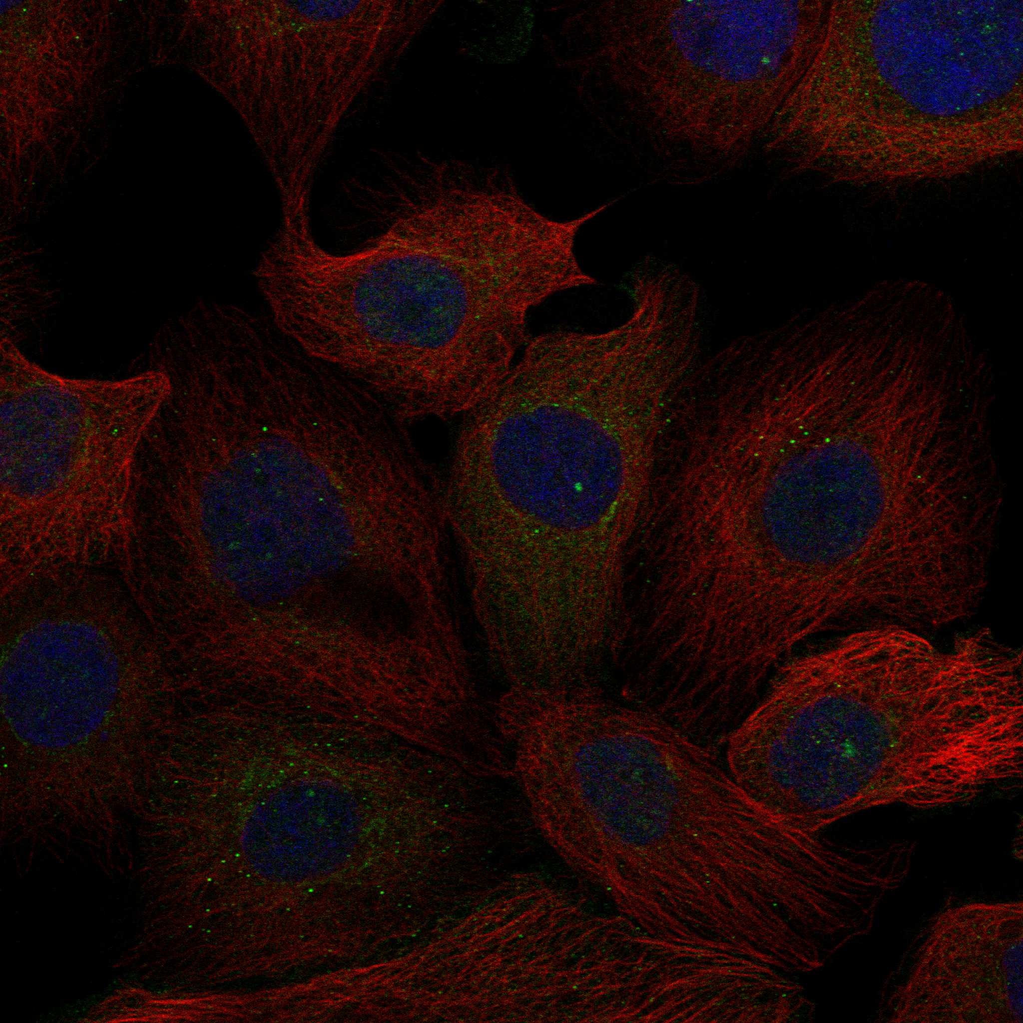
| Does the IF/ICC pattern settle a paraffin-section IHC result? | No. HPA reports an approved intermediate-filament location in ICC-IF images from A-431, U-251MG, and U2OS (HPA subcellular ICC-IF). That observation differs from the predominantly cytoplasmic tissue IHC description and does not establish an IHC compartment rule (HPA tissue IHC). |
| Situation | Likely cause | Next action |
|---|---|---|
| No staining in a section expected to contain lung macrophages. | The expected cells may be absent from the field, or the IHC detection workflow may have failed (HPA: High in lung macrophages; standard IHC practice). | Locate macrophages morphologically; review the paired positive control, antigen retrieval, primary antibody use, and chromogen development (standard IHC practice). |
| Strong signal appears in colon glandular cells. | HPA reports that cell population as Not detected; cross-reactivity or detection background is possible (HPA tissue IHC; standard IHC practice). | Compare a no-primary control and the expected immune-cell pattern; assess the exact stained cells before interpreting the section (standard IHC practice). |
| Diffuse brown haze hides cell boundaries. | Nonspecific staining or endogenous peroxidase activity can obscure a chromogenic IHC readout (standard IHC practice). | Review blocking and washes; include a no-primary control and verify endogenous peroxidase blocking for the detection method (standard IHC practice). |
| Only nuclei or extracellular material stain strongly. | That distribution conflicts with the reported predominance of cytoplasmic PTPRE, though a small nuclear-associated fraction exists (UniProt P23469 subcellular location). | Recheck morphology, expected positive cells, and no-primary control before assigning target-specific staining (standard IHC practice). |
| A prominent membrane rim seems to replace cytoplasmic staining. | PTPRE has a membrane-associated fraction, but predominantly cytoplasmic localisation is reported (UniProt P23469 subcellular location and topology). | Compare cytoplasmic signal in HPA High cells and assess the pattern across controls before treating the rim as specific (HPA tissue IHC; standard IHC practice). |
| A control cell population gives a weaker result than anticipated. | HPA staining levels describe observed cell populations, while its tissue evidence has low consistency with RNA expression (HPA tissue IHC). | Confirm that the same named cells are present and identifiable; compare matched staining controls before changing the interpretation (standard IHC practice). |
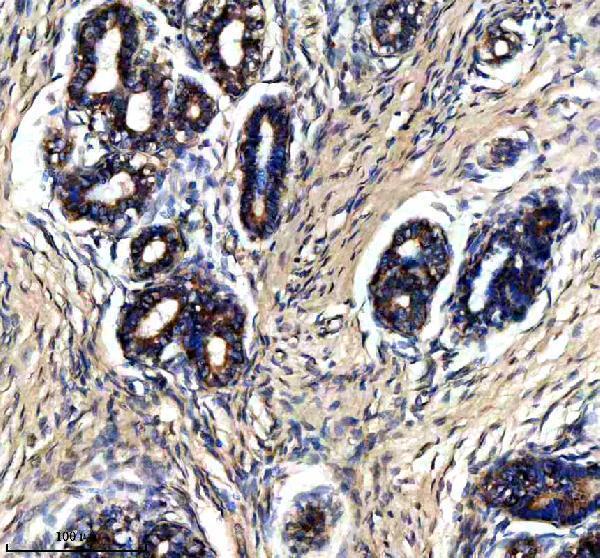
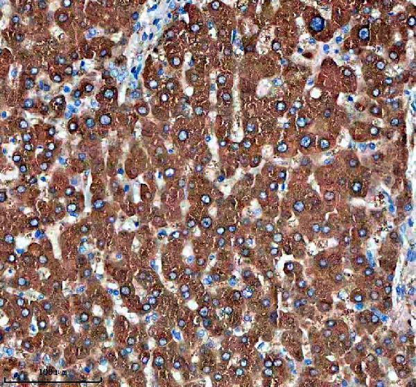
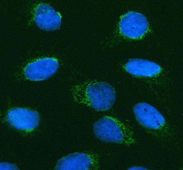
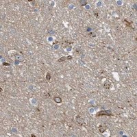


Comprehensive Human Protein Atlas IHC scoring per tissue (reliability: Supported — Low consistency between antibody staining and RNA expression data. Pending external verification.). Rows are taken directly from the HPA tissue chart — click any row's HPA link to view the source.
| Tissue | Cell type | Level | Evidence | Source |
|---|---|---|---|---|
| Adrenal gland | Glandular cells | High | Protein (IHC) | HPA → |
| Appendix | Lymphoid tissue | High | Protein (IHC) | HPA → |
| Epididymis | Glandular cells | High | Protein (IHC) | HPA → |
| Lung | Macrophages | High | Protein (IHC) | HPA → |
| Lymph node | Germinal center cells | High | Protein (IHC) | HPA → |
| Tissue | Cell type | Level | Evidence | Source |
|---|---|---|---|---|
| Breast | Adipocytes | Not detected | Protein (IHC) | HPA → |
| Caudate | Glial cells | Not detected | Protein (IHC) | HPA → |
| Cerebral cortex | Endothelial cells | Not detected | Protein (IHC) | HPA → |
| Cervix | Squamous epithelial cells | Not detected | Protein (IHC) | HPA → |
| Colon | Glandular cells | Not detected | Protein (IHC) | HPA → |
Troubleshoot PTPRE staining in paraffin sections by checking retrieval, cell type, subcellular pattern, and controls before comparing chromogenic signal across samples.
A05574-1 has human paraffin-section IHC images and human cell IF data (catalog image captions); its listed reactivity is human, mouse, and rat (catalog applications/reactivity).
A05574-1 will render with a human breast cancer paraffin-section IHC figure (catalog IHC image caption). The same SKU has IHC images from human liver and liver cancer tissue and an IF image from U2OS cells (catalog image captions).
Which to pick: For paraffin-section IHC, choose A05574-1: its caption documents EDTA pH 8.0 retrieval and 2 μg/ml primary antibody; the fixative is unreported (catalog IHC image caption). For IF/ICC, A05574-1 has a human U2OS cell image at 5 μg/ml (catalog IF image caption). For other species, A05574-1 lists human, mouse, and rat reactivity, while its IHC dilution listing specifies human and rat and its IF listing specifies human (catalog applications/reactivity and dilution listing).