This website uses cookies to ensure you get the best experience on our website.


- Table of Contents
Plan PTPRJ staining in paraffin sections around the cytoplasmic and membranous pattern observed in glandular cells and epithelia (HPA tissue IHC). Start the catalog antibody at 2–5 μg/mL (datasheet A02319-2), using lung macrophages and adipocytes as positive and undetected-cell comparisons (HPA tissue IHC).

Expected localisation, validated protocols, controls and antibodies — the at-a-glance facts below, then the full design guide.
| Expected localisation | Epithelial cytoplasm and membrane; immune-cell cytoplasm (HPA tissue IHC) | |
| Staining pattern | Glandular/epithelial cytoplasmic-membranous staining; immune subsets cytoplasmic (HPA tissue IHC) | |
| Antigen retrieval | EDTA pH 8.0 HIER, heat-mediated (datasheet A02319-2) | |
| Positive control | Appendix+4 more · see all | |
| Negative control | Adipose tissue+4 more · see all |
| Fixation | Keep fixation consistent across sections (standard IHC practice; not target-specific) | |
| Caveat | A secreted variant may complicate RNA–protein agreement (HPA tissue IHC) | |
| Regulation | Enhanced at high cell density (UniProt) | |
| Isoform / epitope | 3 isoforms; check extracellular versus cytoplasmic epitope (UniProt) |
The catalog antibody’s IHC-P protocol (datasheet A02319-2) is accompanied by one published chromogenic IHC protocol for human breast sections (PMC3398958).
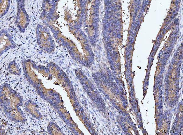
| Sample | Paraffin-embedded human rectal cancer tissue; fixative not specified (datasheet A02319-2) |
| Fixation | Image fixative and duration unreported (datasheet A02319-2); verify before use. |
| Sectioning | 4–5 µm sections on charged slides (standard) |
| Deparaffinisation | Xylene, graded ethanol series to water (standard) |
| Antigen retrieval | Heat retrieval: EDTA pH 8.0 (datasheet A02319-2); 20 min, 95–100 °C (standard) |
| Peroxidase block | 3% H2O2, 10 min, room temperature (standard) |
| Blocking | 10% goat serum (datasheet A02319-2) |
| Primary antibody | Rabbit anti-PTPRJ, 2-5 μg/ml (datasheet A02319-2) |
| Primary incubation | Overnight at 4 °C (datasheet A02319-2) |
| Detection | HRP-conjugated secondary, DAB chromogen (datasheet A02319-2) |
| Counterstain | Hematoxylin, blue, dehydrate and mount (standard) |
| Expected result | PTPRJ-positive staining in glandular cells of appendix (HPA tissue IHC: Medium). HPA tissue profile: Cytoplasmic expression in subsets of immune cells and cytoplasmic membranous expression in most glandular cells and epithelia. No signal in the no-primary control. |
PTPRJ is a cell membrane protein with an extracellular region and cytoplasmic tail; UniProt also lists cell junctions and extracellular space (UniProt Q12913 topology and subcellular location). In tissue IHC, expect cytoplasmic membranous staining in many glandular cells and epithelia, and cytoplasmic staining in subsets of immune cells (HPA tissue IHC profile). HPA rates its tissue staining Approved, with medium consistency against RNA data (HPA tissue IHC reliability).
| Cytoplasmic membranous staining in appendix or endometrial glandular cells. | This fits the glandular pattern; both cell populations have Medium staining in HPA tissue IHC (HPA: appendix and endometrium, glandular cells). Judge the outlined cells and cytoplasm together rather than requiring a thin, uninterrupted membrane rim (HPA tissue IHC profile). |
| Cytoplasmic staining in lung macrophages or bone marrow hematopoietic cells. | These are reported Medium positive populations (HPA: lung macrophages; bone marrow hematopoietic cells). The immune-cell pattern is cytoplasmic, so absence of a crisp membrane outline alone does not make these cells negative (HPA tissue IHC profile). |
| Predominantly nuclear or nucleolar chromogen in tissue sections. | Do not count this as the expected tissue IHC pattern: HPA describes cytoplasmic or cytoplasmic membranous tissue staining (HPA tissue IHC profile). HPA ICC-IF reports uncertain nucleoplasmic and nucleolar locations, which do not establish nuclear staining as the tissue IHC readout (HPA subcellular). |
| Strong color in HPA-negative cell populations, or a uniform haze across unrelated structures. | Staining in adipocytes or skeletal myocytes conflicts with their Not detected HPA calls (HPA: adipocytes; skeletal myocytes). Check morphology and controls before attributing it to PTPRJ; cross-reactivity or endogenous detection activity can produce misleading color (general IHC practice). |
| No signal in a section containing identifiable appendix glandular cells. | The absence conflicts with a reported Medium positive population (HPA: appendix glandular cells). First verify that those cells are present and the detection controls worked; a failed staining run or sample-specific loss remains possible (general IHC practice). HPA approval does not guarantee positivity in every section (HPA tissue IHC reliability). |
| Cell compartment and epitope position | PTPRJ has an extracellular region at residues 36–975, a membrane span at 976–996, and a cytoplasmic region at 997–1337 (UniProt Q12913 topology). The supplied evidence gives no epitope for a particular IHC antibody, so compartment anatomy cannot predict that antibody's staining behavior. |
| Cell population within a tissue | HPA calls glandular cells Medium in appendix, endometrium, epididymis and gallbladder, but salivary gland glandular cells Not detected (HPA tissue IHC). Select and score the named cell population; a tissue name alone is an insufficient positive or negative call (HPA tissue IHC). |
| Protein distribution and variants | UniProt lists 3 isoforms and an extracellular-space location; HPA notes that at least one protein variant is secreted and that RNA–protein correlation is complex (UniProt Q12913 isoforms and subcellular location; HPA tissue IHC reliability). Do not use RNA abundance alone to adjudicate a spatially discordant stain. |
| Evidence strength | The tissue profile is Approved with medium consistency between staining and RNA expression, and antibody HPA006026 is IHC Approved (HPA tissue IHC reliability; HPA antibody validation). These ratings support comparison with the reported pattern; they do not establish that every positive-looking cell is specific. |
| IF/ICC Q&A | What should IF/ICC show? HPA reports supported cell-junction localization, with uncertain nucleoplasm, nucleoli and nuclear bodies, for its ICC-IF data (HPA subcellular). Those cell-image findings answer the IF/ICC question but do not override the tissue IHC pattern or supply an IHC protocol. |
| Antigen retrieval | No PTPRJ-specific retrieval or fixation sensitivity is established by the supplied UniProt and HPA records. If signal is weak, compare retrieval conditions using a known-positive cell population and run controls; treat this as general IHC optimization, not a predicted PTPRJ effect (general IHC practice). |
| Situation | Likely cause | Next action |
|---|---|---|
| Expected glandular cells are blank. | The reported positive population may be absent from the section, or the staining run may have failed (HPA: appendix and endometrium glandular cells; general IHC practice). | Confirm the glandular cells by morphology, then examine the run's positive and detection controls. If controls fail, repeat the run; if they pass, compare another section containing an HPA-reported Medium population (general IHC practice; HPA tissue IHC). |
| Only nuclei appear positive. | Nuclear dominance does not match HPA's cytoplasmic or cytoplasmic membranous tissue profile; ICC-IF nucleoplasmic localization is marked uncertain (HPA tissue IHC profile; HPA subcellular). | Check a no-primary control and inspect whether cytoplasm or cell borders stain in an HPA-reported positive population. Do not score nuclear color alone as the expected tissue IHC result (general IHC practice; HPA tissue IHC profile). |
| The whole section has diffuse brown background. | Non-specific detection, inadequate blocking or excessive development can obscure cell boundaries (general IHC practice). A diffuse haze is not the described population-specific HPA tissue pattern (HPA tissue IHC profile). | Check no-primary and detection controls, review blocking and chromogen development, and reduce background before interpreting localization (general IHC practice). Reassess identifiable glandular or immune cells against the HPA profile. |
| Negative-reference cells show strong color. | Adipocytes, skeletal myocytes and soft-tissue fibroblasts are Not detected in the listed HPA observations (HPA tissue IHC negative populations). Misidentified cells, cross-reactivity or endogenous enzyme activity may explain unexpected color (general IHC practice). | Verify cell identity and compare no-primary and enzyme-block controls. Treat the listed HPA negatives as cell-population references, not proof that every cell in those tissues must be blank (HPA tissue IHC; general IHC practice). |
| Immune cells stain without a clear membrane rim. | HPA describes cytoplasmic expression in subsets of immune cells and reports Medium staining in lung macrophages and bone marrow hematopoietic cells (HPA tissue IHC profile and positive populations). | Score the identified immune-cell population and its cytoplasmic signal; do not require the glandular cytoplasmic membranous appearance in these cells (HPA tissue IHC profile). Use morphology and neighboring cells to assess whether staining is selective (general IHC practice). |
| An RNA-positive area looks IHC-negative. | HPA reports low tissue RNA specificity but only medium RNA–staining consistency; it also flags a secreted variant as a reason RNA and protein locations may differ (HPA tissue IHC reliability and RNA specificity). | Compare the same named cell population in HPA tissue IHC before calling a contradiction. Check the section and detection controls, then report the protein observation separately from the RNA result (HPA tissue IHC; general IHC practice). |


Comprehensive Human Protein Atlas IHC scoring per tissue (reliability: Approved — Medium consistency between antibody staining and RNA expression data. At least one protein variant secreted, tissue location of RNA and protein might differ and correlation is complex.). Rows are taken directly from the HPA tissue chart — click any row's HPA link to view the source.
| Tissue | Cell type | Level | Evidence | Source |
|---|---|---|---|---|
| Appendix | Glandular cells | Medium | Protein (IHC) | HPA → |
| Bone marrow | Hematopoietic cells | Medium | Protein (IHC) | HPA → |
| Endometrium | Glandular cells | Medium | Protein (IHC) | HPA → |
| Epididymis | Glandular cells | Medium | Protein (IHC) | HPA → |
| Gallbladder | Glandular cells | Medium | Protein (IHC) | HPA → |
| Tissue | Cell type | Level | Evidence | Source |
|---|---|---|---|---|
| Adipose tissue | Adipocytes | Not detected | Protein (IHC) | HPA → |
| Cerebral cortex | Endothelial cells | Not detected | Protein (IHC) | HPA → |
| Esophagus | Squamous epithelial cells | Not detected | Protein (IHC) | HPA → |
| Oral mucosa | Squamous epithelial cells | Not detected | Protein (IHC) | HPA → |
| Salivary gland | Glandular cells | Not detected | Protein (IHC) | HPA → |
Troubleshoot PTPRJ staining in paraffin sections by checking retrieval, cell identity, membrane distribution and assay controls before comparing staining intensity.
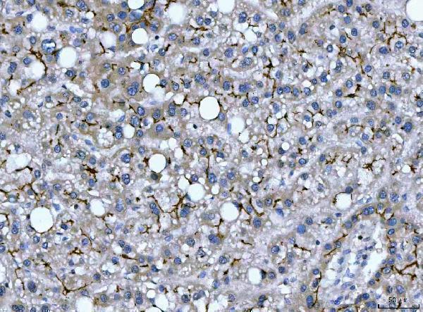
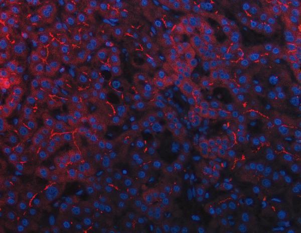
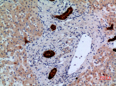
Catalog antibodies A02319-2 and A02319-1 have human paraffin-section IHC images; A02319-2 also has a human liver-cancer IF image (catalog image captions). Listed reactivity spans human, mouse and rat (catalog reactivity).
A02319-2 shows IHC on human rectal and liver cancer paraffin sections and IF on a human liver cancer paraffin section; it lists human and rat reactivity (A02319-2 image captions; catalog reactivity). A02319-1 shows IHC on a human liver paraffin section, lists IF and ICC applications, and lists human, mouse and rat reactivity (A02319-1 image caption; catalog applications and reactivity).
Which to pick: For tissue IHC, A02319-2 offers documented EDTA retrieval at pH 8.0 and a 2 μg/ml incubation on human paraffin sections; its fixative is unreported (A02319-2 image captions). For IF, A02319-2 has a human paraffin-section image at 5 μg/ml; for ICC, A02319-1 lists that application and is described as polyclonal, but has no ICC image in the payload (A02319-2 IF image caption; A02319-1 catalog applications and dilution data). For the broadest listed species coverage, choose A02319-1 for human, mouse and rat, while treating its human liver paraffin-section IHC image as the only pictured IHC evidence for that SKU; its fixative is unreported (A02319-1 catalog reactivity; A02319-1 image caption).