PVR / Poliovirus receptor · IHC design guide

Design Immunohistochemistry for PVR

Plan chromogenic PVR IHC in paraffin sections around membrane and cytoplasmic staining (HPA tissue IHC). Compare high staining in heart cardiomyocytes and placental decidual cells with an undetected tissue, then score cell types and compartments (HPA tissue IHC).

Evidence assembled Oct 2026 · For research use; verify linked source records and product datasheet before use
Immunohistochemistry protocol sheet for PVR (IHC for PVR): expected localisation Membrane and cytoplasmic tissue staining (HPA tissue IHC), antibody M00664-2, validated IHC image, and IHC protocol steps
Printable PVR IHC protocol sheet — expected localisation Membrane and cytoplasmic tissue staining (HPA tissue IHC), antibody M00664-2, controls and protocol steps. Open the full PVR IHC guide →

PVR Immunohistochemistry Experimental Design Guide

Expected localisation, validated protocols, controls and antibodies — the at-a-glance facts below, then the full design guide.

Must know before staining
Expected localisation Membrane and cytoplasmic tissue staining (HPA tissue IHC)
Staining pattern Membrane and cytoplasmic staining in several tissues (HPA tissue IHC)
Antigen retrieval EDTA pH 8.0 HIER, heat-mediated (datasheet M00664-2)
Positive control ⓘ Heart muscle+4 more · see all
Negative control ⓘ Adipose tissue+4 more · see all
Important caveats
Reasons your staining may differ from the expected pattern.
Fixation Keep fixation consistent across sections (standard IHC practice; not target-specific). Selected-image fixative and duration unreported (datasheet M00664-2); verify before use.
Caveat A secreted variant may contribute to plasma positivity (HPA tissue IHC)
Regulation Expression regulation unreported (UniProt)
Isoform / epitope 4 isoforms; check whether the epitope is extracellular or cytoplasmic (UniProt)
Section 1

Recommended PVR IHC & IF Protocols

The catalog antibody protocol and 2 published PVR IHC protocols provide starting points for paraffin sections (datasheet M00664-2; PMC13195874; PMC9352965).

Recommended immunohistochemistry (IHC-P) protocol parameters
SampleParaffin-embedded human colorectal adenocarcinoma tissue; fixative not specified (datasheet M00664-2)
FixationImage fixative and duration unreported (datasheet M00664-2); verify before use.
Sectioning4–5 µm sections on charged slides (standard)
DeparaffinisationXylene, graded ethanol series to water (standard)
Antigen retrievalHeat retrieval: EDTA pH 8.0 (datasheet M00664-2); 20 min, 95–100 °C (standard)
Peroxidase block3% H2O2, 10 min, room temperature (standard)
Blocking10% goat serum (datasheet M00664-2)
Primary antibodyMouse monoclonal (clone 5I13D1) anti-PVR, 2 μg/ml (datasheet M00664-2)
Primary incubationOvernight at 4 °C (datasheet M00664-2)
DetectionHRP-conjugated secondary, DAB chromogen (datasheet M00664-2)
CounterstainHematoxylin, blue, dehydrate and mount (standard)
Expected resultPVR-positive staining in cardiomyocytes of heart muscle (HPA tissue IHC: High). HPA tissue profile: Membrane and cytoplasmic expression in several tissues. Positivity in plasma. No signal in the no-primary control.
💡Decision noteStart with heat-mediated EDTA retrieval at pH 8.0 (datasheet M00664-2). Citrate at pH 6.0 is a published alternative for lung sections (PMC13195874).
Section 2

What Is the Expected PVR Staining Pattern?

PVR is a cell membrane protein with a secreted form; its extracellular region spans residues 21–343 (UniProt P15151 topology and subcellular annotation). In paraffin IHC, expect membrane and cytoplasmic staining in several tissues, including strong staining of cardiomyocytes and placental decidual cells (HPA tissue IHC). HPA rates the tissue pattern Approved, with medium consistency between staining and RNA data; secretion complicates that comparison (HPA tissue IHC).

What am I looking at on my slide?
Distinct membrane staining, with some cytoplasmic staining, in cardiomyocytes or placental decidual cells (HPA tissue IHC).This matches HPA’s tissue profile and its High staining calls for those cells (HPA tissue IHC). Score the named cells and their compartments separately; cytoplasmic signal can be part of the observed pattern (HPA tissue IHC).
Only strong nuclear staining appears in a paraffin section, without a discernible membrane or cytoplasmic pattern.This is discordant with the reported tissue IHC pattern (HPA tissue IHC). Check the antibody control and detection background before calling it PVR; HPA’s nucleoplasm annotation comes from ICC-IF, a different application (HPA subcellular ICC-IF; standard IHC practice).
Prominent staining appears in adipocytes or bone marrow hematopoietic cells.HPA reports PVR as Not detected in those cell populations (HPA tissue IHC). Treat the result as suspect and check for cross-reactivity or endogenous detection activity with appropriate controls (standard IHC practice).
Chromogen spreads diffusely across tissue or appears in compartments without clear cell boundaries.That distribution cannot establish a cell-specific PVR pattern against HPA’s membrane and cytoplasmic tissue profile (HPA tissue IHC). Review no-primary and detection controls, blocking, washes, and exposure time before scoring (standard IHC practice).
No staining is visible in cardiomyocytes or placental decidual cells in a section intended as a positive control.The absence conflicts with HPA’s High calls for those cells (HPA tissue IHC). Verify tissue identity and cellular preservation, then check retrieval, antibody dilution, detection, and counterstain against the validated IHC procedure (standard IHC practice).
💡Expected PVR appearanceA convincing positive is distinct membrane staining, possibly accompanied by cytoplasmic signal, in cardiomyocytes or placental decidual cells at HPA’s High level; diffuse chromogen or isolated nuclear staining is suspect in tissue IHC (HPA tissue IHC; standard IHC practice).
How each factor affects the staining
Membrane topology (UniProt P15151 topology)PVR has an extracellular region at residues 21–343, a membrane segment at 344–367, and a cytoplasmic region at 368–417 (UniProt P15151 topology). Epitope location matters when interpreting accessible staining, but no catalog antibody epitope is supplied.
Secreted protein and tissue distribution (UniProt P15151 subcellular annotation; HPA tissue IHC)UniProt also annotates PVR as secreted, and HPA reports positivity in plasma (UniProt P15151 subcellular annotation; HPA tissue IHC). HPA cautions that a secreted variant can make tissue RNA and protein locations differ (HPA tissue IHC).
Isoforms (UniProt P15151 isoforms)Four isoforms—Alpha, Beta, Gamma, and Delta—are listed (UniProt P15151 isoforms). Without an antibody epitope or isoform validation in the supplied evidence, a stain cannot be assigned to one isoform (UniProt P15151 isoforms).
Evidence strength (HPA tissue IHC; HPA antibody validation)The tissue IHC profile is Approved with medium RNA–protein consistency (HPA tissue IHC). HPA012568 and HPA064739 are individually Approved for IHC; that status does not establish an Enhanced IHC pattern (HPA antibody validation).
Why is my staining missing, weak or wrong?
SituationLikely causeNext action
Positive-control tissue shows no PVR signal.The observed absence conflicts with HPA’s High cardiomyocyte and decidual-cell calls; the technical cause remains undetermined (HPA tissue IHC).Confirm the named cells are present, then review retrieval, catalog antibody dilution, detection reagents, and control results against the IHC procedure (standard IHC practice).
Signal is weak in a tissue chosen as a positive control.Expected levels vary: adrenal gland and colon glandular cells are Medium, while lung macrophages are Low (HPA tissue IHC).Compare the correct cell population and HPA level before changing conditions; use a High population when a stronger benchmark is needed (HPA tissue IHC; standard IHC practice).
Adipocytes or marrow hematopoietic cells stain strongly.Both are listed as Not detected, so cross-reactivity or endogenous detection activity should be investigated (HPA tissue IHC; standard IHC practice).Review no-primary and detection controls, blocking, and whether the signal follows the expected membrane and cytoplasmic profile (HPA tissue IHC; standard IHC practice).
Background obscures cell borders throughout the section.Diffuse chromogen prevents reliable assignment to HPA’s membrane and cytoplasmic tissue pattern (HPA tissue IHC).Check no-primary background, washes, blocking, antibody dilution, and chromogen development under the validated IHC workflow (standard IHC practice).
A nuclear-only IHC result seems to match published PVR localisation.HPA lists nucleoplasm, vesicles, and plasma membrane for ICC-IF, while its tissue IHC profile is membrane and cytoplasmic (HPA subcellular ICC-IF; HPA tissue IHC).Interpret the paraffin section against tissue IHC evidence and check controls before accepting nuclear-only staining (HPA tissue IHC; standard IHC practice).
Q: How should an IF/ICC image with nuclear or vesicular signal be read?HPA approves nucleoplasm and vesicles and supports plasma membrane localisation in ICC-IF images (HPA subcellular ICC-IF).A: Interpret that image using HPA’s ICC-IF localisation and antibody-specific ICC validation; HPA012568 is Uncertain for ICC (HPA subcellular ICC-IF; HPA antibody validation).

Sample controls for PVR IHC & IF

🧪Run heart muscle first; cardiomyocytes should stain (HPA: High in cardiomyocytes). Use adipose tissue as the negative tissue, with adipocytes expected to lack detectable staining (HPA: Not detected in adipocytes); on the heart slide, internal negative cells should show no specific PVR staining, without assuming a particular cell type is negative.
Positive control tissue: Heart muscle (Cardiomyocytes, HPA High)
Negative control tissue: Adipose tissue (HPA Not detected)
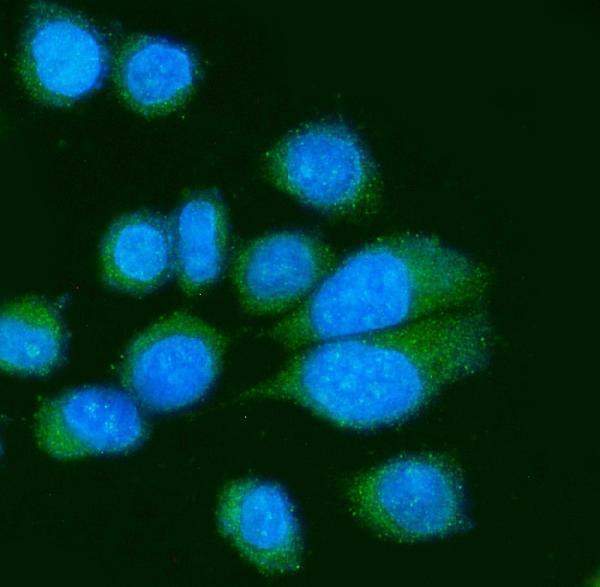
ICC-IF cell lines (HPA subcellular resource): HPA ICC-IF images show PVR in A-431, U-251MG, U2OS, with annotated localisation: Nucleoplasm (approved), Vesicles (approved), Plasma membrane (supported) (HPA subcellular).
Technical controls: Include a no-primary (secondary-only) control, a concentration-matched mouse immunoglobulin isotype control, and a PVR knockout biological control (M00664-2 caption: mouse primary antibody; standard IHC practice). Quench endogenous peroxidase and check heart muscle pigment before scoring DAB signal (standard IHC practice).
⚠️Feasibility: No target-specific fixation window or fixation effect is reported in the supplied evidence, and the selected-SKU caption does not state its fixative (M00664-2 caption). Heat retrieval in EDTA at pH 8.0 was used for a paraffin colorectal adenocarcinoma section, but its performance in heart muscle needs verification (M00664-2 caption); heart pigment can complicate DAB scoring (standard IHC practice). The supplied evidence does not establish that frozen sections or IF are easier than paraffin IHC; HPA does show PVR ICC-IF images (HPA subcellular).

HPA tissue IHC evidence for PVR

Comprehensive Human Protein Atlas IHC scoring per tissue (reliability: Approved — Medium consistency between antibody staining and RNA expression data. At least one protein variant secreted, tissue location of RNA and protein might differ and correlation is complex.). Rows are taken directly from the HPA tissue chart — click any row's HPA link to view the source.

Positive expression · recommended positive controls

TissueCell typeLevelEvidenceSource
Heart muscle Cardiomyocytes High Protein (IHC) HPA →
Placenta Decidual cells High Protein (IHC) HPA →
Adrenal gland Glandular cells Medium Protein (IHC) HPA →
Appendix Glandular cells Medium Protein (IHC) HPA →
Caudate Glial cells Medium Protein (IHC) HPA →

Undetected expression · recommended negative controls

TissueCell typeLevelEvidenceSource
Adipose tissue Adipocytes Not detected Protein (IHC) HPA →
Bone marrow Hematopoietic cells Not detected Protein (IHC) HPA →
Bronchus Basal cells Not detected Protein (IHC) HPA →
Fallopian tube Ciliated cells (cell body) Not detected Protein (IHC) HPA →
Hippocampus Glial cells Not detected Protein (IHC) HPA →
Section 3

Advanced PVR IHC Tips

Use the IHC evidence below to troubleshoot PVR staining in paraffin sections; the IF entry addresses the separate microscopy workflow.

How should I troubleshoot weak PVR staining after antigen retrieval?
Start with heat-mediated retrieval in EDTA at pH 8.0 (datasheet M00664-2). The documented paraffin-section result used 2 μg/ml primary antibody overnight at 4°C, so compare retrieval conditions while holding antibody concentration and detection constant (datasheet M00664-2). If staining remains weak, test another retrieval buffer or heating duration on adjacent sections as a fallback, checking tissue morphology and a no-primary control; these alternatives are unvalidated for this antibody. Compare membrane-associated signal in colorectal adenocarcinoma sections with the documented image (datasheet M00664-2), and retain the condition that improves interpretable staining without increasing diffuse background.
Could fixation explain weak or uneven PVR staining?
The selected paraffin-section caption does not report its fixative, so target-specific fixation sensitivity is unknown (datasheet M00664-2). Record the fixative and fixation duration for each specimen, then compare similarly processed sections before changing antibody concentration or retrieval; those are general IHC controls. For a suspected processing effect, stain adjacent sections together using EDTA at pH 8.0 and 2 μg/ml primary antibody as documented (datasheet M00664-2). Examine morphology and the distribution of staining across each section, and interpret any fixation comparison as an experiment on those specimens rather than an established PVR-specific effect.
Which staining compartments should I expect for PVR in tissue?
Assess membrane staining first because PVR has an extracellular region at residues 21–343, a transmembrane segment at 344–367, and a cytoplasmic tail at 368–417 (UniProt P15151 topology). Cytoplasmic staining can also occur in tissue, and positivity has been reported in plasma (HPA: tissue IHC profile). Thus, record membrane and cytoplasmic scores separately rather than treating every nonmembranous signal as background (HPA: tissue IHC profile). HPA additionally reports nucleoplasm and vesicles in subcellular imaging, with supported plasma-membrane localisation (HPA: subcellular); confirm any predominantly nuclear tissue pattern with an independent antibody or orthogonal assay before assigning it to PVR.
Could isoforms or epitope position change my IHC readout?
PVR has four annotated isoforms, Alpha, Beta, Gamma and Delta, so first check which sequence region the antibody recognizes (UniProt P15151 isoforms). Its extracellular region spans residues 21–343, whereas the cytoplasmic region spans 368–417 (UniProt P15151 topology). The supplied caption does not identify the M00664-2 epitope or establish isoform coverage (datasheet M00664-2); therefore, a negative stain cannot exclude every isoform. When comparing antibodies, document their mapped epitopes and score the same tissue compartments separately; interpret disagreement alongside splice-variant coverage, antigen accessibility and assay controls rather than assigning a specific variant from staining alone.
How can I investigate a PVR pattern with multiplex IF?
For the separate IF workflow, pair PVR with a marker identifying the cell population being assessed; HPA reports staining in cardiomyocytes and decidual cells, among others (HPA: tissue IHC). Choose a fluorophore whose signal can be distinguished from the specimen’s autofluorescence, and include single-stain and no-primary controls to check bleed-through and background (standard IF practice). Match permeabilisation to the antibody epitope: an extracellular epitope may be accessible without it, whereas a cytoplasmic-tail epitope generally requires membrane access (UniProt P15151 topology; standard IF practice). Because the supplied M00664-2 caption documents chromogenic paraffin IHC only, establish IF staining and epitope accessibility independently (datasheet M00664-2).
What should I change when PVR IHC shows diffuse background?
First compare the stained section with a no-primary control, since secondary reagent, endogenous peroxidase and nonspecific chromogen deposition can produce signal in chromogenic IHC (standard IHC practice). The documented image used 10% goat serum block, 2 μg/ml primary overnight at 4°C, and a peroxidase-conjugated secondary followed by DAB (datasheet M00664-2). If background persists, verify an effective peroxidase block and washing, then titrate the primary on adjacent sections while keeping retrieval at EDTA pH 8.0 (standard IHC practice; datasheet M00664-2). Score distinct cellular staining separately from diffuse plasma-associated signal, which HPA reports for PVR (HPA: tissue IHC profile).
How should I quantify heterogeneous PVR staining in sections? ⚠ ANSWER MARKED FOR VERIFICATION
Define the cells of interest on the counterstained section and report membrane and cytoplasmic staining separately, consistent with the tissue staining profile (HPA: tissue IHC). For each compartment, record the percentage of positive cells and an intensity-based H-score from 0–300, using the same threshold across specimens (standard IHC scoring practice). If the endpoint is spatial distribution, count positive cells per mm² of viable tissue and state the regions sampled (standard image-analysis practice). Normalise to the number of eligible cells or viable tissue area, and exclude necrosis, folds and section edges consistently to avoid counting artefact as PVR signal (standard IHC practice).
How do I distinguish genuine PVR staining from artefact?
Look for reproducible cellular staining that fits PVR’s membrane topology, while allowing cytoplasmic signal reported in tissue IHC (UniProt P15151 topology; HPA: tissue IHC profile). Check the cell type: HPA reports high signal in cardiomyocytes and decidual cells, but no detected signal in adipocytes and bone-marrow hematopoietic cells (HPA: tissue IHC). Treat staining restricted to section edges, folds or necrotic areas as suspect, and use a no-primary control to identify endogenous peroxidase or detection background (standard IHC practice). Nuclear signal alone needs independent confirmation because HPA subcellular imaging reports nucleoplasm, while the tissue profile describes membrane and cytoplasmic expression (HPA: subcellular; HPA: tissue IHC).
Boster reagents

Best PVR / Poliovirus receptor IHC Antibodies

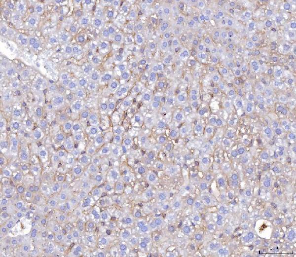
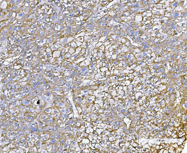
Anti-PVR antibodies have IHC images from human cancer paraffin sections and mouse liver cancer paraffin sections (catalog IHC captions); IF images show A431 and SiHa cells (catalog IF captions).

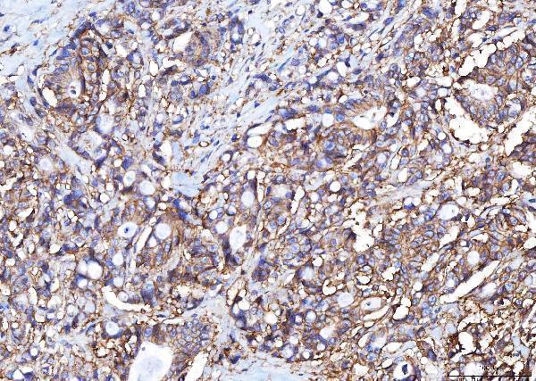
Real IHC data IHC analysis of Poliovirus Receptor/PVR using anti-Poliovirus Receptor/PVR antibody (M00664-2). Poliovirus Receptor/PVR was detected in a paraffin-embedded section of human colorectal adenocarcinoma tissue. Heat mediated antigen retrieval was performed in EDTA buffer (pH 8.0, epitope retrieval solution). The tissue section was blocked with 10% goat serum. The tissue section was then incubated with 2 μg/ml mouse anti-Poliovirus Receptor/PVR Antibody (M00664-2) overnight at 4°C. Peroxidase Conjugated Goat Anti-mouse IgG was used as secondary antibody and incubated for 30 minutes at 37°C. The tissue section was developed using HRP Conjugated Mouse IgG Super Vision Assay Kit (Catalog # SV0001) with DAB as the chromogen.
Anti-Poliovirus Receptor/PVR Antibody ® (monoclonal, 5I13D1)
Cat # M00664-2
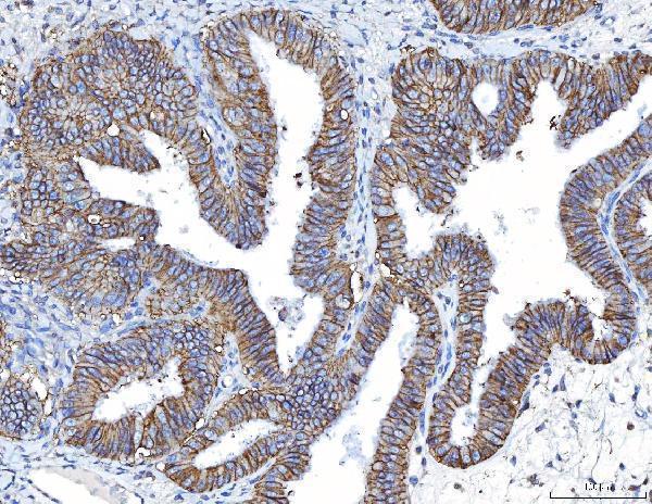
Real IHC data IHC analysis of Poliovirus Receptor/PVR using anti-Poliovirus Receptor/PVR antibody (M00664-1). Poliovirus Receptor/PVR was detected in a paraffin-embedded section of human colorectal adenocarcinoma tissue. Heat mediated antigen retrieval was performed in EDTA buffer (pH 8.0, epitope retrieval solution). The tissue section was blocked with 10% goat serum. The tissue section was then incubated with 2 μg/ml mouse anti-Poliovirus Receptor/PVR Antibody (M00664-1) overnight at 4°C. Peroxidase Conjugated Goat Anti-mouse IgG was used as secondary antibody and incubated for 30 minutes at 37°C. The tissue section was developed using HRP Conjugated Mouse IgG Super Vision Assay Kit (Catalog # SV0001) with DAB as the chromogen.
Anti-Poliovirus Receptor/PVR Antibody ® (monoclonal, 9B9F1)
Cat # M00664-1
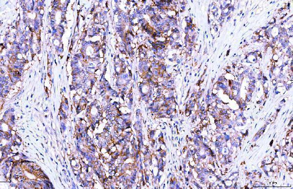
Real IHC data IHC analysis of Poliovirus Receptor/PVR using anti-Poliovirus Receptor/PVR antibody (A00664-2). Poliovirus Receptor/PVR was detected in paraffin-embedded section of human liver cancer tissue. Heat mediated antigen retrieval was performed in EDTA buffer (pH8.0, epitope retrieval solution). The tissue section was blocked with 10% goat serum. The tissue section was then incubated with 2μg/ml rabbit anti-Poliovirus Receptor/PVR Antibody (A00664-2) overnight at 4°C. Biotinylated goat anti-rabbit IgG was used as secondary antibody and incubated for 30 minutes at 37°C. The tissue section was developed using Strepavidin-Biotin-Complex (SABC) (Catalog # SA1022) with DAB as the chromogen.
Anti-Poliovirus Receptor/PVR Antibody ®
Cat # A00664-2
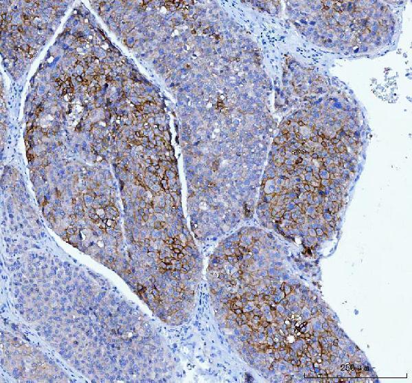
Real IHC data IHC analysis of Poliovirus Receptor/PVR using anti-Poliovirus Receptor/PVR antibody (M00664-3). Poliovirus Receptor/PVR was detected in a paraffin-embedded section of human liver cancer tissue. Heat mediated antigen retrieval was performed in EDTA buffer (pH 8.0, epitope retrieval solution). The tissue section was blocked with 10% goat serum. The tissue section was then incubated with 2 μg/ml mouse anti-Poliovirus Receptor/PVR Antibody (M00664-3) overnight at 4°C. Peroxidase Conjugated Goat Anti-mouse IgG was used as secondary antibody and incubated for 30 minutes at 37°C. The tissue section was developed using HRP Conjugated Mouse IgG Super Vision Assay Kit (Catalog # SV0001) with DAB as the chromogen.
Anti-Poliovirus Receptor/PVR Antibody ® (monoclonal, 3B11E9)
Cat # M00664-3
Real IHC data IHC analysis of Poliovirus Receptor/PVR using anti-Poliovirus Receptor/PVR antibody (A00664-4). Poliovirus Receptor/PVR was detected in a paraffin-embedded section of human liver cancer tissue. Heat mediated antigen retrieval was performed in EDTA buffer (pH 8.0, epitope retrieval solution). The tissue section was blocked with 10% goat serum. The tissue section was then incubated with 2 μg/ml rabbit anti-Poliovirus Receptor/PVR Antibody (A00664-4) overnight at 4°C. Biotinylated goat anti-rabbit IgG was used as secondary antibody and incubated for 30 minutes at 37°C. The tissue section was developed using Strepavidin-Biotin-Complex (SABC) (Catalog # SA1022) with DAB as the chromogen.
Anti-Poliovirus Receptor/PVR Antibody ®
Cat # A00664-4

M00664-1 and M00664-2 have IHC images from human colorectal adenocarcinoma, while M00664-3 has images from human and mouse liver cancer paraffin sections (respective IHC captions). A00664-2 has IHC images from human liver and prostate cancer and an IF image from A431 cells; A00664-4 has a human liver cancer IHC image and a SiHa cell IF image (respective IHC and IF captions).

Which to pick: For human tissue IHC, choose among the five SKUs using the paraffin-section images for the relevant tissue; A00664-2 lists 1–2 μg/ml and A00664-4 lists 2–5 μg/ml for IHC (catalog applications, IHC captions and dilution listings). For IF/ICC, choose A00664-2 or A00664-4: both list IF and ICC, with cell-image examples for A431 and SiHa respectively (catalog applications and IF captions). For human and mouse tissue IHC, M00664-3 has the strongest direct image support, with paraffin-section examples in both species; it is a mouse monoclonal antibody, and the IHC captions do not report the fixative (catalog clone, reactivity and IHC captions).

Each figure is that product's own IHC / IF validation image from its datasheet.

References

  1. UniProt Consortium. UniProt entry P15151 (PVR_HUMAN, Poliovirus receptor).
  2. Human Protein Atlas. PVR tissue IHC expression (reliability: Approved).
  3. Human Protein Atlas. PVR subcellular location (ICC-IF): Localized to the nucleoplasm, plasma membrane and vesicles..
  4. Human Protein Atlas. PVR antibody validation summary (2 antibodies).
  5. TIGIT agonism as a therapeutic strategy to suppress inflammation in hidradenitis suppurativa. Frontiers in immunology 2026 — PMC13079107.
  6. Epithelial CD155-TIGIT/CD226 signaling links CD8⁺ T-cell exhaustion to frequent exacerbations in COPD. Respiratory research 2026 — PMC13195874.
  7. Effect of Methotrexate on an In Vitro Patient-Derived Model of Proliferative Vitreoretinopathy. Investigative ophthalmology & visual science 2017 — PMC5544356.
  8. PVR/TIGIT and PD-L1/PD-1 expression predicts survival and enlightens combined immunotherapy in lung squamous cell carcinoma. Translational oncology 2022 — PMC9352965.
  9. PubMed PMID:2538245 — UniProt-cited evidence.
  10. PubMed PMID:2170108 — UniProt-cited evidence.
  11. PubMed PMID:14702039 — UniProt-cited evidence.