This website uses cookies to ensure you get the best experience on our website.


- Table of Contents
Plan chromogenic PXMP4 IHC in paraffin sections using renal tubules and type II alveolar cells as high-staining references (HPA tissue IHC). The catalog antibody has an IHC dilution range of 1:100–1:300 (datasheet A13147); spleen red-pulp cells are reported as unstained (HPA tissue IHC).

Expected localisation, validated protocols, controls and antibodies — the at-a-glance facts below, then the full design guide.
| Expected localisation | Expected peroxisome membrane (UniProt); observed granular cytoplasm (HPA tissue IHC) | |
| Staining pattern | Granular cytoplasm in renal tubules and type II alveolar cells (HPA tissue IHC) | |
| Antigen retrieval | Tris-EDTA pH 9.0 HIER, heat-mediated (datasheet A13147) | |
| Positive control | Bronchus+4 more · see all | |
| Negative control | Spleen |
| Fixation | Keep fixation consistent across paraffin sections and controls. (selected-SKU IHC image A13147) | |
| Caveat | Tissue staining has medium consistency with RNA expression (HPA tissue IHC) | |
| Regulation | Expression varies with androgen sensitivity (UniProt) | |
| Isoform / epitope | 2 isoforms; epitope side is unannotated (UniProt) |
The catalog antibody’s IHC-P protocol (datasheet A13147) is accompanied by one published PXMP4 IHC protocol in hepatocellular carcinoma tissue (PMC11077113).
| Sample | Paraffin-embedded human tonsil tissue; fixative not specified (datasheet A13147) |
| Fixation | Image fixative and duration unreported (datasheet A13147); verify before use. |
| Sectioning | 4–5 µm sections on charged slides (standard) |
| Deparaffinisation | Xylene, graded ethanol series to water (standard) |
| Antigen retrieval | Heat retrieval: Tris-EDTA pH 9.0 (datasheet A13147); 20 min, 95–100 °C (standard) |
| Peroxidase block | 3% H2O2, 10 min, room temperature (standard) |
| Blocking | 10% normal serum of the secondary host, 30 min, room temperature (standard) |
| Primary antibody | Rabbit anti-PXMP4, 1:100-1:300 (datasheet A13147) |
| Primary incubation | Overnight at 4 °C (standard) |
| Detection | HRP-polymer secondary, DAB chromogen 5–10 min (standard) |
| Counterstain | Hematoxylin, blue, dehydrate and mount (standard) |
| Expected result | PXMP4-positive staining in respiratory epithelial cells of bronchus (HPA tissue IHC: High). HPA tissue profile: Granular cytoplasmic expression in most tissues. No signal in the no-primary control. |
PXMP4 is a peroxisomal membrane protein with 2 transmembrane segments (UniProt Q9Y6I8 topology). In paraffin IHC, expect granular cytoplasmic staining in appropriately expressing cells, including kidney tubular cells, bronchial respiratory epithelial cells and lung type II alveolar cells (HPA tissue IHC). HPA rates the tissue staining Approved, with medium consistency between staining and RNA expression (HPA tissue IHC).
| Discrete cytoplasmic granules in kidney tubular cells or bronchial respiratory epithelial cells, with readily visible chromogen. | This matches the reported granular cytoplasmic pattern and High staining in those cells (HPA tissue IHC). At light-microscope resolution, the granules are consistent with peroxisome-associated PXMP4; IHC alone does not prove each granule is a peroxisome (UniProt Q9Y6I8 subcellular location; HPA tissue IHC). |
| Predominantly uniform nuclear, surface-membrane or extracellular staining, without discernible cytoplasmic granules. | This differs from the expected paraffin IHC pattern (HPA tissue IHC). Consider nonspecific staining or a detection artefact. HPA also reports nucleoli fibrillar-center localization by ICC-IF; that separate observation does not establish uniform nuclear staining as the expected IHC result (HPA subcellular ICC-IF). |
| Strong signal in a cell population where the chosen reference image shows little or no staining. | Check cell identity and morphology before calling it PXMP4: HPA reports low tissue specificity, so an unexpected cell type is not automatically negative (HPA tissue IHC). If the pattern persists without matching granules, investigate antibody cross-reactivity or endogenous detection activity (general IHC practice). |
| Diffuse chromogen over the section, including extracellular spaces or many unrelated cells. | A broad haze obscures the reported granular cytoplasmic pattern (HPA tissue IHC). Review background in a control section and assess blocking, antibody concentration, washing and chromogen development as general IHC variables (general IHC practice). |
| No staining in kidney tubular cells despite preserved tissue morphology and a functioning counterstain. | Kidney tubular cells are reported High by HPA, so this result warrants a technical check (HPA tissue IHC). A counterstain confirms tissue is present, but does not verify primary-antibody binding or detection; check those steps with appropriate assay controls (general IHC practice). |
| Reference tissue and cell population | HPA reports High staining in bronchial respiratory epithelium, Purkinje cells, endometrial and epididymal glandular cells, kidney tubules, lung type II alveolar cells, parathyroid glandular cells and rectal glandular cells (HPA tissue IHC). Score the identified population within its tissue, not the entire section. |
| Low-signal comparator | HPA reports Not detected specifically in spleen red-pulp cells, while several other listed populations are Low (HPA tissue IHC). Use those labels for their named cells only; neither label establishes that every cell in the tissue must be blank. |
| Topology and epitope uncertainty | The protein has 2 transmembrane segments at residues 97–117 and 153–173 (UniProt Q9Y6I8 topology). The supplied record does not identify the IHC antibody's epitope, so topology cannot determine which antigen-retrieval condition will work. |
| Antibody evidence and localization | HPA lists antibody HPA050077 as Approved for IHC and ICC; tissue IHC has medium staining-to-RNA consistency (HPA antibody validation; HPA tissue IHC). Peroxisomes are the main approved ICC-IF location, with an additional nucleoli fibrillar-center location (HPA subcellular ICC-IF). |
| Protein variants and modifications | UniProt lists 2 isoforms and glycosylation sites at residues 57 and 206 (UniProt Q9Y6I8). Without an epitope map or variant-specific staining data, these annotations cannot predict an IHC intensity difference or a missing signal. |
| Situation | Likely cause | Next action |
|---|---|---|
| Known High reference population is blank. | Target-specific fixation effects are not established by the supplied assay evidence. Verify with a matched IHC source before attributing a result to fixation. | Confirm cell identity, run assay controls, and review antigen retrieval, primary-antibody incubation and detection as general IHC steps (general IHC practice). |
| Reference tissue stains weakly or unevenly. | Variable tissue processing or reagent delivery can produce uneven IHC staining (general IHC practice); HPA levels describe observed populations, not guaranteed intensity on every section (HPA tissue IHC). | Compare well-preserved regions and a concurrent reference section; review retrieval consistency, reagent coverage and development time (general IHC practice). |
| Granules disappear into widespread brown haze. | Excess background can conceal the granular cytoplasmic pattern (HPA tissue IHC; general IHC practice). | Inspect a no-primary control, then review blocking, antibody concentration, washing and chromogen development (general IHC practice). |
| Nuclei appear uniformly positive in paraffin IHC. | Uniform nuclear staining is discordant with HPA's granular cytoplasmic tissue pattern; HPA's additional nucleolar localization comes from ICC-IF (HPA tissue IHC; HPA subcellular ICC-IF). | Compare a reference tissue and detection controls; assess nuclear background before treating the IHC signal as specific (general IHC practice). |
| Spleen red-pulp cells stain strongly. | HPA reports PXMP4 Not detected in that specific population (HPA tissue IHC); endogenous detection activity or cross-reactivity may account for unexpected color (general IHC practice). | Verify the stained cells are in red pulp, examine a no-primary control, and compare the pattern with a High reference population (HPA tissue IHC; general IHC practice). |
| Prostate sections differ between samples. | UniProt reports expression in normal prostate epithelial and androgen-sensitive adenocarcinoma cells, but not androgen-insensitive adenocarcinoma cells (UniProt Q9Y6I8 tissue specificity). This is not a guaranteed IHC result for any given specimen. | Document the sampled cell population and assay controls before interpreting a difference; use the HPA tissue images as the staining-pattern reference (HPA tissue IHC; general IHC practice). |


Comprehensive Human Protein Atlas IHC scoring per tissue (reliability: Approved — Medium consistency between antibody staining and RNA expression data.). Rows are taken directly from the HPA tissue chart — click any row's HPA link to view the source.
| Tissue | Cell type | Level | Evidence | Source |
|---|---|---|---|---|
| Bronchus | Respiratory epithelial cells | High | Protein (IHC) | HPA → |
| Cerebellum | Purkinje cells | High | Protein (IHC) | HPA → |
| Endometrium | Glandular cells | High | Protein (IHC) | HPA → |
| Epididymis | Glandular cells | High | Protein (IHC) | HPA → |
| Kidney | Cells in tubules | High | Protein (IHC) | HPA → |
| Tissue | Cell type | Level | Evidence | Source |
|---|---|---|---|---|
| Spleen | Cells in red pulp | Not detected | Protein (IHC) | HPA → |
Troubleshoot PXMP4 staining in paraffin sections by checking retrieval, compartment, cellular context, and controls before comparing signal across samples.
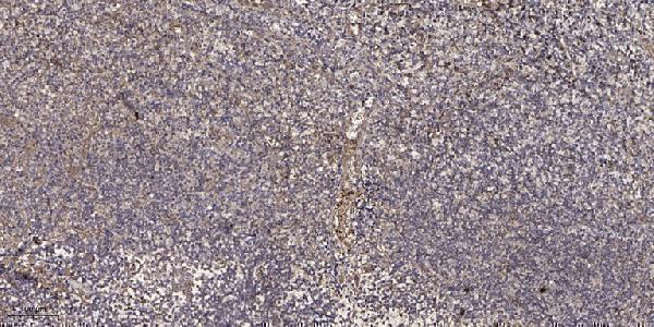
A13147 is listed for human and monkey IHC and IF (catalog: applications, reactivity). Its supplied figure shows paraffin-section IHC of human tonsil (A13147 IHC image caption).
A13147 will render with a human tonsil paraffin-section IHC figure (A13147 IHC image caption). IF is listed as an application for human and monkey samples, but no IF figure is supplied (catalog: applications, reactivity, if_image_alts).
Which to pick: For tissue IHC, choose A13147, a rabbit polyclonal listed for IHC (catalog: host, dilution_raw, applications); its own caption reports 1:200 antibody dilution and Tris-EDTA retrieval at pH 9.0 in paraffin-embedded human tonsil, with fixative unreported (A13147 IHC image caption). For IF, A13147 is listed, but ICC-specific validation and an IF figure are unreported (catalog: applications, if_image_alts). For monkey samples, A13147 lists monkey reactivity, while its supplied IHC figure shows human tissue only (catalog: reactivity; A13147 IHC image caption).