This website uses cookies to ensure you get the best experience on our website.


- Table of Contents
Plan PYCR1 staining in paraffin sections using cytoplasmic tissue staining as the observed pattern (HPA tissue IHC) and mitochondrial location as the molecular expectation (UniProt). The guide covers fixation, controls and interpretation, including the reported low consistency between staining and RNA expression (HPA tissue IHC).

Expected localisation, validated protocols, controls and antibodies — the at-a-glance facts below, then the full design guide.
| Expected localisation | Cytoplasmic staining (HPA tissue IHC); mitochondrial location (UniProt) | |
| Staining pattern | Cytoplasmic in most tissues; high in select glandular cells (HPA tissue IHC) | |
| Antigen retrieval | EDTA pH 8.0 HIER, heat-mediated (datasheet A06018-1) | |
| Positive control | Bronchus+4 more · see all | |
| Negative control | Oral mucosa+2 more · see all |
| Fixation | Keep fixation consistent across sections (standard IHC practice; not target-specific) | |
| Caveat | Staining and RNA expression have low consistency (HPA tissue IHC) | |
| Regulation | Pancreas and salivary gland RNA enhanced (HPA RNA) | |
| Isoform / epitope | 3 isoforms; epitope impact is unknown (UniProt) |
The catalog antibody’s IHC-P protocol is paired with four published PYCR1 staining workflows for renal, osteosarcoma, esophageal, and pancreatic tissues (PMC6641676; PMC13219957; PMC9218025; PMC8965111).
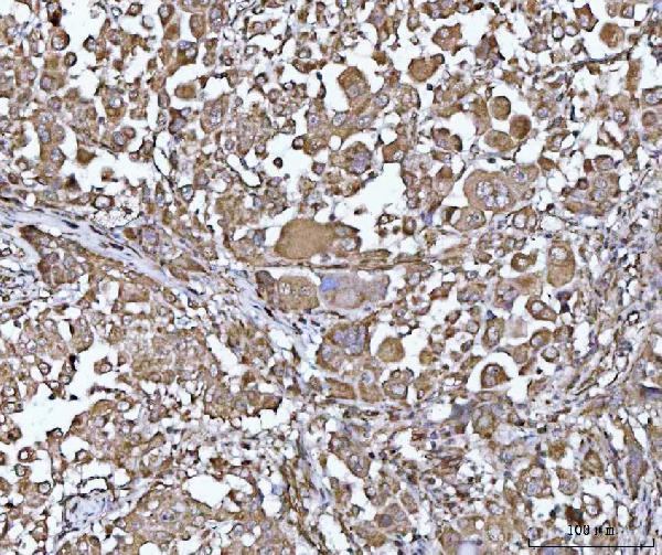
| Sample | Paraffin-embedded human lung cancer tissue; fixative not specified (datasheet A06018-1) |
| Fixation | Image fixative and duration unreported (datasheet A06018-1); verify before use. |
| Sectioning | 4–5 µm sections on charged slides (standard) |
| Deparaffinisation | Xylene, graded ethanol series to water (standard) |
| Antigen retrieval | Heat retrieval: EDTA pH 8.0 (datasheet A06018-1); 20 min, 95–100 °C (standard) |
| Peroxidase block | 3% H2O2, 10 min, room temperature (standard) |
| Blocking | 10% goat serum (datasheet A06018-1) |
| Primary antibody | Rabbit anti-PYCR1, 2-5 μg/ml (datasheet A06018-1) |
| Primary incubation | Overnight at 4 °C (datasheet A06018-1) |
| Detection | HRP-conjugated secondary, DAB chromogen (datasheet A06018-1) |
| Counterstain | Hematoxylin, blue, dehydrate and mount (standard) |
| Expected result | PYCR1-positive staining in basal cells of bronchus (HPA tissue IHC: High). HPA tissue profile: Cytoplasmic expression in most tissues. No signal in the no-primary control. |
PYCR1 localises to mitochondria and has no annotated transmembrane segment (UniProt P32322: location and topology). In paraffin-section IHC, expect cytoplasmic staining in many tissues, including strong staining in pancreatic exocrine glandular cells and bronchial basal cells (HPA: tissue IHC). HPA rates its antibody staining Approved, while reporting low consistency with RNA expression; interpret the pattern alongside controls rather than treating intensity alone as proof of specificity (HPA: reliability).
| Cytoplasmic signal in pancreatic exocrine glandular cells, with little signal in ovarian stroma. | This fits a useful positive–negative comparison: HPA reports High staining in pancreatic exocrine glandular cells and Not detected in ovarian stroma cells (HPA: tissue IHC). A granular cytoplasmic appearance can be compatible with mitochondrial localisation, but chromogenic IHC alone does not resolve individual mitochondria (UniProt P32322: location; general IHC practice). |
| Predominantly nuclear or surface-restricted signal, with little cytoplasmic staining. | That distribution conflicts with the reported mitochondrial location and HPA’s cytoplasmic tissue profile (UniProt P32322: location and topology; HPA: tissue IHC). Check the counterstain and detection controls before assigning it to PYCR1; the pattern could reflect artefact or another antigen (general IHC practice). |
| Strong staining in ovarian stroma cells or oral squamous epithelial cells. | HPA reports PYCR1 as Not detected in those cell populations (HPA: tissue IHC). Reproducible staining there warrants a specificity check, including a no-primary control; cross-reactivity or endogenous detection activity are possibilities, not diagnoses from appearance alone (general IHC practice). |
| Broad, diffuse colour across cells and surrounding tissue without clear cell boundaries. | This is difficult to score as the reported cell-associated cytoplasmic pattern (HPA: tissue IHC). Compare a no-primary control and review blocking, washes and chromogen development for background from the detection workflow (general IHC practice). |
| No signal in pancreatic exocrine glandular cells while the section is otherwise interpretable. | An expected strong reference population is missing (HPA: High in pancreatic exocrine glandular cells). First verify tissue identity and assay controls, then review the antibody’s IHC-P instructions and retrieval and detection steps; a blank slide alone cannot establish absent PYCR1 (general IHC practice). |
| Compartment and optical resolution | PYCR1 is mitochondrial and lacks an annotated transmembrane segment (UniProt P32322: location and topology). HPA describes cytoplasmic tissue staining; a chromogenic section can support that compartment call without proving mitochondrial colocalisation (HPA: tissue IHC; general IHC practice). |
| Reference cell populations | HPA reports High staining in bronchial basal cells, cortical and hippocampal neuronal cells, and pancreatic exocrine glandular cells; oral squamous epithelial cells, ovarian stroma cells and soft-tissue fibroblasts are Not detected (HPA: tissue IHC). Score the named cells within each tissue, rather than the whole section. |
| Strength of antibody evidence | The listed antibody HPA047660 has Approved IHC status; HPA also flags low agreement between antibody staining and RNA expression (HPA: antibody and tissue records). This supports cautious use of the observed pattern, with controls, rather than treating every coloured cell as confirmed PYCR1. |
| Isoforms and processing | UniProt lists three PYCR1 isoforms and a chain spanning residues 2–319 (UniProt P32322: isoforms and processing). These annotations do not identify the catalog antibody’s epitope or establish different staining among isoforms; avoid inferring either from an IHC intensity difference. |
| IF/ICC: what pattern is established? | Mitochondrial localisation predicts intracellular signal (UniProt P32322: location), but HPA supplies no main ICC-IF location or cell-line images, and the listed antibody has no ICC status (HPA: subcellular and antibody records). An IF result needs its own validation; no IF protocol is established here. |
| Situation | Likely cause | Next action |
|---|---|---|
| Pancreatic exocrine glandular cells remain blank. | A reference population reported High by HPA is missing; the slide alone cannot distinguish assay failure from sample variation (HPA: tissue IHC; general IHC practice). | Confirm cell identity and control performance, then check the catalog antibody’s IHC-P instructions, retrieval, dilution and detection steps (general IHC practice). |
| Nuclei dominate the stain. | A nuclear-dominant result conflicts with mitochondrial location and the reported cytoplasmic tissue profile (UniProt P32322: location; HPA: tissue IHC). | Compare the counterstain and no-primary control; review whether chromogen development or nonspecific binding obscures the cytoplasm (general IHC practice). |
| Oral squamous epithelium stains strongly. | HPA reports this cell population as Not detected; cross-reactivity or endogenous detection activity could explain an unexpected signal (HPA: tissue IHC; general IHC practice). | Run a no-primary control and review blocking and detection controls before scoring that staining as PYCR1 (general IHC practice). |
| Colour covers tissue and empty spaces evenly. | A field-wide deposit is inconsistent with a cell-associated cytoplasmic pattern (HPA: tissue IHC); excess detection background is possible (general IHC practice). | Inspect the no-primary control, washing and chromogen development, then rescore only clearly localised cellular signal (general IHC practice). |
| All cell types appear equally intense. | HPA reports different levels across specified cell populations, from High to Not detected (HPA: tissue IHC). Uniform colour can make those differences unreadable (general IHC practice). | Check exposure to chromogen and compare named positive and negative populations on controlled sections before assigning intensity categories (general IHC practice). |
| A granular pattern is hard to distinguish from background. | Mitochondrial location is annotated, while HPA reports tissue staining at the broader cytoplasmic level (UniProt P32322: location; HPA: tissue IHC). | Score reproducible cellular cytoplasmic staining against controls; reserve a mitochondrial colocalisation claim for separately validated IF evidence (general IHC practice). |


Comprehensive Human Protein Atlas IHC scoring per tissue (reliability: Approved — Low consistency between antibody staining and RNA expression data.). Rows are taken directly from the HPA tissue chart — click any row's HPA link to view the source.
| Tissue | Cell type | Level | Evidence | Source |
|---|---|---|---|---|
| Bronchus | Basal cells | High | Protein (IHC) | HPA → |
| Cerebral cortex | Neuronal cells | High | Protein (IHC) | HPA → |
| Cervix | Glandular cells | High | Protein (IHC) | HPA → |
| Hippocampus | Neuronal cells | High | Protein (IHC) | HPA → |
| Pancreas | Exocrine glandular cells | High | Protein (IHC) | HPA → |
Troubleshoot PYCR1 staining in paraffin sections by checking retrieval, controls, cellular distribution and scoring before interpreting chromogenic signal.
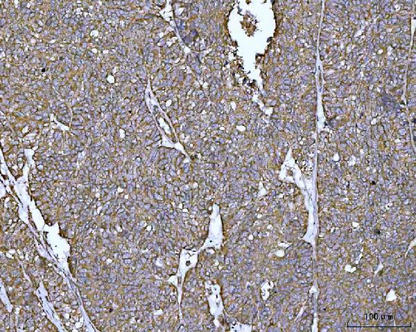
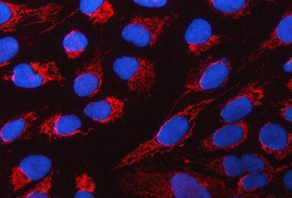
A06018-1 has IHC data from human lung and ovarian cancer paraffin sections and IF data from U2OS cells and human glioma paraffin sections (catalog image captions).
A06018-1 is listed for human IHC and IF (catalog applications and reactivity). Its captions show IHC in human lung and ovarian cancer paraffin sections, plus IF in U2OS cells and human glioma paraffin sections (catalog image captions).
Which to pick: Choose A06018-1 for human tissue IHC on paraffin sections; its IHC captions document those sections, while the fixative is unreported (catalog IHC image captions). For IF/ICC, A06018-1 is listed for both applications and has IF images from U2OS cells and human glioma paraffin sections (catalog applications and IF image captions). No cross-species choice is supported: A06018-1 lists human reactivity only, and its clonality is unreported (catalog reactivity and clone field).