PYY / Peptide YY · IHC design guide

Design Immunohistochemistry for PYY

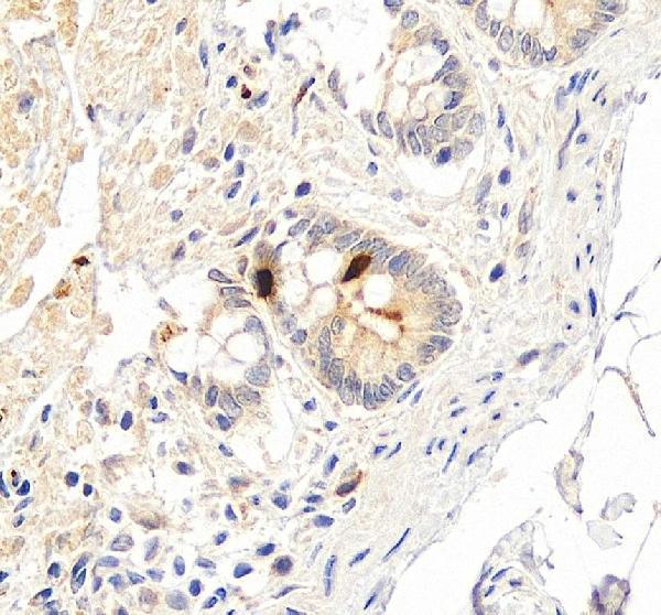
Plan paraffin-section PYY IHC using gastrointestinal endocrine cells as a positive staining reference (HPA tissue IHC). The catalog antibody has documented chromogenic staining in human appendicitis and small intestine sections (datasheet A04223-1); account for PYY secretion when interpreting signal (UniProt).

Evidence assembled Oct 2026 · For research use; verify linked source records and product datasheet before use
Immunohistochemistry protocol sheet for PYY (IHC for PYY): expected localisation Secreted (UniProt); tissue compartment unreported (HPA tissue IHC), antibody A04223-1, validated IHC image, and IHC protocol steps
Printable PYY IHC protocol sheet — expected localisation Secreted (UniProt); tissue compartment unreported (HPA tissue IHC), antibody A04223-1, controls and protocol steps. Open the full PYY IHC guide →

PYY Immunohistochemistry Experimental Design Guide

Expected localisation, validated protocols, controls and antibodies — the at-a-glance facts below, then the full design guide.

Must know before staining
Expected localisation Secreted (UniProt); tissue compartment unreported (HPA tissue IHC)
Staining pattern Selective GI enteroendocrine-cell staining (HPA tissue IHC)
Antigen retrieval Citrate pH 6 HIER, heat-mediated (datasheet A04223-1)
Positive control ⓘ Appendix+4 more · see all
Negative control ⓘ Adipose tissue+4 more · see all
Important caveats
Reasons your staining may differ from the expected pattern.
Fixation Keep fixation consistent across sections. (standard IHC practice; not target-specific)
Caveat Secreted PYY: RNA and protein sites may differ (HPA tissue IHC)
Regulation Regulation unreported (UniProt)
Isoform / epitope 2 isoforms; check epitopes against processing sites (UniProt)
Section 1

Recommended PYY IHC & IF Protocols

The catalog antibody protocol is accompanied by two published chromogenic PYY IHC protocols for paraffin sections (PMC3306780; PMC12834159).

Recommended immunohistochemistry (IHC-P) protocol parameters
SampleParaffin-embedded human appendicitis tissue tissues; fixative not specified (datasheet A04223-1)
FixationImage fixative and duration unreported (datasheet A04223-1); verify before use.
Sectioning4–5 µm sections on charged slides (standard)
DeparaffinisationXylene, graded ethanol series to water (standard)
Antigen retrievalHeat retrieval: Citrate pH 6, 20 min (datasheet A04223-1)
Peroxidase block3% H2O2, 10 min, room temperature (standard)
Blocking10% goat serum (datasheet A04223-1)
Primary antibodyRabbit anti-PYY, 1μg/ml (datasheet A04223-1)
Primary incubationOvernight at 4 °C (datasheet A04223-1)
DetectionStreptavidin-biotin complex (SABC), DAB chromogen (datasheet A04223-1)
CounterstainHematoxylin, blue, dehydrate and mount (standard)
Expected resultPYY-positive staining in endocrine cells of appendix (HPA tissue IHC: High). HPA tissue profile: Selective expression of enteroendocrine cells in the gastrointestinal tract. No signal in the no-primary control.
💡Decision noteStart with heat-mediated citrate retrieval at pH 6 for the catalog antibody (datasheet: A04223-1); neither published excerpt specifies retrieval (PMC3306780; PMC12834159).
Section 2

What Is the Expected PYY Staining Pattern?

PYY is a secreted peptide with no transmembrane segment (UniProt P10082: subcellular location and topology). In paraffin section IHC, expect selective staining of gastrointestinal enteroendocrine cells, with high staining reported in appendix, colon, duodenum, rectum and small intestine (HPA: tissue IHC). HPA rates the tissue pattern Enhanced but notes only medium consistency with RNA expression because a secreted protein’s RNA and protein locations can differ (HPA: reliability description).

What am I looking at on my slide?
Distinct positive cells are scattered among gastrointestinal epithelial cells, while neighboring cells remain largely unstained.This fits selective enteroendocrine expression (HPA: tissue IHC). Assess the pattern by cell identity as well as intensity: HPA reports High staining in endocrine cells at five gastrointestinal sites, rather than uniform staining across the tissue (HPA: tissue IHC).
The strongest signal appears nuclear, membranous or uniformly across many cell types.Recheck compartment and morphology. PYY is secreted and has no transmembrane segment (UniProt P10082: subcellular location and topology); HPA provides no image based ICC-IF subcellular assignment (HPA: subcellular). A dominant nuclear or membrane pattern is therefore unsupported, while even cytoplasmic staining needs the expected cell distribution.
Strong chromogen appears in adipocytes, respiratory epithelium or other cells outside the expected endocrine population.Treat it as a possible false positive and investigate antibody cross-reactivity or endogenous detection activity. HPA reports PYY as Not detected in adipose adipocytes and bronchial respiratory epithelial cells (HPA: tissue IHC). A single unexpected positive site should not redefine the target’s distribution.
Weak color covers most tissue, including spaces between cells and the negative control.This is more consistent with background than selective enteroendocrine staining (HPA: tissue IHC). In chromogenic IHC, nonspecific antibody binding, inadequate blocking or residual endogenous detection activity can produce widespread color (standard IHC practice); compare the slide with matched controls before scoring cells as positive.
No convincing endocrine cell signal appears in an appendix, colon, duodenum, rectum or small intestine section.First confirm that the section contains identifiable endocrine cells and that the run’s controls worked. These are HPA High sites specifically in endocrine cells (HPA: tissue IHC), but their absence on one slide does not establish absent PYY expression; review antibody, retrieval and detection conditions as general IHC troubleshooting.
💡Expected PYY appearanceCall a section positive when discrete gastrointestinal endocrine cells show clear cellular chromogen above neighboring cells (HPA: High in endocrine cells); diffuse tissue color or dominant nuclear or membrane staining is not the supported PYY pattern (HPA: tissue IHC; UniProt P10082: secreted, no transmembrane segment).
How each factor affects the staining
Tissue and cell identityUse the endocrine cell population in appendix, colon, duodenum, rectum or small intestine as the primary pattern reference (HPA: High in endocrine cells). HPA’s Not detected entries describe the specified cells in those tissues, not every cell in an entire organ (HPA: tissue IHC).
Secretion and compartmentPYY is secreted and lacks a transmembrane segment (UniProt P10082: subcellular location and topology). Cellular staining may reflect peptide before secretion; extracellular signal alone needs cautious interpretation. HPA lists no ICC-IF images or assigned intracellular location to establish a finer compartment pattern (HPA: subcellular).
Peptide processing and antibody epitopeThe PYY record annotates a signal peptide at residues 1–28 and a propeptide at 68–97 (UniProt P10082: processing). An antibody’s recognized sequence could affect which PYY form is detected; the supplied records give no epitope, so do not infer mature peptide specificity.
Isoforms and antibody validationUniProt lists 2 isoforms without their antibody epitopes (UniProt P10082: isoforms). HPA rates HPA010973 Enhanced and CAB016734 Supported for IHC (HPA: antibodies). Those ratings support their documented IHC validation status; they do not establish identical recognition of both isoforms.
RNA comparison and preparation limitsHPA reports intestine enriched RNA and warns that secreted protein can occupy a different tissue location from its RNA (HPA: RNA specificity and reliability description). Neither HPA nor UniProt supplies a target specific fixation effect here; evaluate retrieval and fixation changes as general IHC workflow variables only.
Why is my staining missing, weak or wrong?
SituationLikely causeNext action
Known positive gastrointestinal tissue has no endocrine cell staining.The expected population may be missing from the section, or the IHC run may have failed; HPA reports High staining specifically in endocrine cells (HPA: tissue IHC).Check tissue morphology and a working positive control, then review antibody dilution, retrieval and detection steps as general IHC practice. Do not call PYY absent from a single failed section.
Every cell shows similarly strong chromogen.A broad pattern conflicts with HPA’s selective enteroendocrine profile (HPA: tissue IHC); nonspecific binding or endogenous detection activity is possible (standard IHC practice).Compare a no primary control, assess blocking and endogenous activity controls, and adjust detection conditions before scoring. Retain only cell associated signal clearly above surrounding tissue.
Unexpected adipocyte or respiratory epithelial staining appears.Those specified cell types were Not detected in HPA tissue IHC (HPA: adipose tissue and bronchus); cross-reactivity or detection background may explain the discrepancy.Verify the cell type and compare negative controls with a gastrointestinal positive section. Investigate reproducibility with an independently validated antibody before treating the unexpected site as PYY positive.
Signal is predominantly nuclear or membrane outlined.PYY is annotated as secreted with no transmembrane segment (UniProt P10082: location and topology). HPA does not provide an ICC-IF image based finer location (HPA: subcellular).Review morphology, counterstain and controls for localization error or background. Avoid assigning that compartment to PYY from this slide alone; compare with the selective endocrine cell pattern (HPA: tissue IHC).
Staining differs between antibodies or after a preparation change.The record lists 2 isoforms and peptide processing, but supplies no antibody epitope or target specific fixation sensitivity (UniProt P10082: isoforms and processing).Document antibody identity and preparation, then compare matched sections and controls. Interpret a change as assay dependent until the recognized PYY form and tissue pattern are independently supported.
Can IF/ICC use the same localization call?HPA describes PYY as secreted but provides no ICC-IF images, main intracellular location or ICC validation entry for the listed antibodies (HPA: subcellular and antibodies).Use the separate IF/ICC guide for assay design. Here, use HPA tissue IHC only to set an expected gastrointestinal endocrine cell distribution, without treating it as an IF/ICC compartment validation (HPA: tissue IHC; subcellular).

Sample controls for PYY IHC & IF

🧪Run appendix first: its endocrine cells should stain for PYY (HPA: High in appendix endocrine cells). Use adipose tissue as the negative tissue, where adipocytes are not detected, and expect adjacent non-endocrine cells on the appendix slide to show only background (HPA: Not detected in adipose adipocytes; HPA: High in appendix endocrine cells).
Positive control tissue: Appendix (Endocrine cells, HPA High)
Negative control tissue: Adipose tissue (HPA Not detected)
ICC-IF cell lines (HPA subcellular resource): HPA carries no ICC-IF cell line for PYY; derive a cell-line control from the positive tissue's cell type (Endocrine cells) and confirm it by RNA or western blot first.
Technical controls: Include a no-primary (secondary-only) slide, a concentration-matched rabbit isotype control, and a biological specificity control using PYY knockout tissue or peptide competition if available (caption: rabbit primary antibody; standard IHC practice). In appendix sections, check for endogenous peroxidase and biotin background because the reported detection uses a biotinylated secondary, streptavidin–biotin complex, and DAB (caption: SABC/DAB detection; standard IHC practice).
⚠️Feasibility: A target-specific fixation window or fixation effect is unreported, and the selected A04223-1 paraffin-section caption does not state the fixative (caption: fixative not stated). That caption reports citrate retrieval at pH 6 for 20 minutes, but does not establish that retrieval is required for every specimen (caption: heat-mediated citrate retrieval). Comparative ease of frozen-section IHC or IF is unreported; HPA lists no ICC-IF image-bearing cell lines for PYY, and appendix background from endogenous peroxidase or biotin should be checked with the stated detection method (HPA: no ICC-IF cell lines; caption: SABC/DAB detection; standard IHC practice).

HPA tissue IHC evidence for PYY

Comprehensive Human Protein Atlas IHC scoring per tissue (reliability: Enhanced — Medium consistency between antibody staining and RNA expression data. Secreted protein, tissue location of RNA and protein is expected to differ.). Rows are taken directly from the HPA tissue chart — click any row's HPA link to view the source.

Positive expression · recommended positive controls

TissueCell typeLevelEvidenceSource
Appendix Endocrine cells High Protein (IHC) HPA →
Colon Endocrine cells High Protein (IHC) HPA →
Duodenum Endocrine cells High Protein (IHC) HPA →
Rectum Endocrine cells High Protein (IHC) HPA →
Small intestine Endocrine cells High Protein (IHC) HPA →

Undetected expression · recommended negative controls

TissueCell typeLevelEvidenceSource
Adipose tissue Adipocytes Not detected Protein (IHC) HPA →
Adrenal gland Glandular cells Not detected Protein (IHC) HPA →
Bone marrow Hematopoietic cells Not detected Protein (IHC) HPA →
Breast Adipocytes Not detected Protein (IHC) HPA →
Bronchus Respiratory epithelial cells Not detected Protein (IHC) HPA →
Section 3

Advanced PYY IHC Tips

Use matched gastrointestinal controls and cell-level scoring to troubleshoot PYY staining in paraffin sections (HPA: selective enteroendocrine-cell expression).

How should I adjust antigen retrieval when PYY staining is weak?
Start with heat-mediated citrate retrieval at pH 6 for 20 minutes on paraffin sections (datasheet A04223-1). Keep an appendix endocrine-cell section in each run as a positive control (HPA: High in appendix endocrine cells), and compare retrieval changes against that section rather than judging a single specimen. If staining remains weak, adjust heating or cooling conditions one variable at a time while keeping citrate at pH 6; inspect morphology because excessive heating can damage sections (standard IHC practice). The caption used 1 µg/ml primary antibody overnight at 4°C, so check primary incubation and detection before attributing all loss to retrieval (datasheet A04223-1).
Could fixation explain inconsistent PYY staining across paraffin sections?
Target-specific fixation sensitivity is unknown: the paraffin-section caption does not state a fixative (datasheet A04223-1). Record the fixative, fixation duration, tissue thickness and processing history for each specimen, then compare sections prepared alike using the same citrate pH 6 retrieval for 20 minutes (datasheet A04223-1; standard IHC practice). If a weak case has poor morphology or patchy staining, examine matched control tissue before changing antibody concentration; fixation and processing differences can affect antigen access in IHC (standard IHC practice). Do not infer a PYY-specific fixation effect from its secreted annotation, modifications or tissue staining pattern (UniProt P10082; HPA: tissue IHC).
Where should convincing PYY staining appear in a gastrointestinal section?
Look first for staining in gastrointestinal endocrine cells, including those in appendix, colon, duodenum, rectum and small intestine (HPA: High in endocrine cells at these sites). PYY is annotated as secreted and has no transmembrane segment, so a crisp plasma-membrane outline alone is not the expected pattern to use as proof of specificity (UniProt P10082: subcellular location and topology). Compare cell-level signal with morphology and a matched negative-control section; diffuse extracellular colour needs cautious interpretation for a secreted peptide (UniProt P10082; standard IHC practice). HPA rates tissue staining as Enhanced but notes medium RNA–protein consistency and that secretion can separate protein location from RNA location (HPA: reliability description).
Can this stain distinguish PYY isoforms or processed peptide forms?
Do not score this antibody as isoform-specific without epitope mapping or direct validation: PYY has 2 annotated isoforms, while the supplied caption does not identify the antibody epitope (UniProt P10082: isoforms; datasheet A04223-1). The precursor is 97 amino acids long, with a signal segment at residues 1–28 and an annotated propeptide at 68–97 (UniProt P10082: processing). Phosphoserine at 41 and tyrosine amide at 64 are annotated modifications; their effects on antibody binding are unknown here (UniProt P10082: modified residues). If processed forms matter, obtain epitope information and validate specificity against the forms of interest before interpreting differences in chromogenic intensity (standard IHC practice).
How should I adapt PYY detection for multiplex immunofluorescence?
Treat IF as a separate validation exercise because the supplied product evidence describes chromogenic staining of paraffin sections, with no IF result (datasheet A04223-1). Multiplex PYY with a validated endocrine-cell marker and check whether signal falls in the expected gastrointestinal cell population (HPA: selective enteroendocrine-cell expression; standard IF practice). Choose spectrally separated fluorophores, favour a far-red channel if the tissue has substantial shorter-wavelength autofluorescence, and inspect single-stain and unstained controls (standard IF practice). For an intracellular epitope, test gentle permeabilisation; for an accessible extracellular epitope, compare without permeabilisation, since the antibody epitope is unspecified and PYY is secreted without a transmembrane segment (datasheet A04223-1; UniProt P10082).
How can I separate PYY staining from chromogenic background?
The documented paraffin-section method uses 10% goat-serum block, a biotinylated secondary, streptavidin–biotin detection and DAB (datasheet A04223-1). Run a no-primary control through the same detection steps, and include a peroxidase block as a general chromogenic IHC measure before interpreting brown deposits as PYY (standard IHC practice). If background follows the biotin-based detection system, assess endogenous biotin and consider an appropriately validated alternative detection system (standard IHC practice). Compare the result with endocrine cells in appendix as a positive reference and adipocytes in adipose tissue as a negative reference; these HPA patterns guide control selection but do not validate every specimen (HPA: High in appendix endocrine cells; Not detected in adipose adipocytes).
What should I count when comparing PYY staining between sections? ⚠ ANSWER MARKED FOR VERIFICATION
Define the gastrointestinal region and endocrine-cell denominator before scoring, because HPA reports selective PYY expression in enteroendocrine cells rather than uniform tissue staining (HPA: tissue profile). Report the percentage of positive endocrine cells, or an H-score combining their percentage and intensity, using the same threshold and DAB development settings across sections (standard IHC quantification practice). If cell identification is uncertain, report positive-cell density per mm² of evaluable mucosa and document how endocrine cells were identified (standard IHC quantification practice). Normalise to endocrine-cell count or evaluable tissue area, as appropriate, and exclude folds, necrosis and tissue edges by a rule set before scoring (standard IHC practice).
Which apparent PYY-positive patterns should make me question specificity?
A convincing result should localise to gastrointestinal endocrine cells and be reproducible in a matched positive-control section (HPA: selective enteroendocrine-cell expression; standard IHC practice). Treat predominant staining of unrelated cell types, a sharp membrane-only outline, or widespread nuclear colour cautiously: PYY is secreted and lacks a transmembrane segment (UniProt P10082: location and topology). Compare suspect deposits with the no-primary control, especially at section edges, necrotic regions and sites with endogenous peroxidase activity (standard chromogenic IHC practice). Negative staining in a particular section does not by itself prove absent PYY; assess morphology, retrieval and the run control, and remember that RNA and protein locations may differ for a secreted protein (HPA: reliability description; standard IHC practice).
Boster reagents

Best PYY / Peptide YY IHC Antibodies

The IHC-validated anti-PYY antibody has real chromogenic IHC data from human paraffin sections of appendicitis and small intestine tissue (A04223-1 IHC captions).

Real IHC data IHC analysis of Peptide YY/PYY using anti-Peptide YY/PYY antibody (A04223-1). Peptide YY/PYY was detected in paraffin-embedded section of human appendicitis tissue tissues. Heat mediated antigen retrieval was performed in citrate buffer (pH6, epitope retrieval solution) for 20 mins. The tissue section was blocked with 10% goat serum. The tissue section was then incubated with 1μg/ml rabbit anti-Peptide YY/PYY Antibody (A04223-1) overnight at 4°C. Biotinylated goat anti-rabbit IgG was used as secondary antibody and incubated for 30 minutes at 37°C. The tissue section was developed using Strepavidin-Biotin-Complex (SABC)(Catalog # SA1022) with DAB as the chromogen.
Anti-Peptide YY/PYY Antibody
Cat # A04223-1

A04223-1 is listed for human IHC and shown on paraffin sections of human appendicitis tissue (catalog applications/reactivity; A04223-1 IHC caption). A second IHC image shows the same SKU on a paraffin section of human small intestine (A04223-1 IHC caption).

Which to pick: Choose A04223-1 for human paraffin-section IHC: it is a rabbit polyclonal antibody listed for human IHC, with chromogenic images from both tissues (catalog host/dilution_raw/applications/reactivity; A04223-1 IHC captions). The captions do not report the fixative, so fixation and tissue suitability are unconfirmed (A04223-1 IHC captions). The payload provides no validated IF/ICC or cross-species option (catalog applications/reactivity/if_image_alts). The selected A04223-1 tissue-IHC caption documents paraffin sections, but does not specify the fixative (selected-SKU IHC image A04223-1).

Each figure is that product's own IHC / IF validation image from its datasheet.

References

  1. UniProt Consortium. UniProt entry P10082 (PYY_HUMAN, Peptide YY).
  2. Human Protein Atlas. PYY tissue IHC expression (reliability: Enhanced).
  3. Human Protein Atlas. PYY subcellular location (ICC-IF): Secreted.
  4. Human Protein Atlas. PYY antibody validation summary (2 antibodies).
  5. Free fatty acid receptor 1 stimulates cAMP production and gut hormone secretion through Gq-mediated activation of adenylate cyclase 2. Molecular metabolism 2023 — PMC10394103.
  6. Low densities of serotonin and peptide YY cells in the colon of patients with irritable bowel syndrome. Digestive diseases and sciences 2012 — PMC3306780.
  7. Defining G protein-coupled receptor peptide ligand expressomes and signalomes in human and mouse islets. Cellular and molecular life sciences : CMLS 2018 — PMC6061145.
  8. Phascolarctobacterium faecium reduces food intake via PYY signaling, contributing to the mitigation of body weight gain in diet-induced obese mice. Gut microbes 2026 — PMC12834159.
  9. PubMed PMID:8318545 — UniProt-cited evidence.
  10. PubMed PMID:16625196 — UniProt-cited evidence.
  11. PubMed PMID:15489334 — UniProt-cited evidence.