This website uses cookies to ensure you get the best experience on our website.
- Table of Contents
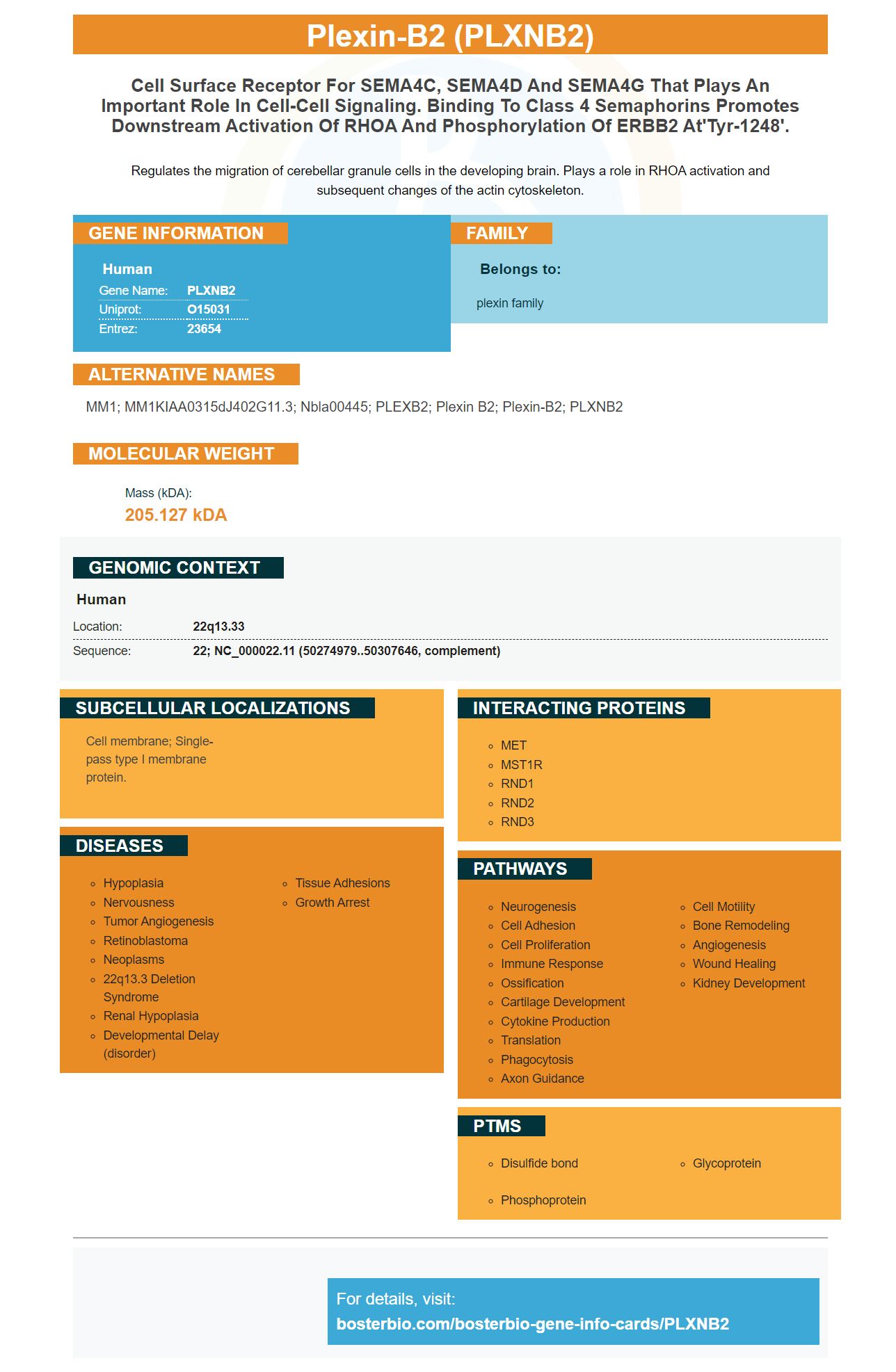
Facts about Plexin-B2.
Regulates the migration of cerebellar granule cells in the developing brain. Plays a role in RHOA activation and subsequent changes of the actin cytoskeleton.
Human | |
---|---|
Gene Name: | PLXNB2 |
Uniprot: | O15031 |
Entrez: | 23654 |
Belongs to: |
---|
plexin family |
MM1; MM1KIAA0315dJ402G11.3; Nbla00445; PLEXB2; Plexin B2; plexin-B2; PLXNB2
Mass (kDA):
205.127 kDA
Human | |
---|---|
Location: | 22q13.33 |
Sequence: | 22; NC_000022.11 (50274979..50307646, complement) |
Cell membrane; Single-pass type I membrane protein.
The PLXNB2 marker is a very valuable tool when it comes to biomarkers. This marker can help you differentiate biological samples from other material. This article will explain the best uses and applications of the PLXNB2 Marker. Then, you'll be well-equipped to perform PLXNB2 experiments in biological samples.
The PLXNB2 marker, which is a protein found both in prostate cancers in mice and humans, is known. A significantly reduced survival rate is associated with the overexpression of this protein in these tumors. PLXNB2 protein has been shown to be associated with angiogenic activity. This can increase the risk of developing glioma. This marker can be used to diagnose prostate cancer.
To test this marker, we used a cell line that was recombinantly engineered with Flag MAb M2. The serum contained 10 mg Hepes, 150 mg NaCl, 3 M EDTA, 0.05% P20, and 3 mM EDTA. We also added a CM5 chip sensor to the solution. The sample was also tagged with the PLXNB2 mark.
The fluorescent protein Lifeact_mScarlet contains a peptide, which specifically binds F-actin. This fluorescent protein is not disrupted by actin dynamics. The OE colonies displayed striking angular cell morphology while the KO colonies had poorly defined junctional borders, filopodia-like protrusions, and poorly defined junctional boundaries. The KO cells also had lower cortical F-actin accumulation and less matrix adhesion.
ANG is expressed in a variety of human cancers and is downregulated in neurological diseases. Supplemental therapy with recombinant ANG protein improved motor function and prolonged life span in SOD1G93A mice. Although it is not known how ANG affects PLXNB2-expressing PLXNB2-expressing cells the receptor of ANG is believed to be involved. Researchers are investigating the role played by PLXNB2 when ANG activity is controlled.
Numerous clinical trials have shown that angiogenic activity is dependent on the PLXNB2 protein. This gene is also implicated in inflammation. ANG-deficient cell are more likely to develop thrombocytopoiesis. There are other ways to identify ANG deficient cells. These include blood-forming cells and the spleen.
There are many methods to assess the function PLXNB2 has in HSPC. One study found that ANG treatment boosted the regeneration WBM and LTHSCs. The mAb17 therapy did not affect LTHSC homing, or CD45.1 expression. These data suggest that cells ang-deficient do not display any changes in cell cycle, self-renewal, or cell homing.
The regulation of ANG is mediated by the ANG binding region. PLXNB2 antibodies in vitro inhibit the activity and formation of xenograft tumours. These results indicate that ang is necessary for the physiological functions PLXNB2, making it a promising therapeutic target. It may also play an important role in neurodegenerative diseases. Although its role in cancer and neurodegenerative disease is not yet clear, it is a promising marker.
We performed a kinetic analyze of mAb17 binding PLXNB2 and determined the receptor's affinity. This was done with the same protocol that was used to analyze ANG. However mAb17 substituted for ANG in different concentrations. Using a CM5 sensors chip, we performed surface plasmon reflection experiments.
We used a lentiviral vector and shRNA system to knock down the PLXNB2 genes. We used two sets PLXNB2-specific vectors, TRCN0000048-188-189 and 189 respectively, and a non-coding ShRNA as a control. We used ViraPower Lentiviral Expression Systems for transient transfection in 293 FT cells.
Additionally, we did microarray analysis on four different glioma patients. We found that all four tumors were elevated for PlexinB2. The PLXNB2 Gene was also expressed in all molecular types of glioblastoma. A poor prognosis was not associated with the two tumors in humans that expressed the most PlexinB2, which indicates that high levels of PlexinB2 expression did no impact on survival.
The mAb used to immunoprecipitate was specifically designed for the recognition of PLXNB2 or RNASE4 proteins. The flag tag was added to the C-terminus ANG peptide. This created the mAb. After expression, the ANG-Flag was mixed with a fraction of LNCaP cells and incubated at 4degC for 30 min. The resulting mixture was divided into three equal fractions. Then, a control mAb (CCL130) was added to the protein A/G-Sepharose (20 ml).
PLXNB2 (a protein expressed in malignant brain cancers) is a human protein. It has been shown to have an angiogenic effect in various cancer cell lines. This protein is immobilized using a CM5 chip sensor chip. HBS-EP+ containing 10 mM Hepes, 150 mN NaCl, 3 mM EDTA, and 0.05% P20 was used as the running buffer.
It has been proven that PLXNB2 expression was present in mouse tumors and human carcinoma cells. PLXNB2-overexpression in human tumors is associated with significantly reduced median survival. It was found in tumors of mice with prostate cancer and brain tumors. It is therefore important to identify the cause for cancer in human tumors, by examining the PLXNB2 level. Recent research indicates that patients with both prostate and glioma are less likely to survive if they over-express this protein.
The prophylactic effect of PLXNb2 markers on PC3 cells was assessed in biological samples. 1.5 x10⁶PC3 cells were inoculated with 50% Matrigel medium. Afterwards, mAb17 was administered to mice for 4 days at a dose of 60 mg daily. Similar results were seen in PLXNB2/mAb17 mice.
Co-IP revealed that PLXNB2 in vivo is associated with ANG. Knockdown of PLXNB2 prevented ang-induced cell proliferation, AKT phosphorylation and 47S rRNA transcript in LNCaP cells. Additionally, mAb17 prevented BCR-ABL-induced CML from immunocompetent mouse models. These findings highlight the therapeutic potential of anti-PLXNB2 antibodies.
Molecular analyses of PLXNB2 indicated that the protein is essential for colony growth and geometry. Mouse hybridoma generated the cDNA. A number of genes correlated with Plexin-B2 levels. This study revealed that 2037 genes were differentially expressed between WT and OE hESCs. You can read the entire paper for more information.
Biological Samples of the PLXNb2 marker have been used to study the angiogenic activity of hESCs in different cell lines. These cells were cultured in RPMI 1640 + 10% FBS, and then were removed with Versene. Three mls complete medium were added in a 10-cm pot. After centrifugation at 800xg, cells were resuspended into cold PBS + 2.2% FBS. Then, 150 ml of cell suspension was prepared for staining. After washing, mAb17 (or an isotype control antibody) were added to the cells at five mg/ml. These antibodies were incubated for 2 hours.
Ang is interconnected with PLXNB2 to regulate each of their brain functions. Ang-induced LTHSCs show a pro-selfrenewal signature. Interestingly, mAb17 didn't inhibit the growth of WT cells or increase cyclin D1 levels. These processes were inhibited by ANG and PLXNB2 mice, but mAb17 had no effect.
PMID: 12183458 by Perrot V., et al. Plexin B regulates Rho through the guanine nucleotide exchange factors leukemia-associated Rho GEF (LARG) and PDZ-RhoGEF.
PMID: 12533544 by Artigiani S., et al. Functional regulation of semaphorin receptors by proprotein convertases.