This website uses cookies to ensure you get the best experience on our website.


- Table of Contents
Plan chromogenic QSOX2 IHC in paraffin sections around the granular cytoplasmic tissue pattern (HPA tissue IHC). Use glandular cells as positive controls (HPA tissue IHC), and interpret membrane staining in light of QSOX2’s membrane and secreted annotations (UniProt).

Expected localisation, validated protocols, controls and antibodies — the at-a-glance facts below, then the full design guide.
| Expected localisation | Granular cytoplasm in tissue sections (HPA tissue IHC) | |
| Staining pattern | Granular cytoplasmic staining in glandular cells (HPA tissue IHC) | |
| Antigen retrieval | EDTA pH 8.0 HIER, heat-mediated (datasheet A10205-1) | |
| Positive control | Adrenal gland+4 more · see all | |
| Negative control | Adipose tissue+3 more · see all |
| Fixation | Keep fixation consistent across sections. (standard IHC practice; not target-specific) | |
| Caveat | Tissue staining has only medium agreement with RNA (HPA tissue IHC) | |
| Regulation | Expression varies by tissue (UniProt) | |
| Isoform / epitope | No isoforms; signal peptide cleaved; map membrane epitope (UniProt) |
Compare the catalog antibody’s IHC-P protocol (datasheet A10205-1) with published QSOX2 IHC methods in the four articles below (PMC5492681; PMC8631333; PMC8326667; PMC13168330).
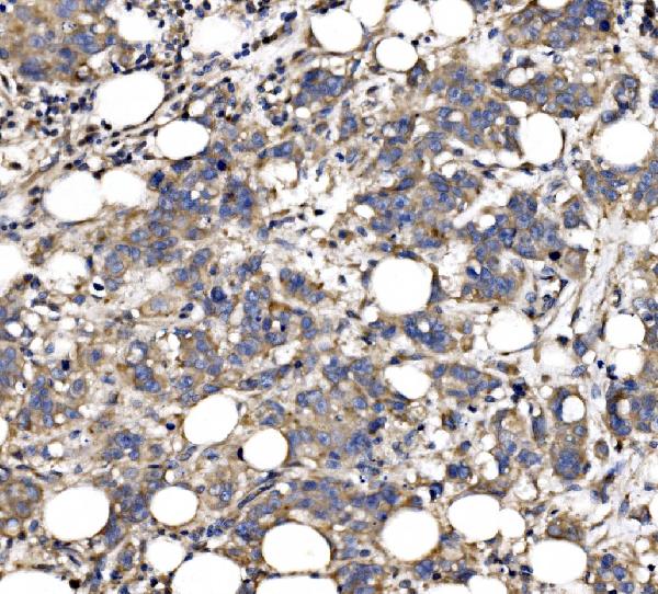
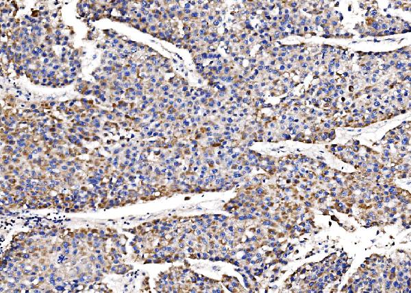
| Sample | Paraffin-embedded human breast cancer tissue; fixative not specified (datasheet A10205-1) |
| Fixation | Image fixative and duration unreported (datasheet A10205-1); verify before use. |
| Sectioning | 4–5 µm sections on charged slides (standard) |
| Deparaffinisation | Xylene, graded ethanol series to water (standard) |
| Antigen retrieval | Heat retrieval: EDTA pH 8.0 (datasheet A10205-1); 20 min, 95–100 °C (standard) |
| Peroxidase block | 3% H2O2, 10 min, room temperature (standard) |
| Blocking | 10% goat serum (datasheet A10205-1) |
| Primary antibody | Rabbit anti-QSOX2, 2-5 μg/ml (datasheet A10205-1) |
| Primary incubation | Overnight at 4 °C (datasheet A10205-1) |
| Detection | Streptavidin-biotin complex (SABC), DAB chromogen (datasheet A10205-1) |
| Counterstain | Hematoxylin, blue, dehydrate and mount (standard) |
| Expected result | QSOX2-positive staining in glandular cells of adrenal gland (HPA tissue IHC: High). HPA tissue profile: Cytoplasmic expression with a granular pattern. No signal in the no-primary control. |
In paraffin-section IHC, expect granular cytoplasmic staining in selected glandular, respiratory epithelial, hematopoietic and neural cell populations (HPA: tissue IHC). UniProt also annotates QSOX2 as secreted and membrane associated, with a transmembrane segment at 662–682; that topology does not establish a distinct membrane pattern on an IHC slide (UniProt Q6ZRP7 topology). Interpret tissue staining cautiously: HPA rates its IHC profile Uncertain, with medium agreement between staining and RNA data (HPA: tissue IHC reliability).
| Granular cytoplasmic signal in colon or endometrial glandular cells, or bronchial respiratory epithelial cells (HPA: High in each listed cell population). | This matches the reported IHC compartment and a high-staining cell population (HPA: tissue IHC profile). Score the relevant cells and their intensity, then check tissue architecture and controls; the overall IHC profile remains Uncertain (HPA: reliability; general IHC practice). |
| Predominantly crisp nuclear, diffuse extracellular, or exclusively plasma-membrane staining replaces the granular cytoplasmic pattern (HPA: tissue IHC profile). | Treat the compartment mismatch as suspect in paraffin IHC and assess background and controls (HPA: tissue IHC profile; general IHC practice). UniProt lists membrane and secreted locations, but those annotations alone cannot validate this slide pattern (UniProt Q6ZRP7 subcellular location). |
| Strong staining appears in adipocytes or cardiomyocytes, while the expected glandular or epithelial cells are unstained (HPA: Not detected in those cells; High in selected glandular and respiratory epithelial cells). | Investigate nonspecific antibody binding or endogenous chromogen activity before calling it QSOX2 (general IHC practice). HPA cell-level observations are comparators, not an absolute exclusion of biological expression (HPA: Uncertain IHC reliability). |
| Brown haze covers stroma, blank regions and many cell types without a discernible granular cytoplasmic pattern (HPA: tissue IHC profile). | Interpret this as background until a clean cell-level pattern emerges (general IHC practice). Review blocking, washes, primary concentration and chromogen exposure, and compare a no-primary control; none of these checks proves QSOX2 specificity alone (general IHC practice). |
| No signal is seen in an adequately represented colon glandular or bronchial respiratory epithelial population (HPA: High in those populations). | Check section quality, retrieval, antibody preparation and detection controls before calling the specimen negative (general IHC practice). Even a reported high-staining population is a reference rather than a guaranteed result for every specimen (HPA: Uncertain IHC reliability). |
| Tissue and cell selection: HPA reports High staining in colon glandular cells and bronchial respiratory epithelial cells, but Not detected in adipocytes and cardiomyocytes (HPA: tissue IHC). | Use the named cell populations as slide-level comparators, including cells of the correct identity and sufficient section area (HPA: tissue IHC; general IHC practice). HPA's Uncertain reliability limits how strongly one discordant specimen can be interpreted (HPA: reliability). |
| Antibody evidence: HPA012716 is IHC Uncertain; HPA017280 has no IHC status in the supplied record (HPA: antibody validation). | Do not describe this IHC pattern as independently antibody-confirmed or IHC Enhanced (HPA: antibody validation). Judge a new stain using cell identity, compartment, controls and replicate sections together (general IHC practice). |
| Topology and processing: QSOX2 has a signal peptide at 1–21 and one transmembrane segment at 662–682 (UniProt Q6ZRP7 topology and processing). | These features inform interpretation of secreted and membrane-associated forms but do not predict which epitope an unspecified antibody detects or establish extracellular IHC staining (UniProt Q6ZRP7 subcellular location; general IHC practice). |
| IF/ICC Q: Should its images define the paraffin IHC compartment? HPA reports supported Golgi localization, with additional nucleoplasmic localization (HPA: subcellular ICC-IF). | A: Use that as context only. ICC-IF has different specimen preparation, and HPA rates ICC Enhanced for both listed antibodies while the supplied IHC status is Uncertain or absent (HPA: subcellular ICC-IF and antibody validation; general IHC practice). |
| Situation | Likely cause | Next action |
|---|---|---|
| A reported high-staining glandular or respiratory epithelial population has no visible chromogen (HPA: High in colon glandular and bronchial respiratory epithelial cells). | The target may be missed because a general IHC step or detection reagent failed; a single blank slide cannot distinguish that from a biological negative (general IHC practice). | Check tissue preservation, retrieval conditions, primary handling and detection with appropriate run controls; repeat with a supported positive cell population (general IHC practice; HPA: High cell populations). |
| Granular cytoplasmic staining is obscured by uniform brown haze (HPA: tissue IHC profile). | Excess primary reagent, inadequate blocking or washing, or excessive chromogen development can raise nonspecific background (general IHC practice). | Compare a no-primary control, optimize primary concentration and washes, and shorten development as needed; retain a counterstain that allows cell identification (general IHC practice). |
| The chromogen appears mainly nuclear or extracellular in paraffin sections (HPA: granular cytoplasmic IHC profile). | A compartment mismatch may reflect background, tissue artefact or an unvalidated staining pattern; UniProt membrane and secreted annotations do not resolve it (general IHC practice; UniProt Q6ZRP7 subcellular location). | Recheck morphology and controls, then repeat using an IHC-validated antibody and the reported positive cell populations before assigning QSOX2 localization (general IHC practice; HPA: tissue IHC). |
| Strong signal occurs in adipocytes or cardiomyocytes (HPA: Not detected in these cell populations). | Nonspecific binding or endogenous detection activity is possible; HPA's Uncertain IHC reliability also limits a categorical biological conclusion (general IHC practice; HPA: reliability). | Assess no-primary and detection controls, inspect which cells contain the deposit, and compare a reported high-staining population on the same run (general IHC practice; HPA: tissue IHC). |
| Bone marrow shows widespread chromogen, making individual hematopoietic cells difficult to score (HPA: High in hematopoietic cells). | Endogenous peroxidase or excessive detection background can complicate chromogenic IHC interpretation (general IHC practice). | Check a no-primary control and the effectiveness of the peroxidase-blocking step; score only interpretable cells against the reported hematopoietic pattern (general IHC practice; HPA: tissue IHC). |
| An ICC-IF Golgi pattern is clearer than the paraffin IHC result (HPA: supported Golgi localization in ICC-IF; Uncertain tissue IHC reliability). | The assays have different preparation and validation evidence; HPA's ICC Enhanced status does not transfer to IHC (general IHC practice; HPA: antibody validation). | Interpret each assay against its own HPA pattern and controls; for this paraffin IHC guide, decide from cell-level granular cytoplasmic staining and IHC controls (HPA: tissue IHC profile; general IHC practice). |


Comprehensive Human Protein Atlas IHC scoring per tissue (reliability: Uncertain — Medium consistency between antibody staining and RNA expression data.). Rows are taken directly from the HPA tissue chart — click any row's HPA link to view the source.
| Tissue | Cell type | Level | Evidence | Source |
|---|---|---|---|---|
| Adrenal gland | Glandular cells | High | Protein (IHC) | HPA → |
| Appendix | Glandular cells | High | Protein (IHC) | HPA → |
| Bone marrow | Hematopoietic cells | High | Protein (IHC) | HPA → |
| Bronchus | Respiratory epithelial cells | High | Protein (IHC) | HPA → |
| Caudate | Glial cells | High | Protein (IHC) | HPA → |
| Tissue | Cell type | Level | Evidence | Source |
|---|---|---|---|---|
| Adipose tissue | Adipocytes | Not detected | Protein (IHC) | HPA → |
| Heart muscle | Cardiomyocytes | Not detected | Protein (IHC) | HPA → |
| Liver | Cholangiocytes | Not detected | Protein (IHC) | HPA → |
| Skeletal muscle | Myocytes | Not detected | Protein (IHC) | HPA → |
Troubleshoot QSOX2 staining in paraffin sections by checking retrieval, tissue handling, compartment patterns and controls before interpreting signal intensity.
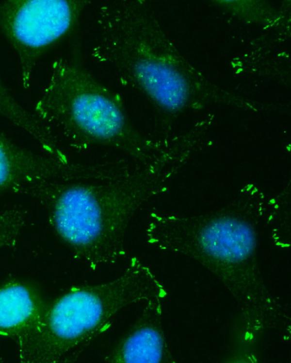
A10205-1 has IHC data from paraffin sections of human breast and liver cancer tissue and IF/ICC data from HeLa cells (A10205-1 IHC/IF image captions).
A10205-1 is listed for human IHC and was shown on paraffin sections of human breast and liver cancer tissue (catalog: applications, reactivity; A10205-1 IHC image captions). A10205-1 is also listed for IF/ICC and was shown in HeLa cells (catalog: applications; A10205-1 IF image caption).
Which to pick: Choose A10205-1 for human paraffin-section IHC; its own captions document EDTA pH 8 retrieval and 2 μg/ml primary antibody, but do not report the fixative (A10205-1 IHC image captions). For IF/ICC, A10205-1 has a HeLa-cell IF example using 5 μg/ml primary antibody (A10205-1 IF image caption). No cross-species choice is supported: A10205-1 lists Human reactivity, a Rabbit host and no clone designation (catalog: reactivity, host, clone).