RAB11A / Ras-related protein Rab-11A · IHC design guide

Design Immunohistochemistry for RAB11A

This guide uses RAB11A’s cytoplasmic and membranous tissue staining to plan paraffin IHC (HPA tissue IHC). It identifies glandular cells with medium staining and adipocytes reported as unstained for tissue control selection (HPA tissue IHC).

Evidence assembled Oct 2026 · For research use; verify linked source records and product datasheet before use
Immunohistochemistry protocol sheet for RAB11A (IHC for RAB11A): expected localisation Cytoplasmic and membranous tissue staining (HPA tissue IHC), antibody M01436-2, validated IHC image, and IHC protocol steps
Printable RAB11A IHC protocol sheet — expected localisation Cytoplasmic and membranous tissue staining (HPA tissue IHC), antibody M01436-2, controls and protocol steps. Open the full RAB11A IHC guide →

RAB11A Immunohistochemistry Experimental Design Guide

Expected localisation, validated protocols, controls and antibodies — the at-a-glance facts below, then the full design guide.

Must know before staining
Expected localisation Cytoplasmic and membranous tissue staining (HPA tissue IHC)
Staining pattern Widespread cytoplasmic and membranous staining (HPA tissue IHC)
Antigen retrieval EDTA pH 8.0 HIER, heat-mediated (datasheet M01436-2)
Positive control ⓘ Adrenal gland+4 more · see all
Negative control ⓘ Adipose tissue+4 more · see all
Important caveats
Reasons your staining may differ from the expected pattern.
Fixation Keep fixation consistent across paraffin sections (standard IHC practice; not target-specific)
Caveat Adipocytes may be unstained despite the broad tissue profile (HPA tissue IHC)
Regulation Expression regulation is not annotated (UniProt)
Isoform / epitope 2 isoforms; mature chain spans residues 2–213. Check epitope coverage (UniProt)
Section 1

Recommended RAB11A IHC & IF Protocols

The catalog antibody’s IHC-P protocol is paired with four published RAB11A IHC methods for lung and colon tissues (PMC12376738; PMC6726494; PMC5438609; PMC6605470).

Recommended immunohistochemistry (IHC-P) protocol parameters
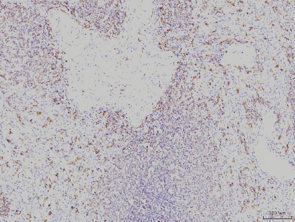
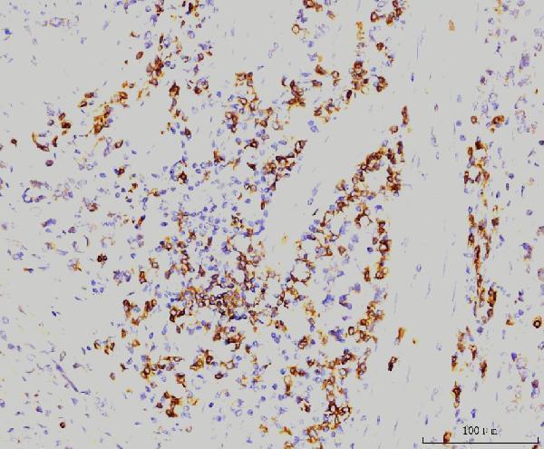
SampleParaffin-embedded human spleen tissue; fixative not specified (datasheet M01436-2)
FixationImage fixative and duration unreported (datasheet M01436-2); verify before use.
Sectioning4–5 µm sections on charged slides (standard)
DeparaffinisationXylene, graded ethanol series to water (standard)
Antigen retrievalHeat retrieval: EDTA pH 8.0 (datasheet M01436-2); 20 min, 95–100 °C (standard)
Peroxidase block3% H2O2, 10 min, room temperature (standard)
Blocking10% goat serum (datasheet M01436-2)
Primary antibodyMouse monoclonal (clone 4H9) anti-RAB11A, 2 μg/ml (datasheet M01436-2)
Primary incubationOvernight at 4 °C (datasheet M01436-2)
DetectionStreptavidin-biotin complex (SABC), DAB chromogen (datasheet M01436-2)
CounterstainHematoxylin, blue, dehydrate and mount (standard)
Expected resultRAB11A-positive staining in glandular cells of adrenal gland (HPA tissue IHC: Medium). HPA tissue profile: Ubiquitous cytoplasmic and membranous expression. No signal in the no-primary control.
💡Decision noteFor the catalog antibody, start with heat-mediated EDTA pH 8.0 retrieval (datasheet: M01436-2). Citrate retrieval is documented with different antibodies (PMC12376738; PMC5438609).
Section 2

What Is the Expected RAB11A Staining Pattern?

RAB11A is associated with recycling endosomes, cytoplasmic vesicles, Golgi membranes and the cell membrane, although it has no transmembrane segment (UniProt P62491 subcellular location; UniProt P62491 topology). In paraffin-section IHC, expect cytoplasmic and membranous staining in selected epithelial cells, including glandular cells in colon and breast (HPA tissue IHC: Medium). HPA describes the tissue pattern as ubiquitous but rates its agreement with RNA expression as medium (HPA tissue IHC: Approved; medium consistency).

What am I looking at on my slide?
Granular cytoplasmic staining, sometimes concentrated near the nucleus, with some membrane staining in glandular cells.This fits the reported cytoplasmic and membranous tissue pattern and RAB11A’s vesicle and recycling-endosome locations (HPA tissue IHC; UniProt P62491 subcellular location). In colon or breast glandular cells, compare intensity with HPA’s Medium category; individual cells need not stain identically (HPA tissue IHC; standard IHC interpretation).
Strong, uniform nuclear staining with little cytoplasmic or membrane signal.A predominantly nuclear result is discordant with the supplied RAB11A locations (UniProt P62491 subcellular location; HPA tissue IHC). Review the antibody and detection controls before treating it as a new location; the slide alone cannot identify the cause (standard IHC practice).
Prominent staining in a cell population reported as not detected, such as soft-tissue fibroblasts or adipocytes.This conflicts with those HPA tissue observations and warrants a specificity check (HPA tissue IHC: fibroblasts and adipocytes, Not detected). Cross-reactivity or endogenous detection activity is possible, but biological variation and sampling also limit a single-slide conclusion (standard IHC interpretation).
A diffuse wash of color covers cells and surrounding tissue without clear cellular boundaries.The distribution is hard to reconcile with HPA’s cellular cytoplasmic and membranous pattern (HPA tissue IHC). Excess background can arise from nonspecific antibody binding or the detection system; evaluate a matched negative control before scoring cells (standard IHC practice).
No RAB11A signal in a colon or breast glandular-cell section expected to be positive.HPA reports Medium staining in these cells, so an entirely blank result calls for a run-level check (HPA tissue IHC). Confirm that the section contains the relevant cells and that positive-control tissue and detection reagents worked; HPA’s medium consistency does not guarantee every specimen will stain (HPA tissue IHC; standard IHC practice).
💡Expected RAB11A appearanceCall positive when glandular epithelial cells show interpretable medium cytoplasmic, often granular, and membranous staining; isolated strong nuclear color or widespread cell-free color should prompt control review (HPA tissue IHC; UniProt P62491 subcellular location; standard IHC practice).
How each factor affects the staining
Tissue and cell selectionColon, breast and duodenum glandular cells provide HPA-reported Medium IHC examples; pancreatic exocrine glandular cells are reported as Not detected (HPA tissue IHC). Select and score the named cell population, since a tissue label alone does not specify which cells should stain (standard IHC practice).
Antibody evidenceHPA lists IHC as Approved for HPA060686 and CAB013097, while its tissue summary reports medium staining–RNA consistency (HPA antibodies; HPA tissue IHC). Treat agreement with an independent IHC-validated antibody as additional evidence if a disputed pattern must be resolved (standard IHC practice).
Membrane association and sequenceRAB11A occupies several membranes despite having no transmembrane segment (UniProt P62491 subcellular location; topology). UniProt lists two isoforms and a short C-terminal propeptide, but the supplied evidence gives no antibody epitope; neither feature predicts this assay’s staining intensity (UniProt P62491 isoforms; processing).
IF/ICC Q: What pattern is expected on its separate guide page?A: HPA supports vesicles and centriolar satellites as the main IF/ICC locations; primary-cilium, basal-body and cytosol assignments are uncertain (HPA subcellular). This localization evidence helps interpret images but does not supply an IF/ICC protocol or establish a paraffin-section staining result (HPA subcellular; HPA tissue IHC).
Why is my staining missing, weak or wrong?
SituationLikely causeNext action
Colon or breast glandular cells and the run’s positive control are both blank.A shared staining or detection failure is plausible; those glandular cells are HPA-reported Medium examples (HPA tissue IHC; standard IHC practice).Check section identity, reagent delivery and detection-control performance, then repeat with the catalog antibody’s documented IHC-P conditions (standard IHC practice).
The positive control stains, but the chosen study cell population is blank.Expression can differ by cell population: HPA reports pancreatic exocrine glandular cells as Not detected despite positive glandular examples elsewhere (HPA tissue IHC).Verify that the scored cells match the HPA comparison and report the negative result for that population; use a separate HPA-positive tissue to assess assay performance (HPA tissue IHC; standard IHC practice).
Staining is widespread, including tissue spaces or areas without intact cells.Nonspecific binding or detection background may obscure the cellular pattern (standard IHC practice).Compare a matched negative control and inspect blocking, washes and detection settings under the documented IHC-P workflow before scoring (standard IHC practice).
Brown signal appears in HPA-reported Not detected cell types.Specificity is uncertain; cross-reactivity or endogenous chromogen-generating activity may contribute, and an HPA category is not an absolute rule for every specimen (HPA tissue IHC; standard IHC practice).Review the no-primary control, cell morphology and signal compartment; if the finding matters, compare an independent IHC-validated antibody (standard IHC practice; HPA antibodies).
Signal is predominantly nuclear rather than cytoplasmic or membranous.The compartment conflicts with the supplied tissue and subcellular annotations; these data do not identify which assay step caused it (HPA tissue IHC; UniProt P62491 subcellular location).Inspect negative controls and repeat with the catalog antibody’s documented IHC-P conditions before assigning a new RAB11A location (standard IHC practice).
Granular cytoplasmic staining is uneven across neighboring epithelial cells.Vesicle-associated localization and differences between cells can produce a nonuniform appearance, but the supplied sources do not define an acceptable cell-by-cell threshold (UniProt P62491 subcellular location; HPA tissue IHC).Score the named cell population and compartment consistently, recording extent and intensity separately; compare the control section from the same run (standard IHC practice).

Sample controls for RAB11A IHC & IF

🧪Run colon first: glandular cells should stain (HPA: Colon glandular cells, Medium). Use smooth muscle as the negative tissue; colonic smooth muscle cells, when present on the positive slide, should show background staining without the glandular-cell signal (HPA: Smooth muscle cells, Not detected).
Positive control tissue: Adrenal gland (Glandular cells, HPA Medium)
Negative control tissue: Adipose tissue (HPA Not detected)
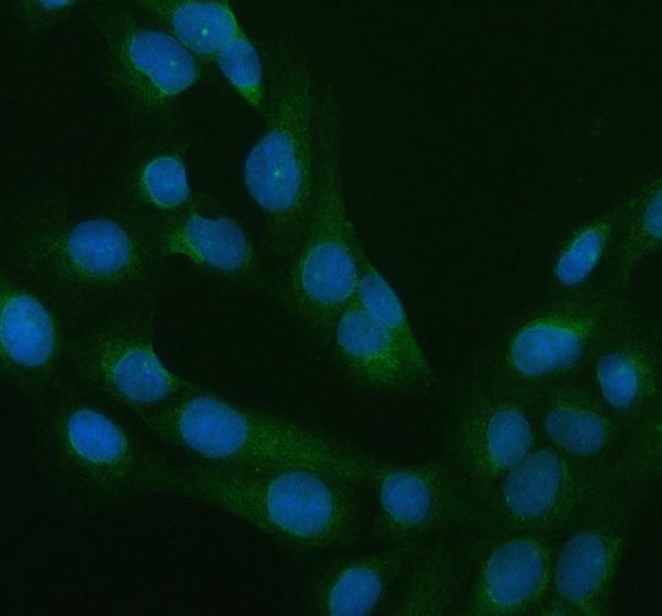
ICC-IF cell lines (HPA subcellular resource): HPA ICC-IF images show RAB11A in A-431, U-251MG, U2OS, ASC52telo, RPTEC/TERT1, hTERT-RPE1 (serum starved), Sperm, HEK293, NIH 3T3, with annotated localisation: Vesicles (supported), Centriolar satellite (supported) (HPA subcellular).
Technical controls: Include a no-primary (secondary-only) control, an isotype control matched to the mouse primary’s immunoglobulin class and clonality, and a RAB11A-knockout specimen if available (selected M01436-2 caption: mouse primary; clonality unreported). Quench endogenous peroxidase and check for endogenous biotin signal in the colon section when using the caption’s SABC/DAB detection (selected M01436-2 caption: SABC with DAB).
⚠️Feasibility: No target-specific fixation window or fixation effect is reported, and the exact M01436-2 paraffin-section caption does not state a fixative (selected M01436-2 caption: fixative unreported). The demonstrated paraffin-section procedure uses heat retrieval in EDTA at pH 8.0; the payload does not establish whether retrieval is essential (selected M01436-2 caption: EDTA heat retrieval). IF/ICC can help assess vesicular localization, but the supplied evidence does not establish that IF or frozen sections are easier than paraffin IHC (HPA subcellular: vesicles supported); endogenous peroxidase or biotin signal could complicate interpretation of SABC/DAB staining (selected M01436-2 caption: SABC with DAB).

HPA tissue IHC evidence for RAB11A

Comprehensive Human Protein Atlas IHC scoring per tissue (reliability: Approved — Medium consistency between antibody staining and RNA expression data.). Rows are taken directly from the HPA tissue chart — click any row's HPA link to view the source.

Positive expression · recommended positive controls

TissueCell typeLevelEvidenceSource
Adrenal gland Glandular cells Medium Protein (IHC) HPA →
Appendix Glandular cells Medium Protein (IHC) HPA →
Breast Glandular cells Medium Protein (IHC) HPA →
Cervix Squamous epithelial cells Medium Protein (IHC) HPA →
Colon Glandular cells Medium Protein (IHC) HPA →

Undetected expression · recommended negative controls

TissueCell typeLevelEvidenceSource
Adipose tissue Adipocytes Not detected Protein (IHC) HPA →
Lymph node Germinal center cells Not detected Protein (IHC) HPA →
Ovary Ovarian stroma cells Not detected Protein (IHC) HPA →
Pancreas Exocrine glandular cells Not detected Protein (IHC) HPA →
Smooth muscle Smooth muscle cells Not detected Protein (IHC) HPA →
Section 3

Advanced RAB11A IHC Tips

Troubleshoot RAB11A staining in paraffin sections by checking retrieval, cellular pattern, controls and scoring before interpreting changes in recycling compartments.

What retrieval conditions should I start with for RAB11A paraffin IHC?
Start with heat-mediated antigen retrieval in EDTA at pH 8.0 (datasheet M01436-2). The selected paraffin-section example used this retrieval before overnight incubation with 2 μg/mL antibody at 4°C (datasheet M01436-2). If staining is weak, compare retrieval duration on adjacent sections while keeping antibody concentration and chromogen development constant (standard IHC practice). Check whether additional heat increases both the expected cytoplasmic or membranous signal and tissue damage or diffuse background (HPA tissue IHC; standard IHC practice). Record the retrieval condition alongside the control section before comparing staining across runs (standard IHC practice).
Could fixation explain weak or uneven RAB11A staining?
RAB11A-specific sensitivity to fixation is unknown from the supplied evidence; the selected paraffin-section caption does not state a fixative (datasheet M01436-2). Record the actual fixative and fixation time for each specimen before attributing weak signal to the antibody or retrieval (standard IHC practice). Compare specimens processed together, because fixation and subsequent tissue processing can change epitope accessibility and tissue morphology (standard IHC practice). Keep the stated EDTA pH 8.0 retrieval and 2 μg/mL primary concentration constant during that comparison (datasheet M01436-2). Assess staining in intact cells away from damaged edges before interpreting a fixation effect (standard IHC practice).
Which RAB11A staining pattern is plausible in chromogenic IHC?
Expect cytoplasmic and membranous staining in tissue sections, with vesicle-associated signal compatible with RAB11A trafficking (HPA tissue IHC; UniProt P62491 subcellular location). Recycling endosomes, endosome membranes and the trans-Golgi network are documented locations, so a perinuclear or punctate component can be plausible (UniProt P62491 subcellular location). RAB11A has no transmembrane segment; apparent membrane association should therefore be interpreted in the context of its documented membrane-associated compartments (UniProt P62491 topology and subcellular location). Compare the pattern with neighboring intact cells and a matched control section before calling uniform nuclear color specific (standard IHC practice; UniProt P62491 subcellular location). Avoid treating one subcellular pattern as universal across cell states (UniProt P62491 subcellular location).
Can this antibody distinguish RAB11A isoforms or reveal epitope masking?
The record lists 2 RAB11A isoforms, but the supplied antibody caption gives no epitope sequence or isoform coverage (UniProt P62491 isoforms; datasheet M01436-2). Consequently, staining alone cannot establish which isoform contributes to the chromogenic signal (UniProt P62491 isoforms; standard IHC interpretation). RAB11A also carries annotated N-terminal glycosylation and modifications near its termini, whose effect on this antibody is unspecified (UniProt P62491 modifications; datasheet M01436-2). If a specimen stains unexpectedly, compare retrieval conditions and use an independently characterized reagent with a disclosed epitope when isoform attribution matters (standard IHC practice). Document antibody identity and retrieval alongside any such comparison (standard IHC practice).
How should I cross-check a RAB11A IHC pattern by IF?
Use IF as an orthogonal check of the chromogenic pattern, with a marker identifying the cell type being compared (standard IF practice). RAB11A is reported mainly in vesicles and centriolar satellites by subcellular imaging, while tissue IHC shows cytoplasmic and membranous expression (HPA subcellular; HPA tissue IHC). Choose spectrally separated fluorophores and check an unstained section for autofluorescence before assigning puncta to RAB11A (standard IF practice). Because RAB11A lacks a transmembrane segment and the antibody epitope is unspecified, optimize permeabilisation to expose intracellular antigen without losing compartment structure (UniProt P62491 topology; datasheet M01436-2; standard IF practice). Interpret overlap at the cellular level before claiming precise compartment colocalisation (standard IF practice).
How can I reduce diffuse brown background without losing RAB11A signal?
First check whether brown signal persists in a section processed without primary antibody, then inspect blocking, washing and chromogen development (standard IHC practice). The selected example used 10% goat serum, a biotinylated secondary and a streptavidin-biotin detection system with DAB (datasheet M01436-2). In that workflow, assess endogenous peroxidase and biotin contributions with appropriate controls before calling diffuse color RAB11A (standard IHC practice; datasheet M01436-2). Keep the documented 2 μg/mL primary condition as a reference while changing one background-control step at a time (datasheet M01436-2; standard IHC practice). Judge improvement against preserved cellular staining and intact morphology (HPA tissue IHC; standard IHC practice).
How should I score RAB11A IHC across specimens? ⚠ ANSWER MARKED FOR VERIFICATION
Define the cell population and tissue region before scoring, because RAB11A staining can vary among cell types (HPA tissue IHC; standard IHC practice). Report the percentage of positive cells and an intensity-based H-score, or count positive cells per mm² when density answers the study question (standard IHC practice). Normalize cell counts to the viable tissue area or the number of eligible cells, and apply one positivity threshold across slides (standard IHC practice). Score cytoplasmic and membranous compartments consistently, since both are reported in tissue IHC (HPA tissue IHC). Include matched controls and flag damaged areas before comparing groups (standard IHC practice).
When is an apparent RAB11A-positive cell likely to be an artefact?
Favor staining in intact cells with cytoplasmic, membranous or vesicle-like distribution over isolated color in an unexpected compartment (HPA tissue IHC; UniProt P62491 subcellular location). Check cell identity: the selected spleen caption reports detection, while HPA lists red-pulp cells as not detected (datasheet M01436-2; HPA tissue IHC). That difference calls for cell-resolved review and controls rather than a tissue-wide positive label (standard IHC practice). Treat staining confined to section edges, necrotic regions or enzyme-rich areas as suspect until no-primary and detection controls are clean (standard IHC practice). Confirm repeatable signal under the documented EDTA pH 8.0 retrieval condition (datasheet M01436-2; standard IHC practice).
Boster reagents

Best RAB11A / Ras-related protein Rab-11A IHC Antibodies

Anti-RAB11A antibodies have human paraffin-section IHC and human-cell IF/ICC images (catalog image captions); all three list human, mouse and rat reactivity (catalog reactivity).

Real IHC data IHC analysis of Rab11A using anti-Rab11A antibody (M01436-2). Rab11A was detected in a paraffin-embedded section of human spleen tissue. Heat mediated antigen retrieval was performed in EDTA buffer (pH 8.0, epitope retrieval solution). The tissue section was blocked with 10% goat serum. The tissue section was then incubated with 2 μg/ml mouse anti-Rab11A Antibody (M01436-2) overnight at 4°C. Biotinylated goat anti-mouse IgG was used as secondary antibody and incubated for 30 minutes at 37°C. The tissue section was developed using Strepavidin-Biotin-Complex (SABC) (Catalog # SA1021) with DAB as the chromogen.
Anti-Rab11A Antibody ® (monoclonal, 4H9)
Cat # M01436-2
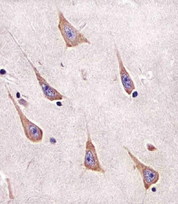
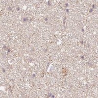
Real IHC data M01436-1 staining RAB11A in human brain sections by Immunohistochemistry (IHC-P -paraformaldehyde-fixed, paraffin-embedded sections). Tissue was fixed with formaldehyde and blocked with 3% BSA for 0. 5 hour at room temperature; antigen retrieval was by heat mediation with a citrate buffer (pH6). Samples were incubated with primary antibody (1/25) for 1 hours at 37°C. A undiluted biotinylated goat polyvalent antibody was used as the secondary antibody.
Anti-RAB11A Antibody
Cat # M01436-1
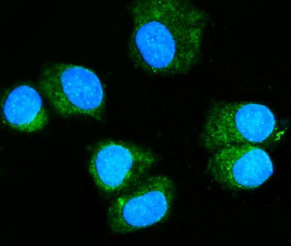
Real IF data IF analysis of Rab11A using anti-Rab11A antibody (PB9789). Rab11A was detected in an immunocytochemical section of U2OS cells. Enzyme antigen retrieval was performed using IHC enzyme antigen retrieval reagent (AR0022) for 15 mins. The cells were blocked with 10% goat serum. And then incubated with 5 μg/mL rabbit anti-Rab11A Antibody (PB9789) overnight at 4°C. DyLight®488 Conjugated Goat Anti-Rabbit IgG (BA1127) was used as secondary antibody at 1:500 dilution and incubated for 30 minutes at 37°C. The section was counterstained with DAPI. Visualize using a fluorescence microscope and filter sets appropriate for the label used.
Anti-Rab11A Antibody ®
Cat # PB9789

M01436-2 has IHC images from human spleen, gastric carcinoma and placenta paraffin sections, plus IF in T-47D cells (M01436-2 image captions); M01436-1 has an IHC-P image from a fixed human brain section (M01436-1 image caption). PB9789 has an IF/ICC image from U2OS cells (PB9789 image caption); all three list human, mouse and rat reactivity (catalog reactivity).

Which to pick: For tissue IHC, choose M01436-2 for paraffin sections with 2 μg/ml primary antibody after EDTA retrieval (M01436-2 IHC captions); its fixative is unreported (M01436-2 IHC captions). M01436-1 is another IHC-P option with a documented paraffin-section human brain section, citrate retrieval and 1:25 primary dilution (M01436-1 image caption); for IF/ICC, choose rabbit PB9789 at 5 μg/ml in the documented U2OS-cell assay (PB9789 image caption). For cross-species planning, all three list human, mouse and rat reactivity (catalog reactivity), while the supplied IHC and IF images show human samples (catalog image captions). The selected M01436-2 tissue-IHC caption documents paraffin sections, but does not specify the fixative (selected-SKU IHC image M01436-2).

Each figure is that product's own IHC / IF validation image from its datasheet.

References

  1. UniProt Consortium. UniProt entry P62491 (RB11A_HUMAN, Ras-related protein Rab-11A).
  2. Human Protein Atlas. RAB11A tissue IHC expression (reliability: Approved).
  3. Human Protein Atlas. RAB11A subcellular location (ICC-IF): Mainly localized to vesicles and centriolar satellites. In addition localized to the cytosol, primary cilium, basal body, equatorial segment, mid piece and principal piece..
  4. Human Protein Atlas. RAB11A antibody validation summary (3 antibodies).
  5. Urinary exosomal RAB11A serves as a novel non-invasive biomarker for diagnosis, treatment response monitoring, and prognosis in small cell lung cancer. Clinical proteomics 2025 — PMC12376738.
  6. Recycling Endosomes in Mature Epithelia Restrain Tumorigenic Signaling. Cancer research 2019 — PMC6726494.
  7. Rab11a promotes proliferation and invasion through regulation of YAP in non-small cell lung cancer. Oncotarget 2017 — PMC5438609.
  8. Effects of miR‑340 overexpression and knockdown on the proliferation and metastasis of NSCLC cell lines. International journal of molecular medicine 2019 — PMC6605470.
  9. PubMed PMID:1704119 — UniProt-cited evidence.
  10. PubMed PMID:9662449 — UniProt-cited evidence.
  11. PubMed PMID:14702039 — UniProt-cited evidence.