This website uses cookies to ensure you get the best experience on our website.


- Table of Contents
Plan RAB20 paraffin-section IHC around cytoplasmic staining reported in several tissues (HPA tissue IHC). Compare positive tissue with controls, while accounting for the low consistency between antibody staining and RNA expression (HPA tissue IHC).

Expected localisation, validated protocols, controls and antibodies — the at-a-glance facts below, then the full design guide.
| Expected localisation | Cytoplasmic staining in several tissues (HPA tissue IHC) | |
| Staining pattern | Cytoplasmic staining in glandular cells and hepatocytes (HPA tissue IHC) | |
| Antigen retrieval | Tris-EDTA pH 9.0 HIER, heat-mediated (datasheet A11018) | |
| Positive control | Duodenum+4 more · see all | |
| Negative control | Adipose tissue+4 more · see all |
| Fixation | Keep fixation consistent across sections. (standard IHC practice; not target-specific) | |
| Caveat | Staining has low consistency with RNA expression (HPA tissue IHC) | |
| Regulation | Stronger in pancreatic adenocarcinomas (UniProt) | |
| Isoform / epitope | No isoforms or processing annotated (UniProt) |
Compare the catalog antibody’s IHC-P protocol (datasheet A11018) with a published RAB20 protocol for paraffin sections of penile squamous cell carcinoma (PMC8909501).
| Sample | Paraffin-embedded human Squamous cell carcinoma of lung tissue; fixative not specified (datasheet A11018) |
| Fixation | Image fixative and duration unreported (datasheet A11018); verify before use. |
| Sectioning | 4–5 µm sections on charged slides (standard) |
| Deparaffinisation | Xylene, graded ethanol series to water (standard) |
| Antigen retrieval | Heat retrieval: Tris-EDTA pH 9.0 (datasheet A11018); 20 min, 95–100 °C (standard) |
| Peroxidase block | 3% H2O2, 10 min, room temperature (standard) |
| Blocking | 10% normal serum of the secondary host, 30 min, room temperature (standard) |
| Primary antibody | Rabbit anti-RAB20, 1:100-1:300 (datasheet A11018) |
| Primary incubation | Overnight at 4 °C (standard) |
| Detection | HRP-polymer secondary, DAB chromogen 5–10 min (standard) |
| Counterstain | Hematoxylin, blue, dehydrate and mount (standard) |
| Expected result | RAB20-positive staining in glandular cells of duodenum (HPA tissue IHC: High). HPA tissue profile: Cytoplasmic expression in several tissues. No signal in the no-primary control. |
RAB20 is associated with the Golgi and cytoplasmic vesicles, including phagosomes (UniProt Q9NX57 subcellular location); it has no transmembrane segment (UniProt Q9NX57 topology). In paraffin tissue IHC, expect cytoplasmic staining in selected glandular cells, hepatocytes and cardiomyocytes (HPA tissue IHC). Treat tissue intensity as provisional: HPA rates the IHC profile Uncertain because antibody staining has low consistency with RNA expression (HPA tissue IHC).
| Cytoplasmic signal in duodenal or small-intestinal glandular cells, hepatocytes, or cardiomyocytes. | This matches cell types scored High by HPA (HPA tissue IHC). Assess signal within the relevant cells and alongside negative controls; HPA calls the overall tissue IHC profile Uncertain (HPA tissue IHC). |
| Predominantly nuclear staining, or a broad surface outline without convincing cytoplasmic staining. | That distribution conflicts with the reported cytoplasmic tissue profile (HPA tissue IHC) and vesicle/Golgi locations (UniProt Q9NX57 subcellular location). Review controls and detection before assigning it to RAB20 (general IHC practice). |
| Strong signal in bronchial respiratory epithelium or adipocytes while the expected positive cells are weak. | Those cells are scored Not detected (HPA tissue IHC). Consider off-target antibody binding or endogenous detection activity (general IHC practice); the Uncertain HPA rating means staining alone cannot settle specificity (HPA tissue IHC). |
| Color spreads across stroma or extracellular spaces with little separation between cells and background. | Diffuse background obscures the expected cellular cytoplasmic pattern (HPA tissue IHC). Check reagent-only controls, blocking, washes and chromogen development as general IHC troubleshooting; do not score haze as positive cells (general IHC practice). |
| No signal in duodenal glandular cells or hepatocytes in an otherwise interpretable section. | Both are scored High (HPA tissue IHC), so inspect the staining run and its controls before calling the sample negative (general IHC practice). The HPA profile is Uncertain, so these are provisional positive comparators (HPA tissue IHC). |
| Tissue and cell selection | HPA scores duodenal and small-intestinal glandular cells, hepatocytes and cardiomyocytes High; bronchial respiratory epithelium and adipocytes are Not detected (HPA tissue IHC). Compare named cell populations, not whole-section color (general IHC practice). |
| IHC evidence strength | HPA rates the tissue profile Uncertain because staining and RNA expression show low consistency; its listed IHC antibody HPA068647 is also Uncertain (HPA tissue IHC; HPA antibodies). Treat agreement with the pattern as support, not independent proof of specificity. |
| Cellular location and topology | Vesicles, phagosomes and Golgi are reported locations (UniProt Q9NX57 subcellular location); no transmembrane segment is annotated (UniProt Q9NX57 topology). A crisp plasma-membrane-only or nuclear-only result therefore needs review. |
| IF/ICC: what should appear? | The separate IF/ICC guide can assess mainly vesicular signal with additional Golgi signal (HPA subcellular ICC-IF, supported). This cellular localisation evidence can inform interpretation, but does not validate a paraffin IHC staining condition. |
| Processing and fixation evidence | UniProt lists one chain spanning residues 1–234, with no signal peptide or propeptide (UniProt Q9NX57 processing). The supplied sources report no RAB20-specific fixation sensitivity; assess retrieval empirically as general IHC practice. |
| Situation | Likely cause | Next action |
|---|---|---|
| Expected positive cells have no visible signal. | A failed staining step is possible (general IHC practice); duodenal glandular cells and hepatocytes are HPA High comparators, though the profile is Uncertain (HPA tissue IHC). | Check the run control, antibody and detection steps, then optimize retrieval and antibody concentration within the validated IHC workflow (general IHC practice). |
| Nuclei stain strongly while cytoplasm remains weak. | Nuclear dominance conflicts with the cytoplasmic tissue profile (HPA tissue IHC) and vesicle/Golgi locations (UniProt Q9NX57 subcellular location). | Compare with reagent-only and suitable positive controls; repeat with a validated staining setup before interpreting nuclear signal as RAB20 (general IHC practice). |
| Bronchial respiratory epithelial cells stain strongly. | These cells are scored Not detected (HPA tissue IHC); off-target binding or endogenous detection activity is possible (general IHC practice). | Review antibody concentration, blocking and detection controls; compare signal with an HPA High cell population on the same run (HPA tissue IHC; general IHC practice). |
| Diffuse chromogen obscures cell boundaries. | Background may reflect nonspecific reagent binding, incomplete washes or excessive development (general IHC practice). | Inspect reagent-only controls and adjust blocking, washes or development within the assay workflow; score only distinguishable cellular staining (general IHC practice). |
| Liver or another highly colored section shows widespread signal. | Endogenous detection activity or tissue pigment can mimic chromogenic staining (general IHC practice); hepatocytes are HPA High, so color alone is insufficient (HPA tissue IHC). | Use appropriate detection and negative controls, examine the expected cellular distribution, and distinguish pigment from reaction product (general IHC practice). |
| A plausible cytoplasmic pattern differs between sections or antibodies. | HPA reports low consistency between antibody staining and RNA expression, and rates tissue IHC Uncertain (HPA tissue IHC). | Compare matched cell types and controls across runs; document the disagreement and seek independent specificity evidence before making a firm biological claim (general IHC practice). |


Comprehensive Human Protein Atlas IHC scoring per tissue (reliability: Uncertain — Low consistency between antibody staining and RNA expression data.). Rows are taken directly from the HPA tissue chart — click any row's HPA link to view the source.
| Tissue | Cell type | Level | Evidence | Source |
|---|---|---|---|---|
| Duodenum | Glandular cells | High | Protein (IHC) | HPA → |
| Heart muscle | Cardiomyocytes | High | Protein (IHC) | HPA → |
| Liver | Hepatocytes | High | Protein (IHC) | HPA → |
| Small intestine | Glandular cells | High | Protein (IHC) | HPA → |
| Adrenal gland | Glandular cells | Medium | Protein (IHC) | HPA → |
| Tissue | Cell type | Level | Evidence | Source |
|---|---|---|---|---|
| Adipose tissue | Adipocytes | Not detected | Protein (IHC) | HPA → |
| Bone marrow | Hematopoietic cells | Not detected | Protein (IHC) | HPA → |
| Breast | Adipocytes | Not detected | Protein (IHC) | HPA → |
| Bronchus | Respiratory epithelial cells | Not detected | Protein (IHC) | HPA → |
| Cerebellum | Cells in granular layer | Not detected | Protein (IHC) | HPA → |
Use the documented paraffin section conditions as a starting point, then verify RAB20 staining by compartment, cell type, and controls.
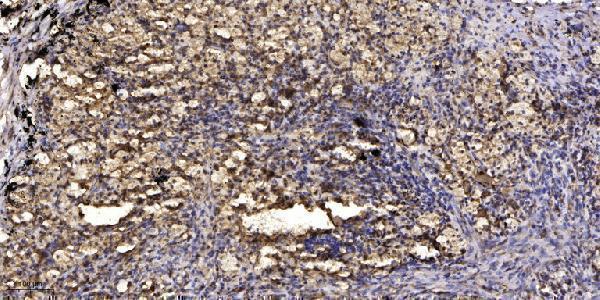
A11018 has a real IHC image from a paraffin-embedded human lung squamous cell carcinoma section (image caption); IF is listed, with Human and Mouse reactivity (catalog).
A11018 will render with its paraffin-section IHC image of human lung squamous cell carcinoma (image caption). The catalog lists IHC and IF applications and Human and Mouse reactivity for A11018 (catalog); no IF image is supplied (catalog).
Which to pick: Choose A11018 for paraffin-section IHC: its image caption reports primary antibody at 1:200 overnight at 4°C and Tris-EDTA retrieval at pH 9.0 (image caption). A11018 is also the listed IF/ICC option because IF appears in its application list, although no IF image is supplied (catalog). For Human or Mouse samples, A11018 is the only listed option (catalog reactivity); its host is Rabbit and clonality is unreported (catalog), while the IHC caption does not report a fixative (image caption).