This website uses cookies to ensure you get the best experience on our website.


- Table of Contents
Plan chromogenic RAB27A IHC on paraffin sections with the catalog antibody at 0.5–1 μg/mL (datasheet: A01608-1). Compare cytoplasmic staining in glandular cells of stomach and colon with the reported absence of staining in cervical glandular cells (HPA tissue IHC).

Expected localisation, validated protocols, controls and antibodies — the at-a-glance facts below, then the full design guide.
| Expected localisation | Cytoplasmic staining in most tissues (HPA tissue IHC) | |
| Staining pattern | Cytoplasmic in most tissues, including immune cells (HPA tissue IHC) | |
| Antigen retrieval | Citrate pH 6 HIER, heat-mediated (datasheet A01608-1) | |
| Positive control | Appendix+4 more · see all | |
| Negative control | Adipose tissue+4 more · see all |
| Fixation | Matched tissue-IHC evidence does not establish the fixation claim. Validate the specimen-specific method before use. (selected-SKU IHC image A01608-1) | |
| Caveat | Glandular staining varies; cervix is undetected (HPA tissue IHC) | |
| Regulation | Low expression in kidney and muscle (UniProt) | |
| Isoform / epitope | Long and Short isoforms; no transmembrane segment; epitope coverage unknown (UniProt) |
Start with the catalog antibody’s IHC-P protocol (datasheet A01608-1), then compare the published RAB27A IHC methods below (PMC3935941; PMC13251207; PMC10079387).
| Sample | Paraffin-embedded human lung cancer tissue; fixative not specified (datasheet A01608-1) |
| Fixation | Image fixative and duration unreported (datasheet A01608-1); verify before use. |
| Sectioning | 4–5 µm sections on charged slides (standard) |
| Deparaffinisation | Xylene, graded ethanol series to water (standard) |
| Antigen retrieval | Heat retrieval: Citrate pH 6, 20 min (datasheet A01608-1) |
| Peroxidase block | 3% H2O2, 10 min, room temperature (standard) |
| Blocking | 10% goat serum (datasheet A01608-1) |
| Primary antibody | Rabbit anti-RAB27A, 0.5-1μg/ml (datasheet A01608-1) |
| Primary incubation | Overnight at 4 °C (datasheet A01608-1) |
| Detection | Streptavidin-biotin complex (SABC), DAB chromogen (datasheet A01608-1) |
| Counterstain | Hematoxylin, blue, dehydrate and mount (standard) |
| Expected result | RAB27A-positive staining in glandular cells of appendix (HPA tissue IHC: High). HPA tissue profile: Cytoplasmic expression in most tissues, including immune cells. No signal in the no-primary control. |
RAB27A is a membrane-associated trafficking protein found on melanosomes, late endosomes, lysosomes and endosomal exocytic vesicles, without a transmembrane segment (UniProt P51159). In paraffin-section IHC, expect chiefly cytoplasmic staining in glandular cells of several digestive tissues and in some immune cells (HPA: tissue IHC profile). HPA rates its tissue staining reliability Enhanced, with medium consistency between antibody staining and RNA expression (HPA: reliability description).
| Cytoplasmic staining is clear in appendix, colon, duodenum, rectum, small-intestine or stomach glandular cells. | This matches HPA's High staining in those specified cell populations (HPA: tissue IHC). Vesicle-associated, sometimes granular cytoplasmic signal is biologically plausible given RAB27A's membrane-trafficking locations (UniProt P51159). Score the named cells and their cytoplasmic compartment; a whole-tissue label can hide differences between cell populations (general IHC interpretation). |
| Signal is predominantly nuclear, or forms a sharp continuous outline around every cell. | Question compartment assignment and staining specificity: HPA describes cytoplasmic tissue expression, while UniProt places RAB27A on intracellular membranes and vesicles and reports no transmembrane segment (HPA: tissue IHC profile; UniProt P51159). Inspect morphology and a matched control before treating an unexpected compartment as RAB27A (general IHC practice). |
| Strong staining appears chiefly in adipocytes, bronchial respiratory epithelium or esophageal squamous epithelium. | Those specified cell populations are Not detected in HPA tissue IHC, so strong signal there is discordant with the reference pattern (HPA: tissue IHC). Consider cross-reactivity or endogenous detection activity; check the antibody and detection controls before concluding that the sample has an altered expression pattern (general IHC practice). |
| Chromogen appears diffusely across the section, including areas without a coherent cell-associated pattern. | Uniform haze obscures the expected cytoplasmic cell pattern (HPA: tissue IHC profile). Possible general IHC causes include inadequate blocking, excessive detection reagent or incomplete washing (general IHC practice). Compare a no-primary control and local tissue morphology; background alone cannot establish RAB27A expression (general IHC practice). |
| A matched known-positive glandular region has no detectable staining. | HPA reports High glandular-cell staining in stomach and several intestinal sites, making those specified cells useful reference regions (HPA: tissue IHC). A blank reference region raises a run-performance or specimen-quality question; review the same-run control, antibody use, retrieval and detection steps before interpreting the test region as negative (general IHC practice). |
| Which tissue and cell population is being read? | HPA reports High glandular-cell staining in appendix, colon, duodenum, prostate, rectum, small intestine and stomach, and Medium staining in bone-marrow hematopoietic cells (HPA: tissue IHC). It reports Not detected for particular populations, including adipocytes and glomerular cells; those calls do not make every cell in their tissues a negative control (HPA: tissue IHC). |
| What does antibody validation establish? | Two listed antibodies, HPA001333 and CAB034046, have Enhanced IHC status (HPA: antibody validation). HPA describes medium consistency between antibody staining and RNA expression overall (HPA: reliability description). Use that evidence to support the reported tissue pattern while assessing any new antibody or unexpected sample on its own merits (general IHC practice). |
| Do protein features change the predicted compartment? | UniProt lists long and short isoforms, no signal peptide or propeptide, and no transmembrane segment (UniProt P51159). Its membrane and vesicle localisation supports intracellular cytoplasmic signal; the supplied record gives no isoform-specific IHC pattern or epitope map, so it cannot predict which isoform an antibody detects (UniProt P51159; supplied record). |
| Where should IF/ICC signal appear? | HPA's ICC-IF entry reports staining in the mid piece and principal piece of sperm (HPA: subcellular ICC-IF). That observation belongs to the separate IF/ICC context; use the tissue IHC profile for paraffin-section interpretation (HPA: tissue IHC; HPA: subcellular ICC-IF). The supplied record gives no basis for an IF/ICC protocol here. |
| Situation | Likely cause | Next action |
|---|---|---|
| The known-positive glandular cells are blank. | The expected High signal in specified glandular cells is absent (HPA: tissue IHC); a failed staining step or compromised section is possible (general IHC practice). | Check that the reference cells are present, review the same-run control, then inspect retrieval, primary-antibody use and chromogenic detection records (general IHC practice). Do not infer RAB27A-specific fixation sensitivity from the supplied evidence. |
| The section is broadly brown with little cellular contrast. | Diffuse background can arise from incomplete blocking or washing, or excessive reagent exposure (general IHC practice). It does not reproduce the cell-associated cytoplasmic profile (HPA: tissue IHC). | Examine a no-primary control and review blocking, reagent exposure and washes; rescore only where cell boundaries and cytoplasm remain interpretable (general IHC practice). |
| Signal is strongest in a population HPA calls Not detected. | Strong adipocyte or bronchial respiratory-epithelial staining conflicts with those HPA cell-specific calls (HPA: tissue IHC). Cross-reactivity or endogenous detection activity is possible (general IHC practice). | Confirm the cell identity and compare no-primary and detection controls. Reassess the antibody with an appropriate positive region before assigning the unexpected signal to RAB27A (general IHC practice). |
| Staining looks mainly nuclear or like a continuous cell-surface rim. | That distribution differs from HPA's cytoplasmic profile and UniProt's intracellular vesicle locations (HPA: tissue IHC; UniProt P51159). | Check focus, counterstain and compartment assignment, then compare control sections. Report the distribution as atypical if it persists rather than scoring it as the expected pattern (general IHC practice). |
| Two samples have different overall staining despite similar target-cell appearance. | HPA scores specific cell populations, and RAB27A staining varies among them: High in specified glandular cells, Medium in bone-marrow hematopoietic cells and Not detected in some others (HPA: tissue IHC). | Compare the same identified cell population and compartment across samples. Record intensity and the fraction of positive target cells separately, with the run control reviewed alongside them (general IHC scoring practice). |
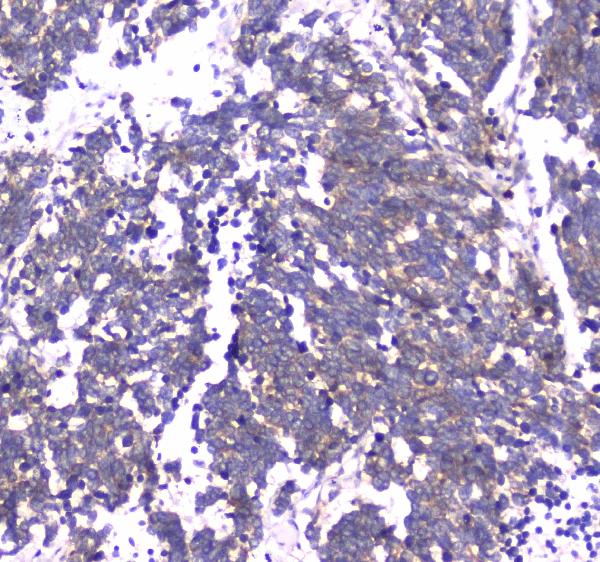
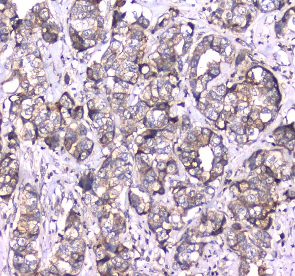
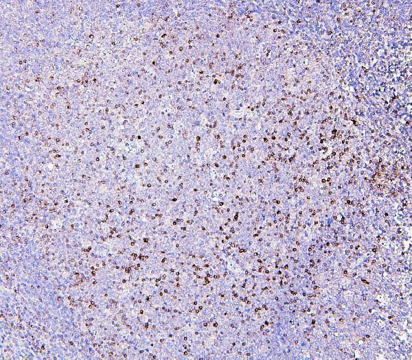
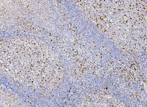


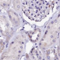


Comprehensive Human Protein Atlas IHC scoring per tissue (reliability: Enhanced — Medium consistency between antibody staining and RNA expression data.). Rows are taken directly from the HPA tissue chart — click any row's HPA link to view the source.
| Tissue | Cell type | Level | Evidence | Source |
|---|---|---|---|---|
| Appendix | Glandular cells | High | Protein (IHC) | HPA → |
| Colon | Glandular cells | High | Protein (IHC) | HPA → |
| Duodenum | Glandular cells | High | Protein (IHC) | HPA → |
| Prostate | Glandular cells | High | Protein (IHC) | HPA → |
| Rectum | Glandular cells | High | Protein (IHC) | HPA → |
| Tissue | Cell type | Level | Evidence | Source |
|---|---|---|---|---|
| Adipose tissue | Adipocytes | Not detected | Protein (IHC) | HPA → |
| Breast | Adipocytes | Not detected | Protein (IHC) | HPA → |
| Bronchus | Respiratory epithelial cells | Not detected | Protein (IHC) | HPA → |
| Cervix | Glandular cells | Not detected | Protein (IHC) | HPA → |
| Endometrium | Cells in endometrial stroma | Not detected | Protein (IHC) | HPA → |
Troubleshoot RAB27A staining in paraffin sections by checking retrieval, cell type, intracellular pattern, and controls before interpreting chromogenic signal.
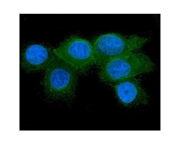
The catalog antibodies show paraffin-section IHC in human, mouse, and rat samples, plus IF/ICC in human MCF7 cells (catalog: A01608-1 and M01608 IHC captions; M01608 IF caption).
A01608-1 will render with IHC images from human lung and mammary cancer and rat small intestine and spleen paraffin sections (A01608-1 IHC captions). M01608 will render with IHC images from human tonsil and prostatic cancer and mouse intestine paraffin sections; its separate IF image shows MCF7 cells (M01608 IHC and IF captions).
Which to pick: For tissue IHC, choose the rabbit antibody A01608-1 for the documented human and rat paraffin sections, or the mouse monoclonal M01608 for the documented human and mouse paraffin sections (catalog: hosts; A01608-1 and M01608 IHC captions). For IF/ICC, choose M01608 because its IF image documents MCF7 cells; A01608-1 lists ICC but has no IF image (catalog: applications and IF captions). Both list human, mouse, and rat reactivity, while their pictured cross-species IHC examples differ; neither IHC caption reports the fixative (catalog: reactivity; A01608-1 and M01608 IHC captions).