This website uses cookies to ensure you get the best experience on our website.


- Table of Contents
Plan RAB27B staining in paraffin sections using glandular, renal tubular and urothelial cells as tissue comparators (HPA tissue IHC). This guide covers fixation consistency, chromogenic detection with the catalog antibody, and interpretation of cytoplasmic and membranous staining (datasheet A04890-1; HPA tissue IHC).

Expected localisation, validated protocols, controls and antibodies — the at-a-glance facts below, then the full design guide.
| Expected localisation | Observed cytoplasmic and membranous staining (HPA tissue IHC) | |
| Staining pattern | Glandular, tubular and urothelial cells: cytoplasmic/membranous (HPA tissue IHC) | |
| Antigen retrieval | EDTA pH 8.0 HIER, heat-mediated (datasheet A04890-1) | |
| Positive control | Colon+4 more · see all | |
| Negative control | Adipose tissue+4 more · see all |
| Fixation | Keep fixation consistent across sections (standard IHC practice; not target-specific) | |
| Caveat | Medium IHC–RNA consistency; verify staining (HPA tissue IHC) | |
| Regulation | Tissue-enhanced RNA in stomach (HPA tissue RNA) | |
| Isoform / epitope | No isoforms annotated; mature chain spans 2–218 (UniProt) |
The catalog antibody protocol is paired with published chromogenic IHC methods for pancreatic and lung cancer tissue (PMC4714634; PMC6360262).
| Sample | Paraffin-embedded human bladder cancer tissue; fixative not specified (datasheet A04890-1) |
| Fixation | Image fixative and duration unreported (datasheet A04890-1); verify before use. |
| Sectioning | 4–5 µm sections on charged slides (standard) |
| Deparaffinisation | Xylene, graded ethanol series to water (standard) |
| Antigen retrieval | Heat retrieval: EDTA pH 8.0 (datasheet A04890-1); 20 min, 95–100 °C (standard) |
| Peroxidase block | 3% H2O2, 10 min, room temperature (standard) |
| Blocking | 10% goat serum (datasheet A04890-1) |
| Primary antibody | Rabbit anti-RAB27B, 2-5 μg/ml (datasheet A04890-1) |
| Primary incubation | Overnight at 4 °C (datasheet A04890-1) |
| Detection | HRP-conjugated secondary, DAB chromogen (datasheet A04890-1) |
| Counterstain | Hematoxylin, blue, dehydrate and mount (standard) |
| Expected result | RAB27B-positive staining in glandular cells of colon (HPA tissue IHC: High). HPA tissue profile: Cytoplasmic and membranous expression mainly in glandular cells of gastrointestinal tract, breast, salivary gland, prostate, cells in renal tubules and urothelial cells. No signal in the no-primary control. |
RAB27B staining in paraffin sections is expected mainly in the cytoplasm and along cell membranes of glandular epithelial cells, renal tubular cells and urothelial cells (HPA: tissue IHC profile). RAB27B is associated with membranes and late endosomes but has no transmembrane segment (UniProt O00194: localization and topology). HPA rates its tissue staining reliability Enhanced, with medium consistency against RNA data and external verification pending (HPA: tissue IHC reliability).
| Glandular cells show cytoplasmic and membranous staining, with strong signal in colon or prostate (HPA: tissue IHC, High). | This matches the reported distribution and supports a positive IHC result (HPA: tissue IHC profile). Compare staining within the identified cell population; adjacent cells need not share its intensity (standard IHC practice). |
| Staining is confined to nuclei, with little cytoplasmic or membranous signal. | A nucleus-only pattern does not match the reported tissue pattern or membrane and late-endosome localization (HPA: tissue IHC; UniProt O00194: localization). Check counterstain and nonspecific detection before scoring it positive (standard IHC practice). |
| Strong signal appears in adipocytes or bronchial respiratory epithelium while expected positive cells are unstained. | Those cell types are reported as Not detected (HPA: tissue IHC). Consider cross-reactivity or endogenous detection activity, then assess controls; a mismatched pattern alone cannot identify the cause (standard IHC practice). |
| Color covers many compartments or the section broadly, obscuring cell boundaries. | Diffuse background prevents a reliable call on the cytoplasmic and membranous pattern (HPA: tissue IHC profile). Inspect the detection-only control and adjust blocking, washing or detection conditions as appropriate (standard IHC practice). |
| No signal appears in colon glandular cells or renal tubular cells, both reported High (HPA: tissue IHC). | The run may have failed, or this specimen may differ from the HPA examples. Verify section integrity, antibody application and detection controls before calling the tissue negative (standard IHC practice). |
| Cell population and tissue | HPA reports High staining in colon, duodenal, small-intestinal and prostate glandular cells, renal tubular cells and urothelial cells; stomach glandular cells are Medium (HPA: tissue IHC). Select and score the specific cell population (standard IHC practice). |
| Subcellular interpretation | The tissue IHC profile is cytoplasmic and membranous (HPA: tissue IHC). Membrane and late-endosome localization is consistent with that pattern, while absence of a transmembrane segment does not require a continuous membrane outline (UniProt O00194: localization and topology). |
| Evidence strength | HPA019849 has Enhanced IHC validation and CAB017712 has Approved IHC validation (HPA: antibodies). The overall tissue profile is also rated Enhanced, but its RNA agreement is only medium and external verification remains pending (HPA: tissue IHC reliability). |
| Choice of comparator | UniProt describes expression primarily in testis, whereas HPA reports staining across several epithelial cell populations (UniProt O00194: tissue specificity; HPA: tissue IHC). Use HPA's cell-level observations when choosing an IHC comparator, and retain this source difference when interpreting it. |
| Situation | Likely cause | Next action |
|---|---|---|
| Known-positive glandular cells have no chromogenic signal. | Colon and prostate glandular cells are reported High, so absence conflicts with the reference pattern (HPA: tissue IHC); a failed staining run remains possible (standard IHC practice). | Check section quality, primary-antibody application and detection controls; repeat alongside a tissue with the expected positive cell population before interpreting the specimen (standard IHC practice). |
| Only nuclei stain strongly. | Nucleus-only staining conflicts with the cytoplasmic and membranous tissue pattern (HPA: tissue IHC) and reported membrane and late-endosome localization (UniProt O00194: localization). | Review the counterstain and detection-only control, then reassess whether convincing cytoplasmic or membranous staining is present in the expected cells (standard IHC practice). |
| Adipocytes or bronchial respiratory epithelial cells stain strongly. | HPA reports RAB27B as Not detected in those cell populations (HPA: tissue IHC). Cross-reactivity or endogenous detection activity are possibilities, not diagnoses from appearance alone (standard IHC practice). | Compare a detection-only control and a reported positive tissue; investigate the detection system before treating the unexpected cells as RAB27B-positive (standard IHC practice). |
| Diffuse color makes glandular or tubular cell boundaries hard to read. | Background can obscure the reported cytoplasmic and membranous pattern (HPA: tissue IHC profile; standard IHC practice). | Check detection-only controls, blocking and wash steps, and evaluate the primary-antibody concentration before scoring intensity (standard IHC practice). No target-specific dilution is supplied here. |
| Stomach glandular cells stain less strongly than colon glandular cells. | This can fit the reference observations: stomach is Medium and colon is High in the listed cell populations (HPA: tissue IHC). | Identify the stained cell types and compare each tissue with its own HPA level; do not require equal intensity across these sites (HPA: tissue IHC; standard IHC practice). |
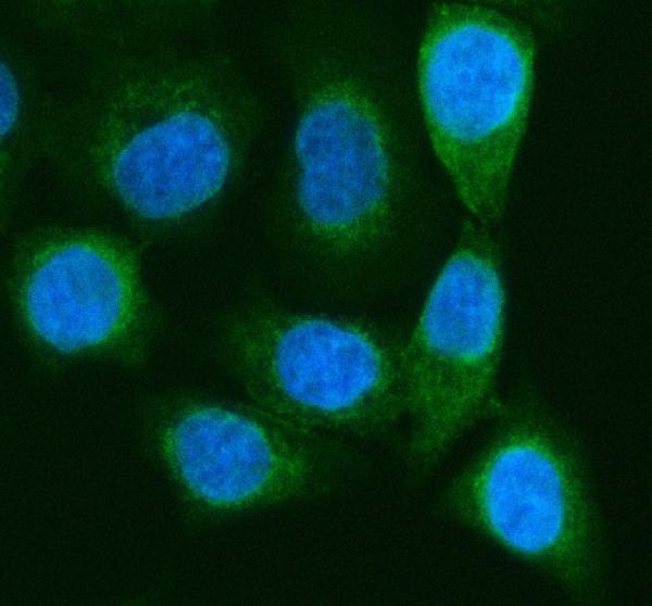
| Q: Is Golgi-enriched signal in IF/ICC compatible with RAB27B? | HPA places IF/ICC signal mainly at the Golgi apparatus and plasma membrane, with additional cytosolic localization (HPA: subcellular ICC-IF). | A: Yes, interpret that pattern against the IF/ICC observations (HPA: subcellular ICC-IF). Use the separate IF/ICC guide for its methods; this IHC-P section supplies no IF/ICC protocol. |
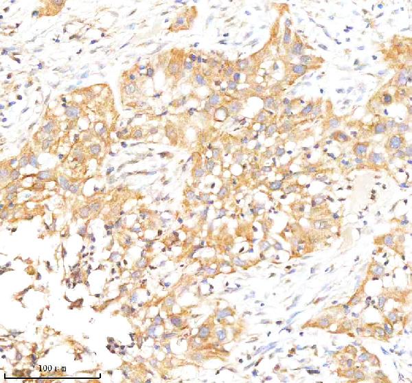


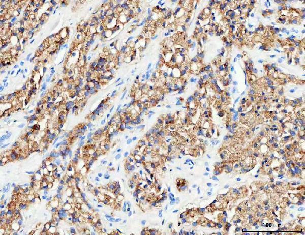
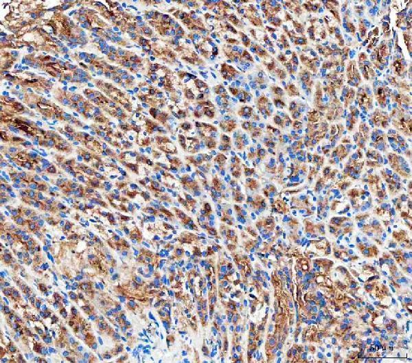

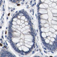


Comprehensive Human Protein Atlas IHC scoring per tissue (reliability: Enhanced — Medium consistency between antibody staining and RNA expression data. Pending external verification.). Rows are taken directly from the HPA tissue chart — click any row's HPA link to view the source.
| Tissue | Cell type | Level | Evidence | Source |
|---|---|---|---|---|
| Colon | Glandular cells | High | Protein (IHC) | HPA → |
| Duodenum | Glandular cells | High | Protein (IHC) | HPA → |
| Kidney | Cells in tubules | High | Protein (IHC) | HPA → |
| Prostate | Glandular cells | High | Protein (IHC) | HPA → |
| Small intestine | Glandular cells | High | Protein (IHC) | HPA → |
| Tissue | Cell type | Level | Evidence | Source |
|---|---|---|---|---|
| Adipose tissue | Adipocytes | Not detected | Protein (IHC) | HPA → |
| Bronchus | Respiratory epithelial cells | Not detected | Protein (IHC) | HPA → |
| Caudate | Glial cells | Not detected | Protein (IHC) | HPA → |
| Cervix | Glandular cells | Not detected | Protein (IHC) | HPA → |
| Heart muscle | Cardiomyocytes | Not detected | Protein (IHC) | HPA → |
Use compartment and cell type controls to troubleshoot RAB27B staining in paraffin sections; interpret IF as a complementary localisation check.
Both anti-RAB27B antibodies have IHC images from human paraffin sections and mouse and rat stomach sections, plus A549 ICC/IF images (catalog image captions: A04890-1, A04890-2).
A04890-1 has IHC images from human bladder and stomach cancer and mouse and rat stomach, plus A549 ICC/IF (catalog A04890-1 image captions). A04890-2 has IHC images from human prostatic cancer and mouse and rat stomach, plus A549 ICC/IF and human bladder paraffin-section IF (catalog A04890-2 image captions).
Which to pick: For tissue IHC, choose A04890-1 if its human bladder or stomach examples match your study; its recommended IHC concentration is 2–5 μg/ml (catalog A04890-1 IHC captions; datasheet: A04890-1). For IF/ICC, choose A04890-2 if you also need a paraffin-section IF example: its images include A549 ICC/IF and human bladder IF, and it is listed as polyclonal (catalog A04890-2 IF captions; datasheet: A04890-2). For cross-species IHC, either SKU has mouse and rat stomach paraffin-section images and listed Human/Mouse/Rat reactivity; neither IHC caption reports the fixative (catalog IHC captions and reactivity: A04890-1, A04890-2).