This website uses cookies to ensure you get the best experience on our website.


- Table of Contents
Plan paraffin section RAB5C IHC around the cytoplasmic tissue pattern (HPA tissue IHC), while treating early endosome membrane localization as a molecular expectation (UniProt). The catalog antibody has an IHC dilution of 1:100–1:300 (datasheet A05148); testis pachytene spermatocytes show high staining (HPA tissue IHC).

Expected localisation, validated protocols, controls and antibodies — the at-a-glance facts below, then the full design guide.
| Expected localisation | Cytoplasmic tissue stain (HPA tissue IHC); early endosome membrane expected (UniProt) | |
| Staining pattern | High cytoplasmic staining in pachytene spermatocytes (HPA tissue IHC) | |
| Antigen retrieval | Tris-EDTA pH 9.0 HIER, heat-mediated (datasheet A05148) | |
| Positive control | Testis+4 more · see all | |
| Negative control | Adipose tissue+1 more · see all |
| Fixation | Keep fixation consistent across sections (standard IHC practice; not target-specific) | |
| Caveat | Staining and RNA expression show low consistency (HPA tissue IHC) | |
| Regulation | No expression regulator annotated (UniProt) | |
| Isoform / epitope | 2 isoforms; no transmembrane segment; check epitope coverage (UniProt) |
Compare the catalog antibody’s IHC-P protocol (datasheet A05148) with published RAB5C protocols for spinal cord sections (PMC4798752) and breast cancer paraffin sections (PMC12183389).
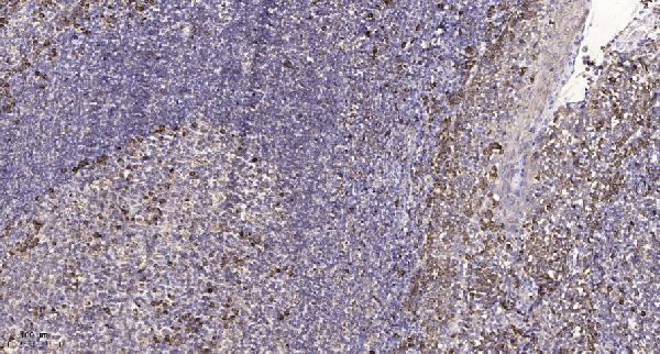
| Sample | Paraffin-embedded human tonsil tissue; fixative not specified (datasheet A05148) |
| Fixation | Image fixative and duration unreported (datasheet A05148); verify before use. |
| Sectioning | 4–5 µm sections on charged slides (standard) |
| Deparaffinisation | Xylene, graded ethanol series to water (standard) |
| Antigen retrieval | Heat retrieval: Tris-EDTA pH 9.0 (datasheet A05148); 20 min, 95–100 °C (standard) |
| Peroxidase block | 3% H2O2, 10 min, room temperature (standard) |
| Blocking | 10% normal serum of the secondary host, 30 min, room temperature (standard) |
| Primary antibody | Rabbit anti-RAB5C, 1:100 - 1:300 (datasheet A05148) |
| Primary incubation | Overnight at 4 °C (standard) |
| Detection | HRP-polymer secondary, DAB chromogen 5–10 min (standard) |
| Counterstain | Hematoxylin, blue, dehydrate and mount (standard) |
| Expected result | RAB5C-positive staining in pachytene spermatocytes of testis (HPA tissue IHC: High). HPA tissue profile: Cytoplasmic expression in several tissues. No signal in the no-primary control. |
RAB5C is an early endosome and cell membrane associated protein without a transmembrane segment (UniProt P51148). In paraffin sections, expect cytoplasmic staining in several tissues, strongest among the supplied examples in testis pachytene spermatocytes (HPA tissue IHC: High). HPA rates the tissue IHC antibody Approved but reports low consistency between staining and RNA expression; interpret intensity alongside morphology and controls (HPA tissue IHC).
| Cytoplasmic staining in pachytene spermatocytes; other listed positive cells stain less strongly. | This matches the reported High signal in testis and Medium signal in several glandular, hematopoietic, respiratory epithelial and neuronal cell populations (HPA tissue IHC). A granular cytoplasmic appearance is plausible for an endosome associated protein, but the HPA tissue profile specifies cytoplasmic expression, not a required punctate IHC pattern (HPA tissue IHC; UniProt P51148). |
| Signal appears predominantly nuclear, or forms a uniform strong rim around most cells. | Those patterns do not match HPA’s cytoplasmic tissue profile or its endosomal IF localization (HPA tissue IHC; HPA subcellular). Review morphology, counterstain and detection controls before scoring them as RAB5C. UniProt also lists the cell membrane, so a localized membrane component alone is not grounds to reject an otherwise credible result (UniProt P51148). |
| Strong staining appears in adipocytes or smooth muscle cells. | HPA reports RAB5C as Not detected in those cell types (HPA tissue IHC). Check whether the color is confined to the expected cells, then compare a reagent control for nonspecific binding or endogenous detection activity (general IHC practice). HPA’s low staining–RNA consistency limits how confidently any single discrepancy can be assigned to cross-reactivity (HPA tissue IHC). |
| Broad, diffuse color obscures cell borders and tissue structure. | This does not provide a scorable cytoplasmic cell pattern (general IHC practice; HPA tissue IHC). Background from antibody binding, detection reagents or residual endogenous activity can mimic widespread positivity in chromogenic IHC (general IHC practice). Assess an appropriate reagent control before comparing tissue intensities; diffuse color alone cannot establish RAB5C localization. |
| No signal appears in the testis pachytene spermatocytes used as a positive reference. | That conflicts with the supplied High HPA observation (HPA tissue IHC). First confirm that the relevant cells are present and preserved, then check the staining run and its controls (general IHC practice). A failed positive reference makes a negative result elsewhere uninterpretable; HPA’s Approved rating does not guarantee that every specimen or run will stain (HPA tissue IHC). |
| Compartment and topology | UniProt places RAB5C at early endosome and cell membranes and annotates no transmembrane segment (UniProt P51148). HPA describes tissue staining as cytoplasmic (HPA tissue IHC). Score the cell and cytoplasmic compartment first; do not require a sharply outlined membrane or individually resolved endosomes in a chromogenic section (general IHC practice). |
| Reference cell intensity | Pachytene spermatocytes are High; the supplied adrenal, appendix and breast glandular cells, bone marrow hematopoietic cells, bronchial respiratory epithelial cells, caudate neuronal cells and cerebellar granular layer cells are Medium (HPA tissue IHC). Adipocytes and smooth muscle cells are Not detected in HPA’s examples; these observations guide comparisons, not universal thresholds (HPA tissue IHC). |
| IHC evidence strength | The listed IHC antibody HPA003426 is Approved, while HPA explicitly reports low consistency between antibody staining and RNA expression (HPA antibodies; HPA tissue IHC). Treat agreement with the observed cell distribution as supportive evidence, and investigate major mismatches with controls and morphology instead of interpreting an Approved label as definitive specificity. |
| IF/ICC cross-check: what should appear? | HPA reports endosomal localization in ICC-IF, with an Enhanced subcellular result, and lists A-549, HEK293, U2OS and HeLa images (HPA subcellular). That supports endosomal IF signal as a localization cross-check. The listed ICC Enhanced antibody is HPA004167, whereas the listed IHC Approved antibody is HPA003426; their validation labels should not be exchanged (HPA antibodies). |
| Antigen retrieval and detection | Target-specific fixation effects are not established by the supplied assay evidence. Verify with a matched IHC source before attributing a result to fixation. |
| Situation | Likely cause | Next action |
|---|---|---|
| Testis positive reference is blank. | The sampled section may lack identifiable pachytene spermatocytes, or the staining run may have failed (HPA tissue IHC; general IHC practice). | Locate the relevant cells on the counterstained section and inspect run controls; then review the established retrieval, primary-antibody and detection steps (general IHC practice). Do not score other blank tissues as negative until the positive reference works. |
| Adipocytes or smooth muscle cells stain strongly. | This conflicts with HPA’s Not detected observations; nonspecific antibody binding or endogenous chromogenic activity is possible (HPA tissue IHC; general IHC practice). | Compare cell morphology with a suitable reagent control and check blocking of endogenous detection activity where the detection system requires it (general IHC practice). Reassess specificity before calling those cells RAB5C positive. |
| A strong nuclear pattern dominates the section. | HPA reports cytoplasmic tissue staining, while its IF localization is endosomal (HPA tissue IHC; HPA subcellular). Nuclear color may reflect background or a scoring error (general IHC practice). | Use the counterstain to distinguish nucleus from adjacent cytoplasm, compare the reagent control, and score only a credible cell associated pattern (general IHC practice). |
| Color is diffuse across tissue and obscures cellular detail. | Nonspecific reagent signal, inadequate washing or endogenous detection activity can create background in chromogenic IHC (general IHC practice). | Inspect the reagent control, tissue preservation and washing steps; adjust the general staining workflow only in light of those controls (general IHC practice). Do not convert diffuse color into a positive RAB5C score. |
| An HPA Medium cell type is negative in a particular specimen. | The HPA values describe observed examples, and the tissue IHC entry reports low staining–RNA consistency (HPA tissue IHC). A local negative result alone does not identify its cause. | Confirm the intended cell population is present, check the testis positive reference and run controls, and document the specimen level discrepancy (HPA tissue IHC; general IHC practice). Avoid assigning a target-specific fixation effect without evidence. |
| Endosomal IF and chromogenic IHC appear different. | IF resolves subcellular structures more clearly than routine chromogenic sections (general IHC practice). The supplied IF and IHC validation labels also belong to different antibodies (HPA antibodies). | Judge paraffin IHC against its cytoplasmic tissue pattern and cell types, and use HPA’s endosomal IF localization as a separate cross-check (HPA tissue IHC; HPA subcellular). Review each assay’s controls before treating the difference as biological. |


Comprehensive Human Protein Atlas IHC scoring per tissue (reliability: Approved — Low consistency between antibody staining and RNA expression data.). Rows are taken directly from the HPA tissue chart — click any row's HPA link to view the source.
| Tissue | Cell type | Level | Evidence | Source |
|---|---|---|---|---|
| Testis | Pachytene spermatocytes | High | Protein (IHC) | HPA → |
| Adrenal gland | Glandular cells | Medium | Protein (IHC) | HPA → |
| Appendix | Glandular cells | Medium | Protein (IHC) | HPA → |
| Bone marrow | Hematopoietic cells | Medium | Protein (IHC) | HPA → |
| Breast | Glandular cells | Medium | Protein (IHC) | HPA → |
Use compartment, cell identity, and processing controls together when assessing RAB5C staining in paraffin sections (UniProt P51148 localization; HPA tissue IHC).
Anti-RAB5C antibodies list IHC and IF/ICC applications in human, mouse, and rat; A05148 includes a human paraffin-section IHC image (catalog: applications/reactivity; A05148 IHC caption).
A05148 lists IHC and IF/ICC for human, mouse, and rat, with an IHC image of paraffin-embedded human tonsil (catalog: applications/reactivity; A05148 IHC caption). M05148-1 lists IHC and ICC/IF for human, mouse, and rat; no IHC or IF image caption is supplied for it (catalog: applications/reactivity; M05148-1 image alts).
Which to pick: For tissue IHC, pick A05148 if a paraffin-section example matters: its human tonsil caption reports Tris-EDTA retrieval at pH 9.0 and primary antibody at 1:200, but does not report the fixative (A05148 IHC caption). For IF/ICC, pick M05148-1 if you prefer clone 28R55; A05148 is polyclonal, and both list IF/ICC without an IF image caption (catalog: clone/applications/dilution_raw; image alts). Both list mouse and rat reactivity, while the supplied tissue IHC image is human only (catalog: reactivity; A05148 IHC caption; M05148-1 image alts).