RAD18 / E3 ubiquitin-protein ligase RAD18 · IHC design guide

Design Immunohistochemistry for RAD18

Plan RAD18 paraffin IHC around selective nuclear staining in specific cell types (HPA tissue IHC). The guide covers the IHC-validated antibody at 2–5 μg/ml (datasheet A01622-2) and cell-level nuclear scoring (standard IHC practice).

Evidence assembled Oct 2026 · For research use; verify linked source records and product datasheet before use
Immunohistochemistry protocol sheet for RAD18 (IHC for RAD18): expected localisation Selective nuclear staining in tissue (HPA tissue IHC), antibody A01622-2, validated IHC image, and IHC protocol steps
Printable RAD18 IHC protocol sheet — expected localisation Selective nuclear staining in tissue (HPA tissue IHC), antibody A01622-2, controls and protocol steps. Open the full RAD18 IHC guide →

RAD18 Immunohistochemistry Experimental Design Guide

Expected localisation, validated protocols, controls and antibodies — the at-a-glance facts below, then the full design guide.

Must know before staining
Expected localisation Selective nuclear staining in tissue (HPA tissue IHC)
Staining pattern Nuclear in germinal center, testis basal and intestinal cells (HPA tissue IHC)
Antigen retrieval EDTA pH 8.0 HIER, heat-mediated (datasheet A01622-2)
Positive control ⓘ Appendix+4 more · see all
Negative control ⓘ Adipose tissue+3 more · see all
Important caveats
Reasons your staining may differ from the expected pattern.
Fixation Keep fixation consistent across sections (standard IHC practice; not target-specific)
Caveat Cell-type staining may differ from tissue RNA levels (HPA tissue IHC)
Regulation DNA damage recruits RAD18 to nuclear foci (UniProt)
Isoform / epitope No annotated isoforms or signal-peptide cleavage (UniProt)
Section 1

Recommended RAD18 IHC & IF Protocols

The catalog antibody uses EDTA pH 8.0 heat retrieval (datasheet A01622-2). Three published RAD18 IHC protocols provide tissue examples (PMC7251712; PMC6558645; PMC9582675).

Recommended immunohistochemistry (IHC-P) protocol parameters
SampleParaffin-embedded human gastric signet ring cell carcinoma tissue; fixative not specified (datasheet A01622-2)
FixationImage fixative and duration unreported (datasheet A01622-2); verify before use.
Sectioning4–5 µm sections on charged slides (standard)
DeparaffinisationXylene, graded ethanol series to water (standard)
Antigen retrievalHeat retrieval: EDTA pH 8.0 (datasheet A01622-2); 20 min, 95–100 °C (standard)
Peroxidase block3% H2O2, 10 min, room temperature (standard)
Blocking10% goat serum (datasheet A01622-2)
Primary antibodyRabbit anti-RAD18, 2-5 μg/ml (datasheet A01622-2)
Primary incubationOvernight at 4 °C (datasheet A01622-2)
DetectionStreptavidin-biotin complex (SABC), DAB chromogen (datasheet A01622-2)
CounterstainHematoxylin, blue, dehydrate and mount (standard)
Expected resultRAD18-positive staining in lymphoid tissue of appendix (HPA tissue IHC: High). HPA tissue profile: Selective nuclear expression in selected cell types including lymphoid germinal center cells, basal cells in testis and in the intestine. No signal in the no-primary control.
💡Decision noteStart with EDTA pH 8.0 heat retrieval for the catalog antibody (datasheet A01622-2); compare citrate retrieval on control sections if optimizing conditions (PMC6558645; PMC9582675).
Section 2

What Is the Expected RAD18 Staining Pattern?

RAD18 should appear predominantly in nuclei in paraffin-section IHC: UniProt places it in the nucleus and on chromatin, and HPA describes selective nuclear staining in particular cell types (UniProt Q9NS91 subcellular; HPA tissue IHC). High staining is reported in appendix lymphoid tissue, colon and rectum glandular cells, and bladder urothelial cells (HPA tissue IHC). HPA rates the tissue pattern Supported, with medium consistency between staining and RNA expression (HPA tissue IHC). RAD18 has no transmembrane segment (UniProt Q9NS91 topology).

What am I looking at on my slide?
Distinct nuclear chromogen in appendix lymphoid cells or colon glandular cells (HPA tissue IHC).This fits RAD18's nuclear location and cell-level staining pattern (UniProt Q9NS91 subcellular; HPA tissue IHC). Compare staining within each cell type; a positive nucleus does not imply that every neighboring cell should stain (HPA tissue IHC).
Predominantly membrane-bound or uniformly cytoplasmic staining, with little nuclear signal.That distribution conflicts with the selective nuclear IHC pattern (HPA tissue IHC). UniProt also lists a centrosomal location, so isolated extranuclear signal alone is inconclusive (UniProt Q9NS91 subcellular). Check morphology and controls before calling it RAD18.
Strong staining in adipocytes or lung type I alveolar cells (HPA tissue IHC: Not detected).This disagrees with those HPA reference observations and warrants a specificity check; possible causes include cross-reactivity or endogenous detection activity (HPA tissue IHC; general IHC practice). HPA's reference pattern does not prove every specimen must remain negative.
Haze spans nuclei, cytoplasm, and extracellular space without a clear cell pattern.Diffuse deposition is less persuasive than discrete nuclear staining (HPA tissue IHC). Check background in a section processed without primary antibody, then review blocking, washing, and detection conditions (general IHC practice).
No nuclear signal in an adequately preserved appendix lymphoid region or colon glandular region (HPA tissue IHC: High).An absent result in an HPA high-staining cell population raises a workflow or antibody-performance question, but does not by itself establish absent RAD18 (HPA tissue IHC; general IHC practice). Verify the cell population and controls before scoring.
💡Expected RAD18 appearanceCall a convincing positive result when nuclei of an HPA high-staining cell population show distinct, cell-associated chromogen (HPA tissue IHC); widespread membrane staining or diffuse section-wide haze is suspect against the nuclear reference pattern (HPA tissue IHC; general IHC practice).
How each factor affects the staining
Cell selection and intensityHPA reports High staining in appendix lymphoid tissue, colon and rectum glandular cells, and bladder urothelial cells; adipocytes are Not detected (HPA tissue IHC). Score the named cells rather than assigning one intensity to an entire organ.
Strength of the tissue referenceThe RAD18 tissue-IHC profile is Supported, with medium staining–RNA consistency; its RNA category is Low tissue specificity (HPA tissue IHC). Treat the listed cell patterns as reference observations, not universal thresholds for every specimen.
Antibody-specific validationIHC is Supported for HPA006716, HPA008752, and CAB080160, and Approved for HPA006724 (HPA antibodies). Those statuses belong to the named antibodies; they do not automatically validate another IHC-validated antibody.
Localization during DNA damageRAD18 associates with chromatin and can relocalize with SLF1 to nuclear foci after DNA damage (UniProt Q9NS91 subcellular). Nuclear puncta can therefore fit its biology, but routine tissue IHC alone cannot identify a DNA-damage response.
IF/ICC Q&A: Where should signal appear?HPA reports mainly nucleoplasm and nuclear bodies, with additional nuclear speckles (HPA subcellular ICC-IF). This supports an IF localization expectation; the paraffin-section IHC cell pattern is assessed from HPA tissue IHC.
Topology and processingRAD18 is a single 1–495 chain with no signal peptide, propeptide, or transmembrane segment (UniProt Q9NS91 processing; topology). These annotations support an intracellular interpretation; they provide no evidence for RAD18-specific fixation sensitivity.
Why is my staining missing, weak or wrong?
SituationLikely causeNext action
Positive control lacks nuclear staining.The selected section may lack the relevant cells, or the staining workflow may have failed (HPA tissue IHC; general IHC practice).Confirm appendix lymphoid or colon glandular cells are present (HPA tissue IHC). Check the IHC-validated antibody's own instructions and run an appropriate staining control (general IHC practice).
Chromogen appears mainly along cell borders.The compartment disagrees with HPA's selective nuclear IHC pattern and UniProt's nuclear localization (HPA tissue IHC; UniProt Q9NS91 subcellular).Recheck the counterstained cell boundaries, compare with a high-staining reference region, and assess the signal against a control lacking primary antibody (HPA tissue IHC; general IHC practice).
Adipocytes stain as strongly as nearby expected-positive cells.Adipocytes are Not detected in HPA's listed adipose and breast observations; nonspecific binding or detection activity is possible (HPA tissue IHC; general IHC practice).Assess a no-primary control, blocking, and wash conditions, then compare nuclear staining in the named positive cell population (HPA tissue IHC; general IHC practice).
Brown signal persists when primary antibody is omitted.Primary-independent signal suggests endogenous detection activity or another reagent-related background source (general chromogenic IHC practice).Address the relevant endogenous activity for the chosen detection system and review blocking and washing before interpreting RAD18 staining (general chromogenic IHC practice).
Nuclei are hard to distinguish from diffuse chromogen.Background or an overly dense reaction can obscure the selective nuclear pattern (HPA tissue IHC; general IHC practice).Review detection development, washing, and counterstain readability; judge RAD18 only where nuclear boundaries and the target cell type remain identifiable (general IHC practice).
Signal differs between two antibodies.HPA assigns IHC validation status per antibody, and its overall tissue profile has medium staining–RNA consistency (HPA antibodies; HPA tissue IHC).Compare each antibody's controls and localization in the same HPA-listed cell population before resolving the discrepancy; do not transfer one antibody's validation status to the other (HPA antibodies; HPA tissue IHC).

Sample controls for RAD18 IHC & IF

🧪Run appendix first: its lymphoid tissue should stain for RAD18 (HPA: High in appendix lymphoid tissue). Use adipose tissue as the negative tissue (HPA: Not detected in adipocytes); on the appendix slide, internal negative cells should lack specific staining above background, but neighboring cell types need validation before being designated RAD18-negative.
Positive control tissue: Appendix (Lymphoid tissue, HPA High)
Negative control tissue: Adipose tissue (HPA Not detected)
ICC-IF cell lines (HPA subcellular resource): HPA ICC-IF images show RAD18 in PC-3, U-251MG, U2OS, SK-MEL-30, SiHa, A-431, HeLa BAC 6618, HeLa , with annotated localisation: Nucleoplasm (supported), Nuclear bodies (supported) (HPA subcellular).
Technical controls: Include a no-primary (secondary-only) control, a concentration-matched rabbit IgG isotype control, and RAD18-knockout material as a biological negative (caption: rabbit primary antibody; standard IHC practice). Quench endogenous peroxidase and assess endogenous biotin when using the reported biotin–SABC/DAB detection system, particularly in appendix lymphoid tissue (caption: biotin–SABC/DAB; HPA: appendix lymphoid tissue).
⚠️Feasibility: No target-specific fixation window or fixation effect is reported, and the selected A01622-2 caption does not state a fixative; its paraffin section does not establish fixation (caption: paraffin section; fixative unreported). Heat retrieval in EDTA at pH 8.0 is reported for that IHC example, but a retrieval dependency and whether frozen sections or IF/ICC are easier are unreported (caption: EDTA heat retrieval). In appendix, endogenous peroxidase in some immune cells could mimic chromogenic signal, so interpret staining against the controls (HPA: appendix lymphoid tissue; standard IHC practice). The selected A01622-2 tissue-IHC caption documents paraffin sections, but does not specify the fixative (selected-SKU IHC image A01622-2).

HPA tissue IHC evidence for RAD18

Comprehensive Human Protein Atlas IHC scoring per tissue (reliability: Supported — Medium consistency between antibody staining and RNA expression data.). Rows are taken directly from the HPA tissue chart — click any row's HPA link to view the source.

Positive expression · recommended positive controls

TissueCell typeLevelEvidenceSource
Appendix Lymphoid tissue High Protein (IHC) HPA →
Colon Glandular cells High Protein (IHC) HPA →
Heart muscle Cardiomyocytes High Protein (IHC) HPA →
Rectum Glandular cells High Protein (IHC) HPA →
Thyroid gland Glandular cells High Protein (IHC) HPA →

Undetected expression · recommended negative controls

TissueCell typeLevelEvidenceSource
Adipose tissue Adipocytes Not detected Protein (IHC) HPA →
Breast Adipocytes Not detected Protein (IHC) HPA →
Liver Cholangiocytes Not detected Protein (IHC) HPA →
Lung Alveolar cells type I Not detected Protein (IHC) HPA →
Section 3

Advanced RAD18 IHC Tips

Troubleshoot RAD18 staining by checking retrieval, nuclear localisation, cell type, and controls before comparing chromogenic IHC results (UniProt Q9NS91; HPA tissue IHC).

How should I retrieve RAD18 in paraffin sections when nuclear staining is weak?
Start with heat-mediated retrieval in EDTA at pH 8.0 for paraffin sections (datasheet A01622-2). The selected image used this retrieval before overnight incubation at 4°C with 2 μg/ml of the catalog antibody, so keep those conditions together for the first comparison (caption A01622-2). If staining remains weak, vary heating time on adjacent sections while holding antibody concentration and detection constant (standard IHC practice). Compare nuclear signal with a no-primary control and inspect section edges for excessive background (standard IHC practice). Nuclear staining is the expected tissue pattern to assess (HPA tissue IHC: selective nuclear expression).
Could fixation explain inconsistent RAD18 staining between paraffin specimens?
RAD18-specific sensitivity to fixation is unknown from the supplied evidence; the selected paraffin-section caption does not state a fixative (caption A01622-2). Record each specimen’s fixative and fixation duration, then compare sections processed together using the same EDTA retrieval and antibody conditions (standard IHC practice; datasheet A01622-2). For an initial comparison, use pH 8.0 retrieval and 2 μg/ml primary antibody, as documented for the selected image (caption A01622-2). Check a no-primary section for detection background before attributing weak nuclear signal to fixation (standard IHC practice; HPA tissue IHC: nuclear expression). Do not assign a RAD18-specific fixation effect without a controlled comparison.
How should I assess cytoplasmic or punctate RAD18 staining in chromogenic IHC?
Prioritise interpretable nuclear staining in paraffin sections because the tissue profile describes selective nuclear expression (HPA tissue IHC). RAD18 can associate with chromatin and relocalise to nuclear foci after DNA damage, but a routine DAB section alone cannot establish those events (UniProt Q9NS91; standard IHC practice). UniProt also lists a centrosomal location, so isolated nonnuclear puncta require validation against controls before being scored as RAD18 (UniProt Q9NS91; standard IHC practice). Compare the same cell type across adjacent sections and inspect morphology and the no-primary control (standard IHC practice). Report nuclear and cytoplasmic staining separately rather than combining them into one score (standard IHC practice).
What should I check if the catalog antibody misses a suspected RAD18 epitope?
The supplied record lists 0 isoforms and a single 1–495 chain, so an isoform-specific explanation is unsupported here (UniProt Q9NS91). RAD18 has no transmembrane segment and includes a SAP domain at residues 248–282, but the catalog antibody’s epitope is not identified in the supplied caption (UniProt Q9NS91 topology and domains; caption A01622-2). The record lists phosphorylated residues, including 99 and 103; their effect on this antibody’s tissue staining is unknown (UniProt Q9NS91 modified residues). Recheck retrieval and section processing with a known staining control before interpreting loss of signal as epitope modification (standard IHC practice). Keep conclusions limited to antibody reactivity in the tested sections (standard IHC practice).
How can IF help verify a disputed RAD18 IHC localisation?
On the separate IF/ICC guide, multiplex RAD18 with a marker for the expected cell type, such as a glandular-cell marker when examining colon, and assess colocalisation cell by cell (HPA tissue IHC: colon glandular cells high; standard IF practice). Choose a fluorophore channel away from the specimen’s strongest autofluorescence and include a no-primary control (standard IF practice). Because RAD18 lacks a transmembrane segment and is mainly nuclear, permeabilise cells sufficiently to access intracellular epitopes while preserving nuclear structure (UniProt Q9NS91 topology; HPA subcellular: nucleoplasm and nuclear bodies; standard IF practice). Nuclear bodies and speckles can guide IF interpretation, but they should not be inferred from diffuse DAB staining (HPA subcellular; standard IHC practice).
What controls help distinguish RAD18 signal from DAB background?
Start with a no-primary section and inspect staining in nuclei, tissue edges, and damaged areas before changing the primary antibody concentration (standard IHC practice). The selected image used 10% goat serum blocking, 2 μg/ml primary antibody, a biotinylated secondary, and DAB development (caption A01622-2). If diffuse brown signal persists, optimise blocking and washes and check endogenous peroxidase activity as part of the general chromogenic workflow (standard IHC practice). With biotin-based detection, assess endogenous biotin as a possible source of signal in the specimen (caption A01622-2; standard IHC practice). Preserve a nuclear positive pattern while reducing background (HPA tissue IHC: selective nuclear expression).
How should RAD18 positivity be scored across paraffin tissue sections? ⚠ ANSWER MARKED FOR VERIFICATION
Define the cell population and score nuclear staining within it, because tissue expression varies by cell type (HPA tissue IHC). Record the percentage of positive nuclei and, where intensity is reproducible, an H-score; for dispersed positive cells, density per mm² can be useful (standard IHC practice). Normalise positive counts to all evaluable nuclei of the same cell type, or density to evaluable tissue area, rather than comparing raw counts across sections (standard IHC practice). Apply one threshold and imaging setting across the comparison set, with blinded review when feasible (standard IHC practice). Report cytoplasmic staining separately and exclude folds, necrosis, and edge artefacts from scoring (standard IHC practice).
Which findings support true RAD18 positivity rather than staining artefact?
A reproducible nuclear pattern in the expected cell population supports interpretation of RAD18 staining (HPA tissue IHC: selective nuclear expression; standard IHC practice). HPA reports high staining in colon glandular cells and no detection in lung type I alveolar cells, but its tissue IHC reliability is only Supported with medium RNA consistency (HPA tissue IHC). Treat isolated cytoplasmic staining cautiously even though UniProt lists a centrosomal location (UniProt Q9NS91; standard IHC practice). Edge-restricted colour, necrotic-area staining, or signal in a no-primary section points toward processing or detection artefact (standard IHC practice). Check endogenous peroxidase and, with biotin-based detection, endogenous biotin before assigning DAB signal to RAD18 (caption A01622-2; standard IHC practice).
Boster reagents

Best RAD18 / E3 ubiquitin-protein ligase RAD18 IHC Antibodies

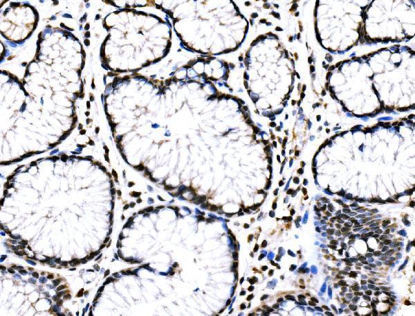
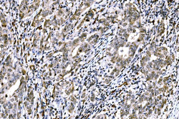
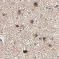
A01622-2 has IHC images from human paraffin sections of gastric signet ring cell carcinoma, renal pelvis squamous metaplasia and tonsil (catalog image captions); no IF image is supplied (catalog payload).

Real IHC data IHC analysis of RAD18 using anti-RAD18 antibody (A01622-2). RAD18 was detected in a paraffin-embedded section of human gastric signet ring cell carcinoma tissue. Heat mediated antigen retrieval was performed in EDTA buffer (pH 8.0, epitope retrieval solution). The tissue section was blocked with 10% goat serum. The tissue section was then incubated with 2 μg/ml rabbit anti-RAD18 Antibody (A01622-2) overnight at 4°C. Biotinylated goat anti-rabbit IgG was used as secondary antibody and incubated for 30 minutes at 37°C. The tissue section was developed using Strepavidin-Biotin-Complex (SABC) (Catalog # SA1022) with DAB as the chromogen.
Anti-RAD18 Antibody ®
Cat # A01622-2

A01622-2 has IHC images from human paraffin sections of gastric signet ring cell carcinoma, renal pelvis squamous metaplasia and tonsil (catalog image captions). Its application list includes IHC and its reactivity list includes human, mouse and rat; no IF validation is listed (catalog applications and reactivity).

Which to pick: For tissue IHC, choose A01622-2: its images document human paraffin sections, with 2 μg/ml primary antibody and EDTA retrieval at pH 8.0 (catalog image captions); the fixative is unreported (catalog image captions). No IF/ICC choice is supported because A01622-2 has no IF application, dilution or image in the payload (catalog payload). For cross-species work, A01622-2 lists mouse and rat reactivity, but its supplied IHC images show only human tissue (catalog reactivity and image captions).

Each figure is that product's own IHC / IF validation image from its datasheet.

References

  1. UniProt Consortium. UniProt entry Q9NS91 (RAD18_HUMAN, E3 ubiquitin-protein ligase RAD18).
  2. Human Protein Atlas. RAD18 tissue IHC expression (reliability: Supported).
  3. Human Protein Atlas. RAD18 subcellular location (ICC-IF): Mainly localized to the nucleoplasm and nuclear bodies. In addition localized to the nuclear speckles..
  4. Human Protein Atlas. RAD18 antibody validation summary (5 antibodies).
  5. RAD18 promotes colorectal cancer metastasis by activating the epithelial‑mesenchymal transition pathway. Oncology reports 2020 — PMC7251712.
  6. RAD18 may function as a predictor of response to preoperative concurrent chemoradiotherapy in patients with locally advanced rectal cancer through caspase-9-caspase-3-dependent apoptotic pathway. Cancer medicine 2019 — PMC6558645.
  7. RAD18 confers radioresistance of esophagus squamous cell carcinoma through regulating p-DNA-PKcs. Cancer medicine 2022 — PMC9582675.
  8. RAS-RAF-miR-296-3p signaling axis increases Rad18 expression to augment radioresistance in pancreatic and thyroid cancers. Cancer letters 2024 — PMC11132429.
  9. PubMed PMID:10884424 — UniProt-cited evidence.
  10. PubMed PMID:10908344 — UniProt-cited evidence.
  11. PubMed PMID:14702039 — UniProt-cited evidence.