This website uses cookies to ensure you get the best experience on our website.


- Table of Contents
Plan RAD23B paraffin IHC using the catalog antibody at 1:100–1:300 (datasheet A02174-2). Assess nuclear and cytoplasmic staining (HPA tissue IHC) while accounting for cell cycle dependent localization (UniProt).

Expected localisation, validated protocols, controls and antibodies — the at-a-glance facts below, then the full design guide.
| Expected localisation | Nuclear and cytoplasmic staining across tissues (HPA tissue IHC) | |
| Staining pattern | Glandular cells: high nuclear and cytoplasmic staining (HPA tissue IHC) | |
| Antigen retrieval | Tris-EDTA pH 8.0 HIER, heat-mediated (datasheet A02174-2) | |
| Positive control | Adipose tissue+4 more · see all | |
| Negative control | None in HPA (detected in all 45 tissues); use no-primary + isotype controls |
| Fixation | Keep fixation consistent across sections (standard IHC practice; not target-specific) | |
| Caveat | Cell cycle can shift the nuclear/cytoplasmic balance (UniProt) | |
| Regulation | Nuclear levels decrease in S phase (UniProt) | |
| Isoform / epitope | 2 isoforms; epitope differences are unspecified (UniProt) |
The catalog antibody protocol uses Tris-EDTA pH 8.0 retrieval (datasheet A02174-2). Published IHC protocols provide colorectal tissue and human brain examples (PMC12573188; PMC13140639).
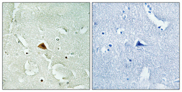
| Sample | Paraffin-embedded Human brain tissue; fixative not specified (datasheet A02174-2) |
| Fixation | Image fixative and duration unreported (datasheet A02174-2); verify before use. |
| Sectioning | 4–5 µm sections on charged slides (standard) |
| Deparaffinisation | Xylene, graded ethanol series to water (standard) |
| Antigen retrieval | Heat retrieval: Tris-EDTA pH 8.0 (datasheet A02174-2); 20 min, 95–100 °C (standard) |
| Peroxidase block | 3% H2O2, 10 min, room temperature (standard) |
| Blocking | 10% normal serum of the secondary host, 30 min, room temperature (standard) |
| Primary antibody | Rabbit anti-RAD23B, 1:100-1:300 (datasheet A02174-2) |
| Primary incubation | Overnight at 4 °C (standard) |
| Detection | HRP-polymer secondary, DAB chromogen 5–10 min (standard) |
| Counterstain | Hematoxylin, blue, dehydrate and mount (standard) |
| Expected result | RAD23B-positive staining in adipocytes of adipose tissue (HPA tissue IHC: High). HPA tissue profile: Nuclear and cytoplasmic expression in all tissues. No signal in the no-primary control. |
RAD23B should show nuclear and cytoplasmic staining across tissues (HPA tissue IHC: nuclear and cytoplasmic expression in all tissues). Nucleoplasmic signal is supported by ICC-IF, with additional cytosolic signal (HPA subcellular: supported). Interpretation should allow both compartments because distribution changes with the cell cycle (UniProt P54727: subcellular location). RAD23B has no transmembrane segment, so a membrane-restricted pattern is unexpected (UniProt P54727: topology). HPA rates the tissue IHC profile Enhanced, while reporting medium consistency with RNA data (HPA tissue IHC: reliability).
| Nuclear staining with some cytoplasmic staining in tissue cells. | This fits the reported distribution (HPA tissue IHC: nuclear and cytoplasmic expression in all tissues). Differences between individual cells can be plausible: RAD23B is nuclear and cytoplasmic in G1, nuclear levels decrease in S phase, and it moves into the cytoplasm during mitosis (UniProt P54727: subcellular location). |
| A sharp cell-surface rim is the dominant pattern, without convincing intracellular staining. | Treat this as a suspected artefact and review the control slide: RAD23B has no transmembrane segment (UniProt P54727: topology), and the observed tissue pattern is nuclear and cytoplasmic (HPA tissue IHC: profile). Cytoplasmic signal alone is less decisive because cell-cycle-dependent redistribution is reported (UniProt P54727: subcellular location). |
| Unexpectedly strong staining is confined to a cell population expected to stain weakly. | Consider cross-reactivity or endogenous detection activity, then compare cell identity and controls (general IHC practice). Hematopoietic cells in bone marrow are reported at a low level, but HPA lists no negative tissue or cell population here; low staining cannot serve as an absolute absence control (HPA tissue IHC: low; negative list empty). |
| Brown color spreads across tissue spaces or coats many cells without discernible nuclei. | This is difficult to score as RAD23B because the reported signal is intracellular (HPA tissue IHC: profile; UniProt P54727: subcellular location). Check reagent-only controls, wash quality and chromogen development for nonspecific background or endogenous detection activity (general chromogenic IHC practice). |
| No convincing staining appears in a tissue and cell population reported as high. | Adipocytes in adipose tissue and neuronal cells in cerebral cortex are examples reported as high (HPA tissue IHC: positive cells). Confirm that the expected cells are present, then review the IHC-validated antibody, detection controls and general retrieval workflow (general IHC practice); the supplied sources do not establish RAD23B-specific fixation sensitivity. |
| Cell cycle and compartment | Nuclear and cytoplasmic signal is reported in G1; nuclear levels fall in S phase, and mitotic RAD23B relocates to the cytoplasm without chromatin association (UniProt P54727: subcellular location). Interpret a mixed field cell by cell before calling a weaker nucleus abnormal. |
| Choice of tissue and cells | The tissue profile spans all tissues, but reported levels differ by cell type (HPA tissue IHC: profile and positive cells). HPA lists high staining in adipocytes, glandular cells, respiratory epithelial cells and selected neural cells, while bone-marrow hematopoietic cells are low (HPA tissue IHC: positive and low cells). |
| IHC evidence strength | The tissue profile is rated Enhanced, with medium consistency between antibody staining and RNA expression (HPA tissue IHC: reliability description). Two listed antibodies have Enhanced IHC validation and one has Supported IHC validation (HPA antibodies: IHC status); assess the actual slide and controls rather than treating validation as proof of every stained structure. |
| Topology and processing | RAD23B has no transmembrane segment, signal peptide or propeptide; the listed chain spans residues 1–409 (UniProt P54727: topology and processing). These annotations support an intracellular interpretation but do not identify the antibody epitope or predict an antigen retrieval condition. |
| Isoforms and epitope uncertainty | Two isoforms are listed (UniProt P54727: isoforms). Their existence alone does not show which form the IHC-validated antibody detects; no epitope location is supplied. Avoid assigning a compartment difference or a missing signal to isoform selectivity without antibody-specific evidence. |
| IF/ICC comparison | Q: Should IF/ICC look identical to paraffin IHC? A: Use it as a compartment check: the main supported location is nucleoplasm, with additional cytosol (HPA subcellular: ICC-IF). The supplied IF record does not establish matching staining intensity or an IF/ICC protocol for this IHC guide. |
| Situation | Likely cause | Next action |
|---|---|---|
| Expected high-staining cells are blank. | The expected cell population may be absent from the section, or the IHC detection workflow may have failed (general IHC practice). | Locate an HPA-listed high cell population, then check the IHC-validated antibody and positive-control slide; review retrieval and detection steps as general IHC workflow checks (HPA tissue IHC: positive cells; general IHC practice). |
| Only a strong membrane rim is visible. | That compartment conflicts with the reported intracellular pattern and lack of a transmembrane segment (HPA tissue IHC: profile; UniProt P54727: topology). | Compare a control slide and examine whether nuclear or cytoplasmic staining is present in identifiable cells; investigate nonspecific edge staining before scoring the rim (general IHC practice). |
| Bone-marrow hematopoietic cells stain as strongly as chosen high controls. | HPA reports these cells as low, although it provides no negative cell population (HPA tissue IHC: low; negative list empty). | Recheck cell identity and use appropriate detection controls to assess cross-reactivity or endogenous activity; do not score low as necessarily zero (HPA tissue IHC: low; general IHC practice). |
| Staining varies markedly among neighboring nuclei. | Nuclear abundance can change during the cell cycle (UniProt P54727: subcellular location). | Inspect accompanying cytoplasmic signal and cell morphology before calling the variation a failure; compare the field with the reported nuclear and cytoplasmic tissue pattern (HPA tissue IHC: profile; UniProt P54727: subcellular location). |
| Diffuse brown haze obscures cellular boundaries. | Background can arise from detection activity, excess chromogen development or inadequate washing (general chromogenic IHC practice). | Review reagent-only controls and the detection and washing steps; score RAD23B only where intracellular staining can be assigned to recognizable cells (general IHC practice; HPA tissue IHC: profile). |
| IF/ICC shows cytosol as well as nucleoplasm. | Both locations are supported by ICC-IF, and distribution can vary with cell cycle (HPA subcellular: supported locations; UniProt P54727: subcellular location). | Treat cytosolic signal as potentially consistent with RAD23B when cellular staining is interpretable; use the separate IF/ICC guide for IF-specific workflow decisions (HPA subcellular: supported locations). |


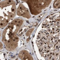


Comprehensive Human Protein Atlas IHC scoring per tissue (reliability: Enhanced — Medium consistency between antibody staining and RNA expression data.). Rows are taken directly from the HPA tissue chart — click any row's HPA link to view the source.
| Tissue | Cell type | Level | Evidence | Source |
|---|---|---|---|---|
| Adipose tissue | Adipocytes | High | Protein (IHC) | HPA → |
| Adrenal gland | Glandular cells | High | Protein (IHC) | HPA → |
| Appendix | Glandular cells | High | Protein (IHC) | HPA → |
| Breast | Glandular cells | High | Protein (IHC) | HPA → |
| Bronchus | Respiratory epithelial cells | High | Protein (IHC) | HPA → |
| Tissue | Cell type | Level | Evidence | Source |
|---|---|---|---|---|
| None in HPA: RAD23B is detected in all 45 scored tissues. Use a no-primary (secondary-only) and an isotype control instead. | ||||
Troubleshoot RAD23B staining in paraffin sections by checking retrieval, compartment patterns, controls and scoring before interpreting chromogenic signal.
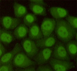
The catalog antibodies have IHC images from human paraffin sections (catalog image captions). M02174 also has a HeLa ICC image; listed reactivity covers human, mouse, rat, and monkey (catalog).
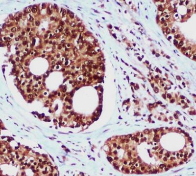
A02174-2 will render with human brain paraffin-section IHC at 1:100 after Tris-EDTA retrieval at pH 8.0 (A02174-2 image caption). M02174 will render with human prostate cancer paraffin-section IHC using citrate retrieval at pH 6.0; its separate image shows HeLa ICC (M02174 image captions).
Which to pick: For tissue IHC, A02174-2 is a rabbit polyclonal option illustrated in human brain, while M02174 is a mouse monoclonal option illustrated in prostate cancer; both captions specify paraffin sections, and neither reports the fixative (catalog; respective IHC image captions). For cell staining, M02174 has a HeLa ICC image, while A02174-2 lists IF with a dilution range of 1:200–1:1000 but has no IF image in this payload (catalog). For listed species coverage, choose M02174 when monkey reactivity is needed; both list human, mouse, and rat reactivity (catalog).