This website uses cookies to ensure you get the best experience on our website.


- Table of Contents
Plan RAD54B staining in paraffin sections with the catalog antibody at 1:100–1:300 (datasheet: IHC dilution). Compare the reported cytoplasmic tissue staining with the annotated nucleoplasmic location, accounting for the uncertain tissue IHC reliability (HPA tissue IHC; UniProt).

Expected localisation, validated protocols, controls and antibodies — the at-a-glance facts below, then the full design guide.
| Expected localisation | Cytoplasmic tissue staining (HPA tissue IHC); nucleoplasmic location (UniProt) | |
| Staining pattern | Cytoplasmic staining in several tissues, including glandular cells (HPA tissue IHC) | |
| Antigen retrieval | Tris-EDTA pH 9.0 HIER, 95–98 °C, 20 min (rule: nuclear antigen) | |
| Positive control | Parathyroid gland+4 more · see all | |
| Negative control | Adrenal gland+3 more · see all |
| Fixation | Keep fixation consistent across sections (standard IHC practice; not target-specific) | |
| Caveat | Cytoplasmic IHC differs from the annotated nucleoplasmic location (HPA tissue IHC; UniProt) | |
| Regulation | Active at DNA double-strand breaks (UniProt) | |
| Isoform / epitope | 2 isoforms; epitope coverage is unspecified (UniProt: isoforms; datasheet: epitope) |
Compare the catalog antibody’s IHC-P protocol with three published RAD54B tissue IHC protocols (PMC12917079; PMC6862887; PMC6072389).
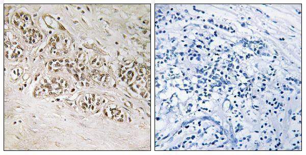
| Sample | Paraffin-embedded human breast carcinoma tissue; fixative not specified (datasheet A06804) |
| Fixation | Image fixative and duration unreported (datasheet A06804); verify before use. |
| Sectioning | 4–5 µm sections on charged slides (standard) |
| Deparaffinisation | Xylene, graded ethanol series to water (standard) |
| Antigen retrieval | Heat-induced epitope retrieval in Tris-EDTA buffer, pH 9.0, 20 min at 95–98 °C (standard rule: nuclear antigen) |
| Peroxidase block | 3% H2O2, 10 min, room temperature (standard) |
| Blocking | 10% normal serum of the secondary host, 30 min, room temperature (standard) |
| Primary antibody | Rabbit anti-RAD54B, 1:100-1:300 (datasheet A06804) |
| Primary incubation | Overnight at 4 °C (standard) |
| Detection | HRP-polymer secondary, DAB chromogen 5–10 min (standard) |
| Counterstain | Hematoxylin, blue, dehydrate and mount (standard) |
| Expected result | RAD54B-positive staining in glandular cells of parathyroid gland (HPA tissue IHC: High). HPA tissue profile: Cytoplasmic expression in several tissues. No signal in the no-primary control. |
RAD54B is assigned to the nucleus and nucleoplasm and is active at DNA double-strand breaks (UniProt Q9Y620). In tissue IHC, however, HPA reports cytoplasmic staining in several tissues, including strong staining of parathyroid glandular cells, testicular Leydig cells and tonsillar squamous epithelial cells (HPA tissue IHC). Treat that compartment difference cautiously: the tissue IHC assessment is Uncertain, and RAD54B has no transmembrane segment (HPA tissue IHC; UniProt Q9Y620).
| Cytoplasmic staining in parathyroid glandular cells, Leydig cells or tonsillar squamous epithelial cells. | This matches the reported High tissue IHC pattern, but is provisional because the IHC assessment is Uncertain (HPA tissue IHC). Record the stained cell type and compartment; intensity alone does not establish that the signal is RAD54B. |
| Predominantly nuclear staining, or staining apparently confined to a tissue lumen or extracellular space. | Nuclear signal is biologically plausible given nucleoplasmic localisation and should not be dismissed solely because HPA tissue IHC describes cytoplasmic staining (UniProt Q9Y620; HPA tissue IHC). Luminal or extracellular signal falls outside the reported intracellular locations; check morphology and staining controls before interpreting it as RAD54B (HPA tissue IHC; HPA ICC-IF). |
| Strong staining in adrenal glandular cells, cardiomyocytes or myocytes. | These cell types are reported as Not detected in the sampled tissues (HPA tissue IHC). Check whether the signal follows the intended cells; discordant staining may reflect cross-reactivity or endogenous detection activity (general IHC practice). A single discordant section does not establish the cause. |
| Broad, diffuse chromogen obscures cell boundaries or appears similarly across most structures. | That distribution cannot support a cell-specific RAD54B call against HPA's varied tissue and cell staining levels (HPA tissue IHC). Compare a no-primary control and assess background from detection reagents or inadequate blocking (general IHC practice). |
| No detectable signal in parathyroid glandular cells, Leyig cells or tonsillar squamous epithelial cells. | These are reported High in tissue IHC, so a blank result warrants a technical check (HPA tissue IHC). Confirm that the selected cells are present, then review the catalog antibody's IHC-P instructions and detection controls (general IHC practice). HPA's Uncertain reliability limits how definitive any one tissue is as a positive control (HPA tissue IHC). |
| Tissue and cell selection | HPA reports High staining in parathyroid glandular cells, Leydig cells and tonsillar squamous epithelial cells; appendix and colon glandular cells are Medium, while adrenal glandular cells and muscle cell types are Not detected (HPA tissue IHC). Use the cell type, not the organ name alone, when judging a field. |
| Compartment evidence | UniProt assigns RAD54B to the nucleoplasm and notes activity at DNA double-strand breaks, whereas HPA tissue IHC describes cytoplasmic staining (UniProt Q9Y620; HPA tissue IHC). Keep the assay and its reliability attached to each observation instead of forcing one compartment call across methods. |
| IF/ICC Q: Where should signal appear? | A: HPA approves both nucleoplasm and cytosol as ICC-IF locations; images are listed for A-431, U-251MG and U2OS (HPA ICC-IF). This answers localisation only: the tissue IHC staining assessment remains Uncertain (HPA tissue IHC). |
| Antibody evidence | HPA007087 is listed as IHC Uncertain and ICC Approved (HPA antibodies). The tissue IHC assessment also reports medium consistency between antibody staining and RNA expression data (HPA tissue IHC). These ratings support cautious scoring of a chromogenic result, especially when its cell type or compartment differs from the reported pattern. |
| Protein forms and topology | UniProt lists two isoforms, a single 1–910 chain, no signal peptide or propeptide, and no transmembrane segment (UniProt Q9Y620). The supplied record gives no antibody epitope, so it cannot establish which isoforms a particular IHC reagent detects or predict an epitope-specific retrieval response. |
| Chromogenic detection background | Endogenous enzyme activity can mimic chromogen staining, depending on the detection system; appropriate blocking and a no-primary control help assess it (general IHC practice). This is a general workflow consideration, not a RAD54B-specific finding or an explanation proven by HPA. |
| Situation | Likely cause | Next action |
|---|---|---|
| Expected High-staining cells are blank. | The target cells may be absent from the section, or an IHC step or detection control may have failed; the tissue IHC result itself is Uncertain (HPA tissue IHC; general IHC practice). | Locate the reported cell type on the counterstained section, verify the detection control, and follow the catalog antibody's IHC-P instructions for retrieval and dilution (general IHC practice). Do not infer RAD54B-specific fixation sensitivity from this result. |
| Staining is strong across reported Not detected cell types. | Cross-reactivity or detection background is possible when adrenal glandular cells or muscle cell types stain strongly despite their HPA calls (HPA tissue IHC; general IHC practice). | Compare matched no-primary and detection controls, examine whether color tracks tissue morphology, and score the discordance explicitly (general IHC practice). A clean control does not by itself prove target specificity. |
| Nuclear signal appears alongside or instead of cytoplasmic signal. | The sources differ by assay: nucleoplasm is assigned by UniProt and approved in HPA ICC-IF, while tissue IHC reports cytoplasmic expression with Uncertain reliability (UniProt Q9Y620; HPA ICC-IF; HPA tissue IHC). | Record nuclear and cytoplasmic scores separately and compare the stained cell types with HPA tissue IHC (general IHC practice; HPA tissue IHC). Avoid calling nuclear staining artefactual solely from the tissue IHC summary. |
| Chromogen is diffuse, granular or hard to assign to cells. | Detection background or tissue-associated pigment can complicate chromogenic interpretation (general IHC practice); the appearance alone does not identify its source. | Review the no-primary section, counterstain and cell boundaries; adjust blocking or detection conditions as appropriate for the assay (general IHC practice). Score RAD54B only where signal can be assigned to cells. |
| A Medium-staining tissue looks weaker than a High-staining tissue. | That ordering is consistent with the HPA levels for appendix or colon glandular cells versus the reported High cell types (HPA tissue IHC). Section-to-section intensity still depends on assay conditions (general IHC practice). | Compare like cell types under matched staining and imaging conditions, then report the observed intensity and distribution (general IHC practice). Do not turn HPA's categorical levels into a quantitative threshold. |
| An IF/ICC result seems inconsistent with tissue IHC. | HPA approves nucleoplasm and cytosol in ICC-IF but describes cytoplasmic tissue IHC with Uncertain reliability (HPA ICC-IF; HPA tissue IHC). The observations come from different assay contexts. | Describe the compartment and assay for each observation, and assess the tissue IHC slide against its reported cell-specific pattern (HPA ICC-IF; HPA tissue IHC). Use the separate IF/ICC guide for that assay's procedure. |
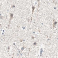


Comprehensive Human Protein Atlas IHC scoring per tissue (reliability: Uncertain — Medium consistency between antibody staining and RNA expression data.). Rows are taken directly from the HPA tissue chart — click any row's HPA link to view the source.
| Tissue | Cell type | Level | Evidence | Source |
|---|---|---|---|---|
| Parathyroid gland | Glandular cells | High | Protein (IHC) | HPA → |
| Testis | Leydig cells | High | Protein (IHC) | HPA → |
| Tonsil | Squamous epithelial cells | High | Protein (IHC) | HPA → |
| Appendix | Glandular cells | Medium | Protein (IHC) | HPA → |
| Bronchus | Respiratory epithelial cells | Medium | Protein (IHC) | HPA → |
| Tissue | Cell type | Level | Evidence | Source |
|---|---|---|---|---|
| Adrenal gland | Glandular cells | Not detected | Protein (IHC) | HPA → |
| Heart muscle | Cardiomyocytes | Not detected | Protein (IHC) | HPA → |
| Skeletal muscle | Myocytes | Not detected | Protein (IHC) | HPA → |
| Smooth muscle | Smooth muscle cells | Not detected | Protein (IHC) | HPA → |
Troubleshoot RAD54B chromogenic staining in paraffin sections by comparing matched controls, compartment patterns and tissue morphology.
A06804 has an IHC image from paraffin-embedded human breast carcinoma (catalog image caption). IF is listed as an application, and human and mouse are listed as reactive species (catalog).
A06804 will render with an IHC image of paraffin-embedded human breast carcinoma and a peptide-blocked comparison (catalog image caption). Its listed applications include IHC and IF, and its listed reactivity covers human and mouse; no IF image is supplied (catalog).
Which to pick: For tissue IHC, choose A06804: its own image caption documents paraffin-embedded human breast carcinoma, but does not report the fixative (catalog image caption). For IF/ICC, A06804 lists IF at 1:50, while ICC validation and an IF image are unreported (catalog: applications, IF dilution, image alts). For cross-species work, A06804 lists human and mouse reactivity; clonality is unreported, and the supplied IHC image documents only human tissue (catalog: reactivity, clone, image caption).