RANBP2 / E3 SUMO-protein ligase RanBP2 · IHC design guide

Design Immunohistochemistry for RANBP2

Plan paraffin-section RANBP2 IHC around nuclear membranous staining, using appendix glandular cells as a positive reference (HPA tissue IHC). The catalog antibody has an IHC range of 2–5 μg/ml (datasheet A00981-1); interpret staining with the reported potential to detect proteins from more than one gene in mind (HPA tissue IHC).

Evidence assembled Oct 2026 · For research use; verify linked source records and product datasheet before use
Immunohistochemistry protocol sheet for RANBP2 (IHC for RANBP2): expected localisation Nuclear membrane in tissue (HPA tissue IHC), antibody A00981-1, validated IHC image, and IHC protocol steps
Printable RANBP2 IHC protocol sheet — expected localisation Nuclear membrane in tissue (HPA tissue IHC), antibody A00981-1, controls and protocol steps. Open the full RANBP2 IHC guide →

RANBP2 Immunohistochemistry Experimental Design Guide

Expected localisation, validated protocols, controls and antibodies — the at-a-glance facts below, then the full design guide.

Must know before staining
Expected localisation Nuclear membrane in tissue (HPA tissue IHC)
Staining pattern Widespread nuclear membranous staining (HPA tissue IHC)
Antigen retrieval EDTA pH 8.0 HIER, heat-mediated (datasheet A00981-1)
Positive control ⓘ Adrenal gland+4 more · see all
Negative control ⓘ None in HPA (detected in all 45 tissues); use no-primary + isotype controls
Important caveats
Reasons your staining may differ from the expected pattern.
Fixation Keep fixation consistent across sections (standard IHC practice; not target-specific). Selected-image fixative and duration unreported (datasheet A00981-1); verify before use.
Caveat Staining may include proteins from another gene (HPA tissue IHC)
Regulation No expression regulator annotated (UniProt)
Isoform / epitope No isoforms or cleavage annotated; one 1–3224 chain (UniProt)
Section 1

Recommended RANBP2 IHC & IF Protocols

The catalog antibody’s IHC-P protocol is accompanied by published RANBP2 staining protocols for paraffin sections (PMC8017305) and synovial sarcoma and ganglioneuroma tissues (PMC9158115).

Recommended immunohistochemistry (IHC-P) protocol parameters
SampleParaffin-embedded human appendix adenocarcinoma tissue; fixative not specified (datasheet A00981-1)
FixationImage fixative and duration unreported (datasheet A00981-1); verify before use.
Sectioning4–5 µm sections on charged slides (standard)
DeparaffinisationXylene, graded ethanol series to water (standard)
Antigen retrievalHeat retrieval: EDTA pH 8.0 (datasheet A00981-1); 20 min, 95–100 °C (standard)
Peroxidase block3% H2O2, 10 min, room temperature (standard)
Blocking10% goat serum (datasheet A00981-1)
Primary antibodyRabbit anti-RANBP2, 2-5 μg/ml (datasheet A00981-1)
Primary incubationOvernight at 4 °C (datasheet A00981-1)
DetectionHRP-conjugated secondary, DAB chromogen (datasheet A00981-1)
CounterstainHematoxylin, blue, dehydrate and mount (standard)
Expected resultRANBP2-positive staining in glandular cells of adrenal gland (HPA tissue IHC: High). HPA tissue profile: Ubiquitous nuclear membranous expression. No signal in the no-primary control.
💡Decision noteStart with EDTA pH 8.0 heat retrieval (datasheet A00981-1); citrate pH 6.0 is a published comparison condition (PMC9158115).
Section 2

What Is the Expected RANBP2 Staining Pattern?

RANBP2 is a nuclear pore protein on the nuclear envelope, with cytoplasmic filaments and reported intranuclear foci (UniProt P49792). In paraffin sections, expect broadly distributed nuclear membranous staining, including strong staining in several epithelial and neuronal cell types (HPA tissue IHC). HPA rates the tissue profile Supported, but reports medium agreement with RNA data and cautions that the antibody may detect proteins from more than one gene (HPA tissue IHC).

What am I looking at on my slide?
A defined rim outlines nuclei in glandular or neuronal cells.This fits RANBP2 at the nuclear pore and nuclear envelope (UniProt P49792). HPA reports High staining in colon glandular cells, cerebellar Purkinje cells, and cerebral cortex neuronal cells (HPA tissue IHC). Score the rim in identifiable cells rather than treating all brown signal as target staining (general IHC practice).
Nuclei stain uniformly, or cytoplasm dominates while the nuclear rim is absent.Reassess specificity and localization: the expected tissue pattern is nuclear membranous (HPA tissue IHC). Intranuclear foci are reported, and HPA lists nucleoplasm as an additional supported IF location, so a non-rim component alone is inconclusive (UniProt P49792; HPA subcellular IF). HPA considers cytosol uncertain in IF (HPA subcellular IF).
Only an unexpected cell population stains strongly.Compare its compartment with nearby cells. HPA describes ubiquitous nuclear membranous expression, though intensity varies by cell type (HPA tissue IHC). A discrepant cell-restricted pattern can reflect nonspecific antibody binding or endogenous detection activity (general IHC practice); the HPA antibody-specificity caution strengthens the need for controls (HPA tissue IHC).
Color spreads across tissue without clear nuclear boundaries.Diffuse background cannot establish RANBP2 localization. Excess primary or detection reagent, inadequate blocking, or incomplete washing can obscure a specific rim (general IHC practice). Compare a no-primary control and examine whether any nuclear membranous pattern remains (general IHC practice; HPA tissue IHC).
A known high-staining tissue shows no nuclear rim.Absence in a positive reference raises concern about section processing, retrieval, antibody performance, or detection (general IHC practice). HPA reports High staining in adrenal glandular cells and bronchial respiratory epithelial cells (HPA tissue IHC). Their observed profile is a reference, not a guarantee for every antibody or preparation (HPA tissue IHC).
💡Expected RANBP2 appearanceCall a result positive when identifiable cells show a distinct nuclear membranous rim, with High staining possible in HPA-listed epithelial and neuronal cells; diffuse, unstructured color or dominant cytoplasmic staining without a rim is suspect (HPA tissue IHC; UniProt P49792; general IHC practice).
How each factor affects the staining
Compartment and topologyRANBP2 belongs to the nuclear pore complex and has no transmembrane segment (UniProt P49792). Interpret a nuclear envelope rim as a localization pattern, not evidence that the protein spans a membrane (UniProt P49792).
Reference tissue and cell typeHPA reports High staining in appendix and duodenal glandular cells, among others, but Low staining in liver cholangiocytes, soft-tissue fibroblasts, and splenic red-pulp cells (HPA tissue IHC). Select and score controls by the named cell type (general IHC practice).
Specificity of the tissue profileThe tissue IHC profile is Supported, with medium staining–RNA consistency, a multi-gene targeting caution, and external verification pending (HPA tissue IHC). Treat matching morphology as supportive rather than definitive identification of RANBP2 (HPA tissue IHC).
IF/ICC Q: What pattern should appear?A: Mainly nuclear membrane; supported additional nucleoplasmic localization is reported. Vesicular and cytosolic locations are uncertain (HPA subcellular IF). IF/ICC is assessed separately from this paraffin-section IHC guide.
Why is my staining missing, weak or wrong?
SituationLikely causeNext action
No rim in a high-staining reference.The staining run or chosen reference may have failed; HPA's High designation describes observed cell types, not guaranteed signal in every run (HPA tissue IHC; general IHC practice).Confirm the named cells are present, then review retrieval, primary antibody, detection reagents, and a concurrently stained positive control (general IHC practice).
Weak rim with otherwise clean tissue.Signal may be near the detection threshold, or the sampled cell type may have lower observed staining; HPA records Low staining in some cell types (HPA tissue IHC; general IHC practice).Compare the same cell type in a HPA-listed high-staining reference and optimize the antibody and detection conditions within the assay's validated range (HPA tissue IHC; general IHC practice).
Strong uniform nuclear staining masks the rim.Excess background or a non-rim antibody signal may obscure the expected nuclear membranous pattern; some intranuclear localization is reported (HPA tissue IHC; UniProt P49792; general IHC practice).Inspect nuclear edges at higher magnification, compare a no-primary control, and review antibody concentration and washing before calling the pattern discordant (general IHC practice).
Brown signal is strongest in unexpected cells.Nonspecific binding or endogenous chromogenic detection activity is possible (general IHC practice). HPA also cautions that its tissue antibody targets protein from more than one gene (HPA tissue IHC).Check whether those cells show a nuclear rim, use no-primary and detection controls, and compare an independently validated antibody when available (general IHC practice).
Diffuse color covers tissue and obscures cell borders.Background from blocking, reagent concentration, washing, or chromogen development can prevent compartment-level scoring (general IHC practice).Review the no-primary control, blocking and wash steps, reagent concentrations, and chromogen development; rescore only where nuclei and rims remain distinguishable (general IHC practice).
Cytoplasmic signal appears without a nuclear rim.The result does not match HPA's dominant tissue pattern, although UniProt reports cytoplasmic filaments and HPA labels cytosolic IF localization uncertain (HPA tissue IHC; UniProt P49792; HPA subcellular IF).Avoid calling the cytoplasmic signal alone a positive IHC result. Check controls and seek a reproducible nuclear membranous pattern in a HPA-listed high-staining cell type (HPA tissue IHC; general IHC practice).

Sample controls for RANBP2 IHC & IF

🧪Run appendix first and assess its glandular cells, which have High RANBP2 staining (HPA: appendix glandular cells, High). HPA detects RANBP2 in all 45 scored tissues, so no negative tissue is available; use no-primary and isotype controls, and expect any unstained neighboring cells to show background only rather than treating them as validated biological negatives (HPA: no negative tissue rows).
Positive control tissue: Adrenal gland (Glandular cells, HPA High)
Negative control tissue: None in HPA: RANBP2 is detected in all 45 scored tissues. Use a no-primary (secondary-only) and an isotype control instead.
ICC-IF cell lines (HPA subcellular resource): HPA ICC-IF images show RANBP2 in A-431, U-251MG, U2OS, HAP1, K-562, CACO-2, Rh30, RT-4, with annotated localisation: Nuclear membrane (supported) (HPA subcellular).
Technical controls: Include a no-primary, secondary-only slide, a concentration-matched rabbit IgG isotype control, and RANBP2-knockout material as a biological negative (caption: rabbit primary antibody; standard IHC controls). Quench endogenous peroxidase before HRP–DAB detection, particularly where appendix sections contain inflammatory cells (caption: HRP–DAB detection; standard IHC practice).
⚠️Feasibility: The selected A00981-1 paraffin-section caption does not report a fixative, and no target-specific fixation window or fixation effect is supplied. Its EDTA retrieval at pH 8.0 demonstrates a workable retrieval condition, but does not establish that retrieval is required (A00981-1 caption). Paraffin IHC has a reported example, while the supplied ICC–IF evidence supports nuclear-membrane localization without showing that frozen sections or IF are easier; endogenous peroxidase in inflammatory cells can complicate appendix DAB interpretation (A00981-1 caption; HPA: nuclear membrane supported; standard IHC practice).

HPA tissue IHC evidence for RANBP2

Comprehensive Human Protein Atlas IHC scoring per tissue (reliability: Supported — Medium consistency between antibody staining and RNA expression data. Caution, targets protein from more than one gene. Pending external verification.). Rows are taken directly from the HPA tissue chart — click any row's HPA link to view the source.

Positive expression · recommended positive controls

TissueCell typeLevelEvidenceSource
Adrenal gland Glandular cells High Protein (IHC) HPA →
Appendix Glandular cells High Protein (IHC) HPA →
Bronchus Respiratory epithelial cells High Protein (IHC) HPA →
Cerebellum Purkinje cells High Protein (IHC) HPA →
Cerebral cortex Neuronal cells High Protein (IHC) HPA →

Undetected expression · recommended negative controls

TissueCell typeLevelEvidenceSource
None in HPA: RANBP2 is detected in all 45 scored tissues. Use a no-primary (secondary-only) and an isotype control instead.
Section 3

Advanced RANBP2 IHC Tips

Troubleshoot RANBP2 chromogenic IHC by checking nuclear envelope staining, processing controls, and the limits of the available antibody evidence.

How should I retrieve RANBP2 in paraffin sections with weak staining?
Start with heat-mediated retrieval in EDTA at pH 8.0 for the catalog antibody (datasheet A00981-1). The pictured paraffin section used that retrieval before incubation with 2 μg/ml primary antibody overnight at 4°C (datasheet A00981-1). If staining remains weak, compare a modestly longer heating interval on matched sections while keeping cooling, detection, and section thickness consistent (standard IHC practice). Score recovery at the nuclear rim, since RANBP2 is associated with nuclear pores and the nuclear envelope (UniProt P49792; HPA subcellular). Include an identically processed no-primary control to distinguish recovered signal from retrieval-related background (standard IHC practice).
Could fixation explain weak or uneven RANBP2 staining?
Target-specific fixation sensitivity is unknown: the selected paraffin-section caption does not state a fixative (datasheet A00981-1). Do not infer a preferred fixation condition from the nuclear envelope location or phosphorylation annotations (UniProt P49792). Compare sections with documented fixation histories using the same EDTA pH 8.0 retrieval and antibody concentration before attributing a signal difference to fixation (datasheet A00981-1; standard IHC practice). Inspect morphology and section edges alongside staining, because uneven processing can alter antigen accessibility across a section (standard IHC practice). Record fixation duration and processing conditions for each specimen so that an apparent biological difference can be checked against handling (standard IHC practice).
Where should convincing RANBP2 staining appear in chromogenic IHC?
Prioritize a nuclear rim pattern: RANBP2 is a nuclear pore component, and tissue IHC reports ubiquitous nuclear membranous expression (UniProt P49792; HPA tissue). Some intranuclear signal is plausible because discrete and diffuse intranuclear foci are reported, while HPA also supports nucleoplasmic localization (UniProt P49792; HPA subcellular). Assess the rim against a nuclear counterstain and adjacent unstained structures before calling diffuse cytoplasmic DAB positive (standard IHC practice). Cytoplasmic filaments are annotated, but broad cytoplasmic staining alone is less persuasive than the expected envelope pattern (UniProt P49792; HPA subcellular). Compare cells within the same section under identical development conditions to judge whether the pattern is reproducible (standard IHC practice).
Could epitope position change the staining pattern I expect?
The supplied record lists 0 isoforms and one 1–3224 chain, so it provides no isoform-specific staining prediction (UniProt P49792). RANBP2 has no annotated transmembrane segment, but its association with nuclear pores and cytoplasmic filaments still makes subcellular access relevant (UniProt P49792). Its annotated Ran-binding and cyclophilin-type regions, together with numerous modified residues, do not identify the catalog antibody's epitope (UniProt P49792; datasheet A00981-1). If staining changes with retrieval, document the pattern and compare matched sections before assigning the change to epitope masking (standard IHC practice). Do not interpret a compartment shift as an isoform switch without independent epitope information (UniProt P49792; standard IHC practice).
How can IF help assess an ambiguous IHC pattern?
Use IF as a separate localization check, since HPA supports nuclear membrane staining and also reports supported nucleoplasmic localization (HPA subcellular). Multiplex RANBP2 with a marker for the expected cell type, such as a glandular epithelial marker when examining appendix glandular cells (HPA tissue; standard IF practice). Choose spectrally separated fluorophores and favor a longer-wavelength channel for the weaker signal when tissue autofluorescence obscures shorter wavelengths (standard IF practice). Optimize permeabilisation for antibody access to the relevant side of the nuclear envelope; RANBP2 is associated with cytoplasmic pore filaments, but the catalog epitope's orientation is unspecified (UniProt P49792; datasheet A00981-1). Compare rim and nucleoplasmic signal with the nuclear counterstain and single-channel controls (standard IF practice).
How do I reduce diffuse brown staining without losing the nuclear rim?
Run a no-primary section and check whether brown deposit persists at the nuclear rim or appears throughout the tissue (standard IHC practice). The pictured workflow used 10% goat serum blocking, a peroxidase-conjugated secondary antibody, and DAB development (datasheet A00981-1). Include a peroxidase-blocking step before HRP detection and control DAB development time; these are general chromogenic IHC measures, not evidence of a RANBP2-specific problem (standard IHC practice). If background rises with signal, titrate the primary around the documented 2 μg/ml condition using matched sections (datasheet A00981-1; standard IHC practice). Judge improvements by preserved nuclear envelope contrast and clean no-primary controls (HPA subcellular; standard IHC practice).
What should I score when comparing RANBP2 IHC sections? ⚠ ANSWER MARKED FOR VERIFICATION
Define positivity at the nuclear envelope before scoring, because HPA describes a ubiquitous nuclear membranous tissue pattern (HPA tissue). Report the percentage of eligible cells with rim staining and, if intensity is reproducible, an H-score derived from the same predefined cell population (standard IHC practice). For spatial comparisons, report rim-positive cell density per mm² of viable tissue and normalize to the number of evaluable nuclei or viable area (standard IHC practice). Keep retrieval, the documented 2 μg/ml primary condition, imaging, and DAB development consistent across compared sections (datasheet A00981-1; standard IHC practice). Record excluded necrotic or damaged areas so they cannot drive an apparent difference (standard IHC practice).
When should I doubt an apparent RANBP2-positive result?
Treat a crisp nuclear envelope pattern as more convincing than diffuse staining in an unrelated compartment, given RANBP2's nuclear pore association (UniProt P49792; HPA subcellular). Check cell identity: HPA reports high staining in appendix glandular cells, but its tissue assessment is only Supported and cautions that its antibody targets protein from more than one gene (HPA tissue). Discount signal restricted to section edges or necrotic areas until it reproduces in intact interior tissue (standard IHC practice). Brown deposit in a no-primary section can indicate detection background, including endogenous enzyme activity in an HRP workflow (standard IHC practice). Require consistent morphology, compartment, and control results before interpreting staining as RANBP2-specific (standard IHC practice).
Boster reagents

Best RANBP2 / E3 SUMO-protein ligase RanBP2 IHC Antibodies

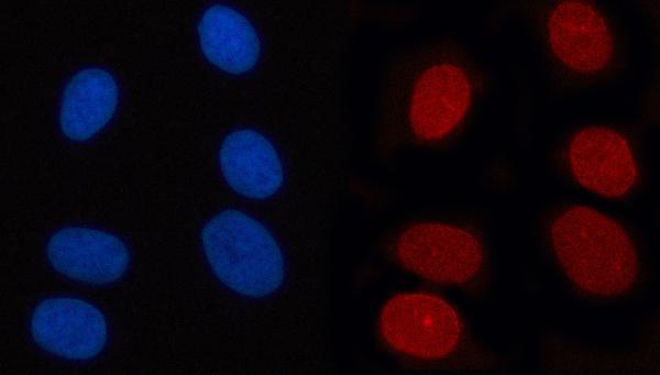
The catalog antibody has IHC data from human paraffin sections and IF/ICC data from U2OS cells (catalog: A00981-1 image captions); human is the only listed reactive species (catalog: A00981-1 reactivity).

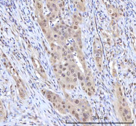
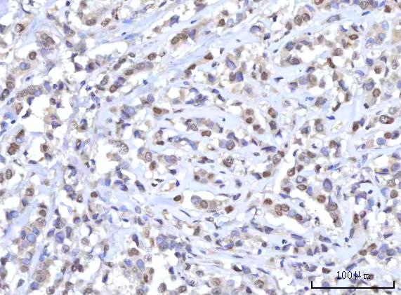
Real IHC data IHC analysis of RANBP2 using anti-RANBP2 antibody (A00981-1). RANBP2 was detected in a paraffin-embedded section of human appendix adenocarcinoma tissue. Heat mediated antigen retrieval was performed in EDTA buffer (pH 8.0, epitope retrieval solution). The tissue section was blocked with 10% goat serum. The tissue section was then incubated with 2 μg/ml rabbit anti-RANBP2 Antibody (A00981-1) overnight at 4°C. Peroxidase Conjugated Goat Anti-rabbit IgG was used as secondary antibody and incubated for 30 minutes at 37°C. The tissue section was developed using HRP Conjugated Rabbit IgG Super Vision Assay Kit (Catalog # SV0002) with DAB as the chromogen.
Anti-RANBP2 Antibody ®
Cat # A00981-1

A00981-1 will render with an IHC figure from a human appendix adenocarcinoma paraffin section (catalog: A00981-1 IHC image caption). The same SKU is listed for IF/ICC and has a U2OS cell IF image (catalog: A00981-1 applications and IF image caption).

Which to pick: Choose A00981-1 for human paraffin-section IHC: its own image caption documents that preparation, while the fixative is unreported (catalog: A00981-1 IHC image caption). For IF/ICC, the same rabbit antibody is listed for both applications and has U2OS cell IF data (catalog: A00981-1 host, applications and IF image caption). No cross-species choice is supported because A00981-1 lists human reactivity only (catalog: A00981-1 reactivity).

Each figure is that product's own IHC / IF validation image from its datasheet.

References

  1. UniProt Consortium. UniProt entry P49792 (RBP2_HUMAN, E3 SUMO-protein ligase RanBP2).
  2. Human Protein Atlas. RANBP2 tissue IHC expression (reliability: Supported).
  3. Human Protein Atlas. RANBP2 subcellular location (ICC-IF): Mainly localized to the nuclear membrane. In addition localized to the nucleoplasm, vesicles and cytosol..
  4. Human Protein Atlas. RANBP2 antibody validation summary (7 antibodies).
  5. YTHDF1 Aggravates the Progression of Cervical Cancer Through m(6)A-Mediated Up-Regulation of RANBP2. Frontiers in oncology 2021 — PMC8017305.
  6. Systems-Level Mapping of Cancer Testis Antigen 1b/a to Sarcoma Pathways Identifies Activated Ran Binding-2 E3 SUMO-Protein Ligase and Transducin-Like Enhancer Protein 1. Frontiers in genetics 2022 — PMC9158115.
  7. RANBP2 Activates O-GlcNAcylation through Inducing CEBPα-Dependent OGA Downregulation to Promote Hepatocellular Carcinoma Malignant Phenotypes. Cancers 2021 — PMC8304650.
  8. Nuclear pore pathology underlying multisystem proteinopathy type 3-related inclusion body myopathy. Annals of clinical and translational neurology 2024 — PMC10963302.
  9. PubMed PMID:7775481 — UniProt-cited evidence.
  10. PubMed PMID:7603572 — UniProt-cited evidence.
  11. PubMed PMID:15815621 — UniProt-cited evidence.