RANGAP1 / Ran GTPase-activating protein 1 · IHC design guide

Design Immunohistochemistry for RANGAP1

Plan chromogenic RANGAP1 IHC in paraffin sections with the catalog antibody at 2–5 μg/ml (datasheet A02771-2). Assess nuclear, nuclear membrane and cytoplasmic staining (HPA tissue IHC), allowing for mitotic redistribution to spindles and kinetochores (UniProt).

Evidence assembled Oct 2026 · For research use; verify linked source records and product datasheet before use
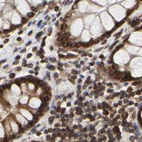
Immunohistochemistry protocol sheet for RANGAP1 (IHC for RANGAP1): expected localisation Nuclear, nuclear membrane and cytoplasmic staining (HPA tissue IHC), antibody A02771-2, validated IHC image, and IHC protocol steps
Printable RANGAP1 IHC protocol sheet — expected localisation Nuclear, nuclear membrane and cytoplasmic staining (HPA tissue IHC), antibody A02771-2, controls and protocol steps. Open the full RANGAP1 IHC guide →

RANGAP1 Immunohistochemistry Experimental Design Guide

Expected localisation, validated protocols, controls and antibodies — the at-a-glance facts below, then the full design guide.

Must know before staining
Expected localisation Nuclear, nuclear membrane and cytoplasmic staining (HPA tissue IHC)
Staining pattern All cells: nuclear, nuclear membrane and cytoplasmic (HPA tissue IHC)
Antigen retrieval EDTA pH 8.0 HIER, heat-mediated (datasheet A02771-2)
Positive control ⓘ Adrenal gland+4 more · see all
Negative control ⓘ None in HPA (detected in all 45 tissues); use no-primary + isotype controls
Important caveats
Reasons your staining may differ from the expected pattern.
Fixation Keep fixation consistent across sections (standard IHC practice; not target-specific); Matched tissue-IHC evidence does not establish the fixation claim. Validate the specimen-specific method before use. (selected-SKU IHC image A02771-2)
Caveat Mitotic cells may show spindle and kinetochore staining (UniProt)
Regulation Testis-enhanced RNA expression (HPA tissue RNA)
Isoform / epitope 0 annotated isoforms; no signal peptide (UniProt)
Section 1

Recommended RANGAP1 IHC & IF Protocols

The catalog antibody has an IHC-P protocol (datasheet A02771-2). These published RANGAP1 IHC protocols provide conditions for lymphoma, spinal cord, and brain tissue (PMC3819250; PMC5427168; PMC5595097).

Recommended immunohistochemistry (IHC-P) protocol parameters
SampleParaffin-embedded human colorectal cancer tissue; fixative not specified (datasheet A02771-2)
FixationImage fixative and duration unreported (datasheet A02771-2); verify before use.
Sectioning4–5 µm sections on charged slides (standard)
DeparaffinisationXylene, graded ethanol series to water (standard)
Antigen retrievalHeat retrieval: EDTA pH 8.0 (datasheet A02771-2); 20 min, 95–100 °C (standard)
Peroxidase block3% H2O2, 10 min, room temperature (standard)
Blocking10% goat serum (datasheet A02771-2)
Primary antibodyRabbit anti-RANGAP1, 2-5 μg/ml (datasheet A02771-2)
Primary incubationOvernight at 4 °C (datasheet A02771-2)
DetectionHRP-conjugated secondary, DAB chromogen (datasheet A02771-2)
CounterstainHematoxylin, blue, dehydrate and mount (standard)
Expected resultRANGAP1-positive staining in glandular cells of adrenal gland (HPA tissue IHC: High). HPA tissue profile: Distinct expression in the nuclei, nuclear membrane and cytoplasm in all cells. No signal in the no-primary control.
💡Decision noteStart with heat-mediated EDTA pH 8.0 retrieval for the catalog antibody (datasheet A02771-2); adjust retrieval using the published conditions for the relevant tissue (PMC3819250; PMC5427168; PMC5595097).
Section 2

What Is the Expected RANGAP1 Staining Pattern?

RANGAP1 staining should appear in the nucleus, nuclear membrane and cytoplasm across many cell types (HPA: tissue IHC profile; reliability Supported). A nuclear rim is consistent with its nuclear envelope location; interphase cytoplasmic staining is also expected (UniProt P46060: subcellular location). RANGAP1 has no transmembrane segment, so a membrane-like rim here refers to the nuclear envelope, not the cell surface (UniProt P46060: topology).

What am I looking at on my slide?
Nuclear rim with nuclear and cytoplasmic staining in glandular or neuronal cells.This fits the reported tissue pattern (HPA: tissue IHC profile). High staining is documented in colon glandular cells and cerebral cortex neuronal cells (HPA: High). Judge the rim in relation to the counterstained nucleus; cytoplasmic staining alone does not rule out RANGAP1 (UniProt P46060: interphase location).
Strong cell-surface outlines without a nuclear rim or convincing intracellular staining.Treat this as a compartment mismatch requiring review, since the reported membrane location is the nuclear envelope and RANGAP1 lacks a transmembrane segment (UniProt P46060: subcellular location; topology). Compare with a known-positive tissue and the detection controls before interpreting it as specific staining (general IHC practice).
The strongest signal is in adipocytes, while expected positive cells stain weakly.This distribution warrants a specificity check: adipocytes are listed as low, whereas several glandular, neuronal and hematopoietic cell types are high (HPA: tissue IHC levels). It does not by itself prove cross-reactivity; endogenous detection activity can also mislead chromogenic interpretation (general IHC practice).
Diffuse color covers cells and spaces between them, obscuring nuclear borders.A nuclear-rim call is unreliable when background masks the compartment pattern (HPA: tissue IHC profile). Review blocking, antibody concentration, washes and chromogen development, then compare a control without primary antibody for detection-related background (general IHC practice).
No signal appears in a tissue and cell type listed as High.A negative result in that setting needs a technical check before biological interpretation (HPA: High in named cell types). Confirm that the expected cells are present and assess retrieval, antibody dilution and detection controls (general IHC practice). HPA reports no negative cell type here, so absence alone is not a reference pattern (HPA: tissue IHC record).
💡Expected RANGAP1 appearanceCall a convincing positive when identifiable cells show nuclear-rim staining with nuclear and/or cytoplasmic signal, especially in an HPA High cell type; isolated cell-surface outlines or diffuse deposits are suspect (HPA: tissue IHC profile and levels; UniProt P46060: location and topology; general IHC practice).
How each factor affects the staining
Cell-cycle locationDuring interphase, RANGAP1 is cytoplasmic and occurs at the nuclear envelope; during mitosis it associates with spindles and kinetochores, with little detection at spindle poles (UniProt P46060: subcellular location). Score a mitotic figure in context rather than requiring its pattern to match an interphase nuclear rim (general IHC practice).
Cell type and tissueHPA reports distinct nuclear, nuclear-membrane and cytoplasmic expression across cells, with High levels in the listed glandular, neuronal and hematopoietic cells and Low levels in adipocytes (HPA: tissue IHC profile and levels). These are reference observations, not a guarantee that every section or cell stains equally.
IHC antibody evidenceThe HPA tissue pattern has Supported reliability, described as consistency between staining and RNA expression (HPA: tissue IHC reliability). Two listed antibodies have Supported IHC status, HPA050110 and CAB004293; HPA062034 has no listed IHC status (HPA: antibody validation). Match interpretation to the antibody actually used.
SUMO-dependent targetingSumoylation targets RANGAP1 to nuclear pores and is required for its reported mitotic location (UniProt P46060: subcellular location). A weak rim in one preparation does not establish altered sumoylation: this stain alone does not measure the modification (general IHC interpretation).
Retrieval and epitope informationNo antibody epitope or RANGAP1-specific fixation sensitivity is supplied (UniProt P46060 and HPA: supplied records). Antigen retrieval is a general paraffin-IHC variable; evaluate it with the chosen antibody's controls, without assigning a target-specific fixation effect (general IHC practice).
Why is my staining missing, weak or wrong?
SituationLikely causeNext action
The positive-control section is blank.The supplied records identify High-staining cells but do not establish the cause of a failed run (HPA: tissue IHC levels).Check that the named cells are present, then review retrieval, antibody dilution, detection reagents and the run control (general IHC practice). Avoid calling the test section biologically negative until the control works.
Nuclear rim and cytoplasm are both faint.Weak staining may prevent reliable compartment assessment; the expected pattern includes both locations (HPA: tissue IHC profile; UniProt P46060: interphase location).Compare an HPA High cell type on the same run, review the counterstain and optimize the chosen IHC workflow using controls (HPA: tissue IHC levels; general IHC practice).
There is diffuse brown background or staining without primary antibody.Detection-system background, including endogenous enzyme activity, can produce misleading chromogenic signal (general IHC practice).Review the no-primary control, endogenous-activity blocking, reagent handling, washes and chromogen development (general IHC practice). Reassess localization only after nuclear borders are readable.
Cell surfaces stain more strongly than nuclei or cytoplasm.A cell-surface outline conflicts with the supplied nuclear-envelope and cytoplasmic locations and the lack of a transmembrane segment (UniProt P46060: location; topology).Check section morphology, controls and antibody specificity; require a plausible intracellular pattern in known-positive cells before scoring the outline as RANGAP1 (HPA: tissue IHC profile; general IHC practice).
Adipocytes dominate the signal in a mixed tissue.Adipocytes are listed as Low, so a strong adipocyte-only pattern is discordant with the supplied tissue reference (HPA: Low in adipocytes).Compare neighboring identifiable cells and an HPA High control; inspect no-primary staining for endogenous activity before considering cross-reactivity (HPA: tissue IHC levels; general IHC practice).
Q: Should IF/ICC show the same pattern as chromogenic tissue IHC?IF/ICC images mainly localize RANGAP1 to the nuclear membrane, with additional vesicle, cytosol and flagellar-centriole locations (HPA: subcellular ICC-IF).A: Use the nuclear membrane as the shared reference, while interpreting additional IF compartments against the IF/ICC evidence and its own guide (HPA: subcellular ICC-IF; tissue IHC profile).

Sample controls for RANGAP1 IHC & IF

🧪Run bone marrow first; hematopoietic cells should stain (HPA: High in bone marrow hematopoietic cells). HPA detects RANGAP1 in all 45 scored tissues, so there is no validated negative tissue or internal negative cell population; use no-primary and isotype controls to define background, and interpret cells without specific staining cautiously (HPA: no negative tissue rows; RANGAP1 detected in all 45 scored tissues).
Positive control tissue: Adrenal gland (Glandular cells, HPA High)
Negative control tissue: None in HPA: RANGAP1 is detected in all 45 scored tissues. Use a no-primary (secondary-only) and an isotype control instead.
ICC-IF cell lines (HPA subcellular resource): HPA ICC-IF images show RANGAP1 in A-431, U-251MG, U2OS, HEK293, MCF-7, RPTEC/TERT1, hTERT-RPE1 (serum starved), Sperm, KOLF2.1J, NIH 3T3, with annotated localisation: Nuclear membrane (supported) (HPA subcellular).
Technical controls: Include a no-primary, secondary-only slide and a concentration-matched rabbit IgG isotype control, alongside a matched RANGAP1 knockout specimen if available (caption: rabbit primary and anti-rabbit secondary; standard IHC control practice). Quench endogenous peroxidase and check the no-primary slide for residual marrow signal before interpreting DAB staining (HPA: bone marrow hematopoietic cells High; standard chromogenic IHC practice).
⚠️Feasibility: A target-specific fixation window or fixation effect is unreported, and the selected A02771-2 tissue-IHC caption does not state its fixative (selected tissue-IHC caption). The caption reports heat retrieval in EDTA at pH 8.0 for a paraffin section; this is a starting condition, not evidence that retrieval is required for bone marrow (selected tissue-IHC caption). The supplied evidence does not establish whether frozen sections or IF are easier; in bone marrow, endogenous peroxidase can complicate chromogenic interpretation (HPA: bone marrow hematopoietic cells High; standard IHC practice).

HPA tissue IHC evidence for RANGAP1

Comprehensive Human Protein Atlas IHC scoring per tissue (reliability: Supported — High consistency between antibody staining and RNA expression data.). Rows are taken directly from the HPA tissue chart — click any row's HPA link to view the source.

Positive expression · recommended positive controls

TissueCell typeLevelEvidenceSource
Adrenal gland Glandular cells High Protein (IHC) HPA →
Bone marrow Hematopoietic cells High Protein (IHC) HPA →
Cerebellum Purkinje cells High Protein (IHC) HPA →
Cerebral cortex Neuronal cells High Protein (IHC) HPA →
Cervix Glandular cells High Protein (IHC) HPA →

Undetected expression · recommended negative controls

TissueCell typeLevelEvidenceSource
None in HPA: RANGAP1 is detected in all 45 scored tissues. Use a no-primary (secondary-only) and an isotype control instead.
Section 3

Advanced RANGAP1 IHC Tips

Use the catalog antibody’s paraffin-section result as a starting point, then judge staining against RANGAP1’s expected compartments and tissue patterns.

What retrieval should I try first if RANGAP1 staining is weak in paraffin sections?
Start with heat-mediated retrieval in EDTA at pH 8.0 (datasheet A02771-2). The selected paraffin-section image used that retrieval before staining with 2 μg/ml primary antibody overnight at 4°C (caption A02771-2). If signal is weak, compare a modest range of heating times on adjacent sections while keeping antibody concentration and detection constant (standard IHC practice). Include a section that stained reliably in the same run, so a failed retrieval can be distinguished from a detection failure (standard IHC practice). Check nuclear-envelope and cytoplasmic signal before increasing retrieval intensity, since both are expected locations (UniProt P46060 subcellular; HPA tissue IHC).
Could fixation explain weak or uneven RANGAP1 staining?
The selected image documents a paraffin-embedded section, but does not state its fixative (caption A02771-2). RANGAP1-specific fixation sensitivity is therefore unknown from the supplied evidence; tissue staining patterns and protein features do not establish one (caption A02771-2; HPA tissue IHC; UniProt P46060). Record fixative, fixation duration, section thickness, and processing history before comparing specimens (standard IHC practice). Test specimens with different recorded processing histories in the same staining run, using EDTA at pH 8.0 and a shared detection workflow (datasheet A02771-2; standard IHC practice). Judge changes in both staining intensity and compartment pattern, particularly at the nuclear envelope (UniProt P46060 subcellular; standard IHC practice).
Which RANGAP1 staining compartments should I expect in tissue sections?
Expect nuclear-membrane, nuclear, and cytoplasmic staining across cells in tissue sections (HPA tissue IHC). RANGAP1 is cytoplasmic during interphase and is detected at the nuclear envelope; sumoylation targets it to nuclear pores (UniProt P46060 subcellular). Mitotic cells can show spindle and kinetochore association, with little detection at spindle poles (UniProt P46060 subcellular). Examine intact cells at high magnification before calling a thin perinuclear rim positive, and compare that rim with cytoplasmic and nuclear staining in the same field (standard IHC practice; HPA tissue IHC). Record each compartment separately, because a single whole-cell intensity score can conceal a change in distribution (standard IHC practice).
How should epitope uncertainty affect interpretation of variable RANGAP1 staining?
No alternative isoforms or annotated domains are supplied for RANGAP1, and its record lists no transmembrane segment (UniProt P46060 isoforms, domains, topology). The record does list phosphorylation and SUMO-dependent localisation, so compartment shifts can reflect protein state without establishing a change in total abundance (UniProt P46060 modified residues, subcellular). The selected antibody’s epitope is unspecified in the supplied caption; avoid assigning a particular modification-dependent staining effect to it (caption A02771-2). Compare serial sections stained with the same retrieval, 2 μg/ml primary concentration, and detection conditions before interpreting differences (caption A02771-2; standard IHC practice). If an epitope-dependent claim matters, seek direct epitope or modification validation rather than inferring it from staining alone (standard IHC practice).
How can I assess RANGAP1 by IF alongside a cell-type marker?
Treat IF/ICC as a separate assay requiring its own antibody validation and controls; the selected image documents chromogenic staining of a paraffin section (caption A02771-2; standard IF practice). Multiplex with a validated marker for the cell type under study, and assess RANGAP1’s nuclear-envelope and cytoplasmic distribution within marker-positive cells (HPA subcellular; standard IF practice). Choose spectrally separated fluorophores and inspect an unstained tissue section for autofluorescence before assigning a faint signal (standard IF practice). Because RANGAP1 occurs on the cytoplasmic side of nuclear-pore-associated structures and in cytoplasm, optimise permeabilisation for access to intracellular epitopes (UniProt P46060 subcellular; standard IF practice). Check single-stain controls for bleed-through and compare the compartment pattern with the chromogenic result cautiously (standard IF practice).
What should I adjust when RANGAP1 IHC has diffuse background?
The selected paraffin-section workflow used 10% goat-serum block, 2 μg/ml rabbit primary overnight at 4°C, and peroxidase detection with DAB (caption A02771-2). Run a no-primary control and apply a peroxidase block to distinguish nonspecific detection or endogenous enzyme activity from antibody signal (standard IHC practice). If background persists, titrate primary concentration downward and improve washing while holding retrieval constant at EDTA pH 8.0 (datasheet A02771-2; standard IHC practice). Assess background outside tissue and at cut edges separately from staining in intact cells (standard IHC practice). Do not dismiss all diffuse cellular colour as background: cytoplasmic RANGAP1 is expected, alongside nuclear-envelope staining (UniProt P46060 subcellular; HPA tissue IHC).
How should I score RANGAP1 across sections with different cell mixtures? ⚠ ANSWER MARKED FOR VERIFICATION
Define the cell population and compartment before scoring, because RANGAP1 can appear at the nuclear membrane, in nuclei, and in cytoplasm (HPA tissue IHC). For a defined population, report percent positive cells and an H-score using intensity grades 0–3, giving a possible range of 0–300 (standard IHC practice). If cell abundance itself is the question, report positive-cell density per mm² of viable tissue as well (standard IHC practice). Normalise counts to the eligible cells or viable area examined, and exclude necrosis, folds, and poorly preserved edges by a prespecified rule (standard IHC practice). Keep retrieval, exposure to DAB, and scoring thresholds consistent across the compared sections (standard IHC practice).
How do I distinguish true RANGAP1 signal from staining artefacts?
A credible result should include staining in expected compartments: nuclear membrane, nucleus, or cytoplasm, with mitotic patterns interpreted in their cell-cycle context (HPA tissue IHC; UniProt P46060 subcellular). High staining in colon glandular cells and low staining in adipocytes provide supplied tissue-pattern reference points, though neither alone proves antibody specificity (HPA tissue IHC; standard IHC practice). Be cautious with colour confined to section edges, folds, or necrotic regions rather than intact cells (standard IHC practice). Compare a no-primary control for endogenous peroxidase or detection background, especially when DAB appears widespread (standard IHC practice). Confirm that the localisation survives a consistent EDTA pH 8.0 workflow before interpreting between-section intensity differences biologically (datasheet A02771-2; standard IHC practice).
Boster reagents

Best RANGAP1 / Ran GTPase-activating protein 1 IHC Antibodies

A02771-2 has IHC data from human colorectal cancer paraffin sections and IF data from A431 cells (catalog image captions); the catalog lists human, mouse and rat reactivity (catalog: A02771-2).

Real IHC data IHC analysis of RANGAP1 using anti-RANGAP1 antibody (A02771-2). RANGAP1 was detected in a paraffin-embedded section of human colorectal cancer tissue. Heat mediated antigen retrieval was performed in EDTA buffer (pH 8.0, epitope retrieval solution). The tissue section was blocked with 10% goat serum. The tissue section was then incubated with 2 μg/ml rabbit anti-RANGAP1 Antibody (A02771-2) overnight at 4°C. Peroxidase Conjugated Goat Anti-rabbit IgG was used as secondary antibody and incubated for 30 minutes at 37°C. The tissue section was developed using HRP Conjugated Rabbit IgG Super Vision Assay Kit (Catalog # SV0002) with DAB as the chromogen.
Anti-RANGAP1 Antibody ®
Cat # A02771-2

A02771-2 has IHC data from human colorectal cancer paraffin sections and IF data from A431 cells (catalog image captions). M02771-1 lists IHC and ICC/IF applications and human, mouse and rat reactivity, but has no IHC or IF image caption in the payload (catalog: M02771-1).

Which to pick: Choose A02771-2 for paraffin-section IHC because its own caption documents staining human colorectal cancer tissue; the fixative is unreported (A02771-2 IHC image caption). For IF/ICC, A02771-2 has an A431 cell image, while M02771-1 is a rabbit monoclonal, clone 19R47, with ICC/IF listed but no image caption (A02771-2 IF image caption; catalog: M02771-1). Both list human, mouse and rat reactivity, but the supplied images document human samples only (catalog: A02771-2 and M02771-1; A02771-2 image captions).

Each figure is that product's own IHC / IF validation image from its datasheet.

References

  1. UniProt Consortium. UniProt entry P46060 (RAGP1_HUMAN, Ran GTPase-activating protein 1).
  2. Human Protein Atlas. RANGAP1 tissue IHC expression (reliability: Supported).
  3. Human Protein Atlas. RANGAP1 subcellular location (ICC-IF): Mainly localized to the nuclear membrane. In addition localized to vesicles, cytosol and flagellar centriole..
  4. Human Protein Atlas. RANGAP1 antibody validation summary (3 antibodies).
  5. The Ran GTPase-activating protein (RanGAP1) is critically involved in smooth muscle cell differentiation, proliferation and migration following vascular injury: implications for neointima formation and restenosis. PloS one 2014 — PMC4079658.
  6. Ran GTPase-activating protein 1 is a therapeutic target in diffuse large B-cell lymphoma. PloS one 2013 — PMC3819250.
  7. Mutant TDP-43 within motor neurons drives disease onset but not progression in amyotrophic lateral sclerosis. Acta neuropathologica 2017 — PMC5427168.
  8. Mutant Huntingtin Disrupts the Nuclear Pore Complex. Neuron 2017 — PMC5595097.
  9. PubMed PMID:7878053 — UniProt-cited evidence.
  10. PubMed PMID:11347906 — UniProt-cited evidence.
  11. PubMed PMID:12168954 — UniProt-cited evidence.