This website uses cookies to ensure you get the best experience on our website.


- Table of Contents
Plan RAP1GAP chromogenic IHC in paraffin sections using the catalog antibody’s documented conditions (datasheet A03561-2). Compare staining with the cytoplasmic tissue pattern and cell-level controls reported by HPA (HPA tissue IHC).

Expected localisation, validated protocols, controls and antibodies — the at-a-glance facts below, then the full design guide.
| Expected localisation | Cytoplasmic staining in most tissues (HPA tissue IHC) | |
| Staining pattern | Cytoplasmic signal in most tissues, including brain (HPA tissue IHC) | |
| Antigen retrieval | EDTA pH 8.0 HIER, heat-mediated (datasheet A03561-2) | |
| Positive control | Adrenal gland+4 more · see all | |
| Negative control | Adipose tissue+4 more · see all |
| Fixation | Keep fixation consistent across paraffin sections (standard IHC practice; not target-specific); Matched tissue-IHC evidence does not establish the fixation claim. Validate the specimen-specific method before use. (selected-SKU IHC image A03561-2) | |
| Caveat | Antibody staining and RNA expression have medium consistency (HPA tissue IHC) | |
| Regulation | Cortex expression exceeds spinal cord (UniProt) | |
| Isoform / epitope | Four isoforms; verify antibody epitope coverage (UniProt) |
Compare the catalog antibody’s IHC-P protocol with 4 published RAP1GAP IHC protocols (datasheet: A03561-2; PMC12098257; PMC2822891; PMC5352382; PMC3531979).
| Sample | Paraffin-embedded human gastric adenocarcinoma tissue; fixative not specified (datasheet A03561-2) |
| Fixation | Image fixative and duration unreported (datasheet A03561-2); verify before use. |
| Sectioning | 4–5 µm sections on charged slides (standard) |
| Deparaffinisation | Xylene, graded ethanol series to water (standard) |
| Antigen retrieval | Heat retrieval: EDTA pH 8.0 (datasheet A03561-2); 20 min, 95–100 °C (standard) |
| Peroxidase block | 3% H2O2, 10 min, room temperature (standard) |
| Blocking | 10% goat serum (datasheet A03561-2) |
| Primary antibody | Rabbit anti-RAP1GAP, 2-5 μg/ml (datasheet A03561-2) |
| Primary incubation | Overnight at 4 °C (datasheet A03561-2) |
| Detection | HRP-conjugated secondary, DAB chromogen (datasheet A03561-2) |
| Counterstain | Hematoxylin, blue, dehydrate and mount (standard) |
| Expected result | RAP1GAP-positive staining in glandular cells of adrenal gland (HPA tissue IHC: High). HPA tissue profile: Cytoplasmic expression in most tissues, including thryoid gland and brain. No signal in the no-primary control. |
In paraffin-section IHC, expect RAP1GAP mainly in the cytoplasm, including High staining in cerebral cortex neurons and selected glandular or epithelial cells (HPA tissue IHC). UniProt places RAP1GAP at the Golgi apparatus membrane and reports no transmembrane segment (UniProt P47736). HPA rates its tissue IHC reliability Enhanced, while noting medium consistency between antibody staining and RNA expression (HPA tissue IHC).
| Cerebral cortex neurons show clear cytoplasmic chromogen, with distinguishable cell outlines and limited background. | This fits a strong positive tissue and the reported cytoplasmic pattern (HPA: High in cerebral cortex neuronal cells; HPA tissue IHC profile). Score the neurons separately from surrounding cells; the tissue-level label does not assign every cell the same intensity. |
| Staining appears predominantly nuclear, with little or no cytoplasmic signal in otherwise positive cells. | A nuclear-only result conflicts with the reported IHC profile and approved ICC-IF cytosolic location (HPA tissue IHC; HPA subcellular). Treat it as a localisation concern and review controls, antibody specificity and image interpretation before assigning it to RAP1GAP. |
| Cells reported as undetected stain strongly, such as adipocytes beside other tissue elements. | HPA reports RAP1GAP as Not detected in adipocytes, specifically, rather than in every cell of adipose tissue (HPA: Not detected in adipocytes). Check cell identification and background controls; staining in an unexpected cell population may reflect cross-reactivity or endogenous chromogenic activity (general IHC practice). |
| Color is spread across tissue, including extracellular spaces, and cell boundaries are hard to resolve. | That distribution is less interpretable than cell-associated cytoplasmic staining (HPA tissue IHC profile). Uneven background can arise during blocking, washing or chromogenic detection (general IHC practice); compare a reagent control and review the entire section before scoring cells. |
| A cerebral cortex section has no detectable staining in neurons. | The absence conflicts with the reported High neuronal staining (HPA: cerebral cortex neuronal cells). It does not by itself establish biological absence. Check tissue identification, the IHC-validated antibody, antigen retrieval, primary-antibody application and detection controls (general IHC practice). |
| Tissue and cell selection | HPA reports High staining in cerebral cortex neurons, adrenal gland glandular cells and bronchial respiratory epithelial cells (HPA tissue IHC). It reports Not detected in skeletal-muscle myocytes and adipocytes (HPA tissue IHC). Choose and score the named cell population, since a tissue can contain several cell types. |
| Compartment interpretation | HPA describes cytoplasmic staining across most tissues and approved cytosolic localisation by ICC-IF; UniProt lists the Golgi apparatus membrane and no transmembrane segment (HPA tissue IHC; HPA subcellular; UniProt P47736). These records support a cytoplasmic call, but do not require a visibly Golgi-restricted chromogenic pattern. |
| IHC antibody evidence | HPA marks the tissue IHC assessment Enhanced and lists HPA001922 as IHC Enhanced; its summary notes medium agreement between staining and RNA data (HPA tissue IHC; HPA antibodies). That supports the published pattern while leaving an unexpected result to be checked with appropriate controls. |
| Isoforms and epitope coverage | UniProt lists 4 RAP1GAP isoforms (UniProt P47736). The supplied records give no antibody epitope or isoform coverage, so a difference between specimens cannot be attributed to a particular isoform from these data alone. |
| Processing and modifications | UniProt describes one chain spanning residues 1–663, no signal peptide, no propeptide and no listed glycosylation sites; it lists several phosphoserines (UniProt P47736). These annotations do not establish whether retrieval or fixation changes staining, because no target-specific sensitivity data are supplied. |
| IF/ICC Q: where should signal appear? | A: HPA approves cytosol as the main ICC-IF location and shows images for A-431 and U-251MG; CAB003851 has an Uncertain ICC assessment (HPA subcellular; HPA antibodies). Use the separate IF/ICC guide for that application; these data do not turn its images into an IHC protocol. |
| Situation | Likely cause | Next action |
|---|---|---|
| No signal in cortex neurons on a nominal positive section. | Possible tissue misidentification or an IHC workflow failure; HPA reports High neuronal staining there (HPA: cerebral cortex). | Confirm the neurons and section identity, then review retrieval, primary-antibody application, chromogen and a detection control in sequence (general IHC practice). |
| Only nuclei stain in cells expected to be positive. | The compartment differs from HPA's cytoplasmic IHC profile and approved cytosolic ICC-IF location (HPA tissue IHC; HPA subcellular). | Inspect the counterstain and no-primary control, then repeat with the IHC-validated antibody and score cytoplasmic signal separately (general IHC practice). |
| Adipocytes or skeletal-muscle myocytes stain strongly. | Those named cell populations are reported Not detected (HPA: adipocytes; HPA: skeletal-muscle myocytes). Cell misidentification, cross-reactivity or endogenous detection activity are possibilities (general IHC practice). | Recheck morphology and compare no-primary and detection-only controls; record staining by cell type rather than calling the whole tissue positive (general IHC practice). |
| Diffuse brown deposit obscures cellular localisation. | The slide cannot be compared reliably with HPA's cell-associated cytoplasmic profile (HPA tissue IHC). Blocking, washing or detection background may contribute (general IHC practice). | Review reagent controls and staining uniformity; optimise general blocking, washing and detection conditions before interpreting intensity (general IHC practice). |
| Staining differs between the catalog antibody and an HPA image. | HPA's Enhanced assessment describes its evaluated IHC evidence; the supplied records do not establish epitope equivalence or matching isoform coverage for the catalog antibody (HPA tissue IHC; UniProt P47736). | Verify the antibody's IHC-P validation and compare the same tissue, cell population and compartment before treating intensity differences as biological (general IHC practice). |
| A result changes after a retrieval or fixation adjustment. | Target-specific fixation effects are not established by the supplied assay evidence. Verify with a matched IHC source before attributing a result to fixation. | Keep processing and scoring consistent across compared sections, and use matched controls to determine whether the workflow change explains the result (general IHC practice). |
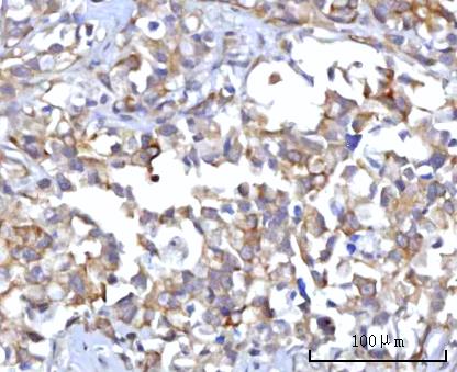
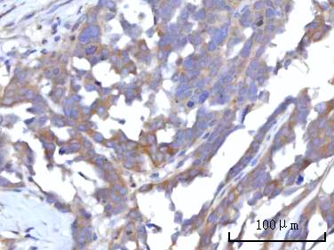
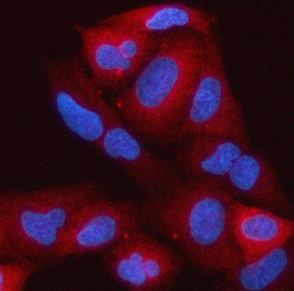


Comprehensive Human Protein Atlas IHC scoring per tissue (reliability: Enhanced — Medium consistency between antibody staining and RNA expression data.). Rows are taken directly from the HPA tissue chart — click any row's HPA link to view the source.
| Tissue | Cell type | Level | Evidence | Source |
|---|---|---|---|---|
| Adrenal gland | Glandular cells | High | Protein (IHC) | HPA → |
| Appendix | Goblet cells | High | Protein (IHC) | HPA → |
| Bronchus | Respiratory epithelial cells | High | Protein (IHC) | HPA → |
| Caudate | Neuronal cells | High | Protein (IHC) | HPA → |
| Cerebral cortex | Neuronal cells | High | Protein (IHC) | HPA → |
| Tissue | Cell type | Level | Evidence | Source |
|---|---|---|---|---|
| Adipose tissue | Adipocytes | Not detected | Protein (IHC) | HPA → |
| Bone marrow | Hematopoietic cells | Not detected | Protein (IHC) | HPA → |
| Liver | Cholangiocytes | Not detected | Protein (IHC) | HPA → |
| Lung | Alveolar cells type I | Not detected | Protein (IHC) | HPA → |
| Oral mucosa | Squamous epithelial cells | Not detected | Protein (IHC) | HPA → |
Troubleshoot RAP1GAP staining in paraffin sections by checking retrieval, cell identity, subcellular pattern, and assay controls before scoring chromogenic signal.
A03561-2 has paraffin-section IHC images from human carcinomas and mouse and rat brain, plus an IF image from A549 cells (catalog image captions). M03561 lists IHC and IF/ICC applications (catalog applications).
A03561-2 has IHC images from human gastric adenocarcinoma, human ovary serous carcinoma, mouse brain and rat brain paraffin sections, plus an IF image from A549 cells (catalog image captions). M03561 lists IHC and ICC/IF applications and human, mouse and rat reactivity, but provides no IHC or IF image caption (catalog applications, reactivity and image captions).
Which to pick: Choose A03561-2 for paraffin-section IHC: its own captions show staining in human, mouse and rat tissue, and list 2 μg/ml primary antibody with EDTA retrieval at pH 8.0; the fixative is unreported (A03561-2 IHC image captions). Choose A03561-2 for IF/ICC when an image example matters: its own caption shows A549 cells stained at 5 μg/ml (A03561-2 IF image caption). For cross-species work, both SKUs list human, mouse and rat reactivity, but A03561-2 supplies IHC images across all three species; M03561 is a rabbit monoclonal, clone 21R48, with IHC and ICC/IF listed but no image examples supplied (catalog reactivity, clone, applications and image captions).