This website uses cookies to ensure you get the best experience on our website.


- Table of Contents
Plan RAPH1 paraffin IHC around the cytoplasmic staining reported in most tissues (HPA tissue IHC). Compare documented positive and negative cell populations, and interpret intensity with the reported low consistency between staining and RNA expression in mind (HPA tissue IHC).

Expected localisation, validated protocols, controls and antibodies — the at-a-glance facts below, then the full design guide.
| Expected localisation | Cytoplasmic staining in most tissues (HPA tissue IHC) | |
| Staining pattern | Cytoplasmic signal across many tissue cell types (HPA tissue IHC) | |
| Antigen retrieval | EDTA pH 8.0 HIER, heat-mediated (datasheet A06130-1) | |
| Positive control | Appendix+4 more · see all | |
| Negative control | Adipose tissue+4 more · see all |
| Fixation | Keep fixation consistent across sections (standard IHC practice; not target-specific); Matched tissue-IHC evidence does not establish the fixation claim. Validate the specimen-specific method before use. (selected-SKU IHC image A06130-1) | |
| Caveat | Staining has low consistency with RNA expression (HPA tissue IHC) | |
| Regulation | RMO1 expression is highest in liver (UniProt) | |
| Isoform / epitope | 9 isoforms; check epitope coverage across variants (UniProt) |
The catalog antibody uses EDTA pH 8.0 heat retrieval (datasheet A06130-1). One published RAPH1 IHC protocol provides staining conditions for wounded skin sections (PMC3542609).
| Sample | Paraffin-embedded human breast cancer tissue; fixative not specified (datasheet A06130-1) |
| Fixation | Image fixative and duration unreported (datasheet A06130-1); verify before use. |
| Sectioning | 4–5 µm sections on charged slides (standard) |
| Deparaffinisation | Xylene, graded ethanol series to water (standard) |
| Antigen retrieval | Heat retrieval: EDTA pH 8.0 (datasheet A06130-1); 20 min, 95–100 °C (standard) |
| Peroxidase block | 3% H2O2, 10 min, room temperature (standard) |
| Blocking | 10% goat serum (datasheet A06130-1) |
| Primary antibody | Rabbit anti-RAPH1, 2-5 μg/ml (datasheet A06130-1) |
| Primary incubation | Overnight at 4 °C (datasheet A06130-1) |
| Detection | HRP-conjugated secondary, DAB chromogen (datasheet A06130-1) |
| Counterstain | Hematoxylin, blue, dehydrate and mount (standard) |
| Expected result | RAPH1-positive staining in glandular cells of appendix (HPA tissue IHC: Medium). HPA tissue profile: Cytoplasmic expression in most tissues. No signal in the no-primary control. |
RAPH1 is a membrane-associated, cytoskeletal protein found at lamellipodia and filopodia (UniProt Q70E73; no transmembrane segment). In paraffin-section IHC, expect mainly cytoplasmic staining in many tissues (HPA tissue IHC). Examples with medium staining include appendix and breast glandular cells and cerebellar Purkinje cells (HPA tissue IHC). HPA rates its tissue IHC as Approved but reports low consistency between antibody staining and RNA expression (HPA tissue IHC).
| Cytoplasmic staining in appendix or breast glandular cells, or cerebellar Purkinje cells. | This matches cell populations scored at medium intensity by HPA (HPA tissue IHC). Compare intensity within the same cell type and staining run; a positive result need not fill every cell uniformly (general IHC practice). |
| Staining near a cell edge, alongside cytoplasmic staining. | A peripheral component is plausible: RAPH1 is recruited to membranes and localizes to lamellipodia and filopodia (UniProt Q70E73). In tissue sections, cell projections may be difficult to resolve, so edge staining alone is weak evidence of specificity (general IHC practice). |
| Strong, predominantly nuclear staining with little cytoplasmic or peripheral signal. | Question the IHC result because HPA describes tissue staining as cytoplasmic (HPA tissue IHC). Nuclear bodies appear in HPA ICC-IF data, but that location is marked uncertain (HPA subcellular). Check controls before calling a nuclear IHC pattern specific. |
| Strong staining in a cell type HPA lists as not detected, such as adipocytes. | The result conflicts with the listed tissue observation (HPA tissue IHC: adipocytes not detected). Possible explanations include cross-reactivity or endogenous detection activity (general IHC practice). Check morphology, the no-primary control, and a second antibody before assigning RAPH1. |
| Diffuse color across cells and extracellular space, or no signal in an expected-positive tissue. | Diffuse color suggests background rather than the reported cell-associated pattern (HPA tissue IHC; general IHC practice). No signal in appendix glandular cells may reflect an assay failure, but HPA's Approved rating and low RNA concordance limit how definitive one sample can be (HPA tissue IHC). |
| Tissue and cell-type selection | Appendix and breast glandular cells and cerebellar Purkinje cells are medium-staining examples; adipocytes and heart cardiomyocytes are listed as not detected (HPA tissue IHC). Score named cell types, not an entire tissue as uniformly positive or negative. |
| Evidence strength | HPA labels tissue IHC Approved while reporting low consistency with RNA expression (HPA tissue IHC). Treat its staining map as a comparison point, and corroborate surprising results with controls or an independent antibody (general IHC practice). |
| Membrane association and topology | RAPH1 reaches the membrane through its PH domain and has no transmembrane segment (UniProt Q70E73). Cytoplasmic staining with a peripheral component is therefore plausible; a continuous cell-surface outline is not required to recognize the reported IHC pattern (HPA tissue IHC). |
| Isoform-dependent expression | UniProt reports nine isoforms; RMO1-RAPH1 is broadly expressed with highest levels in brain, heart, ovary and developing embryo, whereas RMO1 is highest in liver (UniProt Q70E73). Those expression summaries do not override HPA's cell-level IHC observations, including cardiomyocytes listed as not detected (HPA tissue IHC). |
| IF/ICC: What localization should I expect? | HPA reports supported plasma-membrane localization, with nuclear bodies and cytosol marked uncertain in ICC-IF (HPA subcellular). UniProt also places RAPH1 at cell projections and the cytoskeleton (UniProt Q70E73). Interpret the uncertain IF locations separately from the predominantly cytoplasmic tissue IHC pattern (HPA tissue IHC). |
| Situation | Likely cause | Next action |
|---|---|---|
| No color in appendix glandular cells. | A positive cell population is missing from this run (HPA tissue IHC: medium in appendix glandular cells); the assay may have failed (general IHC practice). | Check tissue preservation, retrieval and detection controls, then repeat with an IHC-validated antibody (general IHC practice). Do not infer target-specific fixation sensitivity from these sources. |
| Color covers the section without cell boundaries. | Diffuse chromogen can reflect nonspecific binding or detection background (general IHC practice); HPA reports a cytoplasmic cellular pattern (HPA tissue IHC). | Inspect the no-primary control and review blocking, washes and detection reagent exposure (general IHC practice). |
| Unexpected cells stain strongly while the expected cell type does not. | Cross-reactivity or endogenous detection activity is possible (general IHC practice); compare the exact cell types with HPA's tissue entries (HPA tissue IHC). | Check morphology and a no-primary control; seek agreement with an independent antibody before assigning RAPH1 (general IHC practice). |
| Only nuclei stain strongly. | Nuclear-dominant tissue IHC conflicts with HPA's cytoplasmic summary (HPA tissue IHC); nuclear bodies in ICC-IF are marked uncertain (HPA subcellular). | Review counterstain versus chromogen and compare controls and an independent antibody (general IHC practice). |
| Brown color appears in a no-primary control. | The signal can arise from endogenous enzyme activity or the detection system without primary antibody (general IHC practice). | Review the appropriate endogenous-activity block and detection reagents, then repeat the control alongside the sample (general IHC practice). |
| A heart section appears negative despite reported high expression. | UniProt reports high expression of one isoform in heart, while HPA lists cardiomyocytes as not detected by tissue IHC (UniProt Q70E73; HPA tissue IHC). | Use a documented medium-staining cell population to check assay performance before interpreting the heart result (HPA tissue IHC; general IHC practice). |
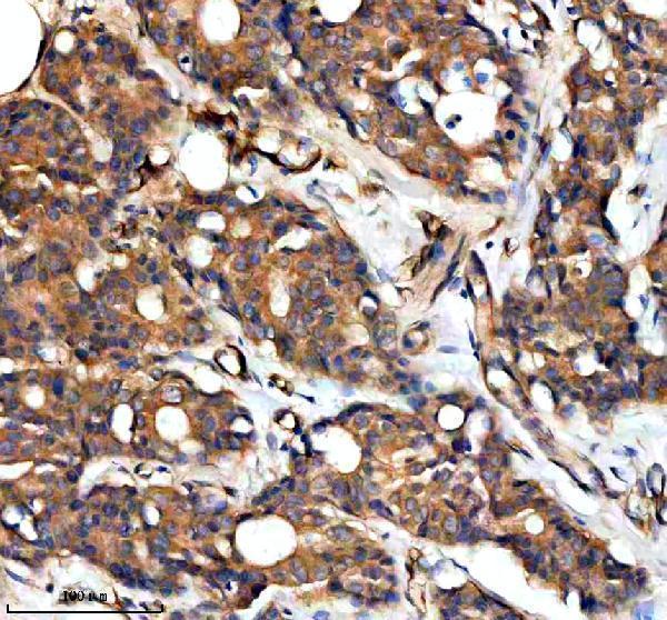
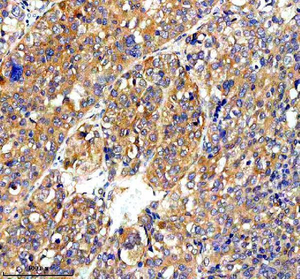


Comprehensive Human Protein Atlas IHC scoring per tissue (reliability: Approved — Low consistency between antibody staining and RNA expression data.). Rows are taken directly from the HPA tissue chart — click any row's HPA link to view the source.
| Tissue | Cell type | Level | Evidence | Source |
|---|---|---|---|---|
| Appendix | Glandular cells | Medium | Protein (IHC) | HPA → |
| Breast | Glandular cells | Medium | Protein (IHC) | HPA → |
| Cerebellum | Purkinje cells | Medium | Protein (IHC) | HPA → |
| Cerebral cortex | Endothelial cells | Medium | Protein (IHC) | HPA → |
| Colon | Endothelial cells | Medium | Protein (IHC) | HPA → |
| Tissue | Cell type | Level | Evidence | Source |
|---|---|---|---|---|
| Adipose tissue | Adipocytes | Not detected | Protein (IHC) | HPA → |
| Bronchus | Respiratory epithelial cells | Not detected | Protein (IHC) | HPA → |
| Caudate | Glial cells | Not detected | Protein (IHC) | HPA → |
| Esophagus | Squamous epithelial cells | Not detected | Protein (IHC) | HPA → |
| Fallopian tube | Glandular cells | Not detected | Protein (IHC) | HPA → |
These checks help distinguish RAPH1 staining from section preparation, detection, and interpretation artefacts in chromogenic IHC.
Real RAPH1 IHC images show human paraffin sections (A06130-1 image captions). The catalog lists human and mouse reactivity, but provides no IF/ICC images (A06130-1 catalog).
A06130-1 will render with its human breast cancer paraffin-section IHC image (A06130-1 figure caption). Its catalog also documents human liver cancer paraffin-section IHC and lists human and mouse reactivity (A06130-1 image captions; A06130-1 catalog).
Which to pick: Choose A06130-1 for human paraffin-section IHC; its image captions document EDTA retrieval at pH 8.0 and primary antibody at 2 μg/ml, while the catalog recommends 2–5 μg/ml for human IHC (A06130-1 image captions; A06130-1 catalog). The captions do not report the fixative (A06130-1 image captions). No IF/ICC option is validated here, and although A06130-1 lists mouse reactivity, its documented IHC examples are human, so mouse tissue IHC would require validation (A06130-1 applications/reactivity; A06130-1 image captions).